ORIGINAL RESEARCH
FUNCTIONAL
Different Functional and Microstructural Changes Depending
on Duration of Mild Cognitive Impairment in Parkinson Disease
XN.-Y. Shin, X Y.S. Shin, XP.H. Lee, XU. Yoon, XS. Han, XD.J. Kim, and XS.-K. Lee
ABSTRACT
BACKGROUND AND PURPOSE: The higher cortical burden of Lewy body and Alzheimer disease–type pathology has been reported to be associated with a faster onset of cognitive impairment of Parkinson disease. So far, there has been a few studies only about the changes of gray matter volume depending on duration of cognitive impairment in Parkinson disease. Therefore, our aim was to evaluate the different patterns of structural and functional changes in Parkinson disease with mild cognitive impairment according to the duration of parkinsonism before mild cognitive impairment.
MATERIALS AND METHODS: Fifty-nine patients with Parkinson disease with mild cognitive impairment were classified into 2 groups on the basis of shorter (⬍1 year,n⫽16) and longer (ⱖ1 year,n⫽43) durations of parkinsonism before mild cognitive impairment. Fifteen drug-naïve patients with de novo Parkinson disease with intact cognition were included for comparison. Cortical thickness, Tract-Based Spatial Statistics, and seed-based resting-state functional connectivity analyses were performed. Age, sex, years of education, age at onset of parkinsonism, and levodopa-equivalent dose were included as covariates.
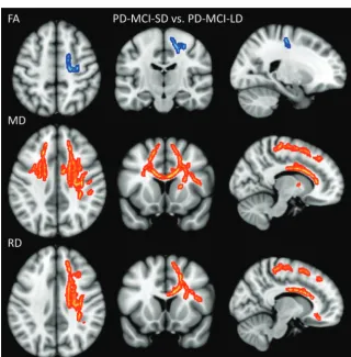
RESULTS:The group with shorter duration of parkinsonism before mild cognitive impairment showed decreased fractional anisotropy and increased mean and radial diffusivity values in the frontal areas compared with the group with longer duration of parkinsonism before mild cognitive impairment (correctedP⬍.05). The group with shorter duration of parkinsonism before mild cognitive impairment showed decreased resting-state functional connectivity in the default mode network area when the left or right posterior cingulate was used as a seed, and in the dorsolateral prefrontal areas when the left or right caudate was used as a seed (correctedP⬍.05). The group with longer duration of parkinsonism before mild cognitive impairment showed decreased resting-state functional connectivity mainly in the medial prefrontal cortex when the left or right posterior cingulate was used as a seed, and in the parieto-occipital areas when the left or right caudate was used as a seed (correctedP⬍.05). No differences in cortical thickness were found in all group contrasts.
CONCLUSIONS: Resting-state functional connectivity and WM alterations might be useful imaging biomarkers for identifying changes in patients with Parkinson disease with mild cognitive impairment according to the duration of parkinsonism before mild cognitive impair-ment. The functional and microstructural substrates may topographically differ depending on the rate of cognitive decline in these patients.
ABBREVIATIONS:AD⫽Alzheimer disease; DMN⫽default mode network; MCI⫽mild cognitive impairment; PCC⫽posterior cingulate cortex; PD⫽Parkinson disease; PD-IC⫽PD with intact cognition; PD-MCI⫽PD with mild cognitive impairment; PD-MCI-LD⫽PD-MCI withⱖ1 year of parkinsonism prior to MCI; PD-MCI-SD⫽ PD-MCI with⬍1 year of parkinsonism prior to MCI; RSFC⫽resting-state functional connectivity
P
arkinson disease (PD) has been considered, until recently, primarily a motor disorder. It is now recognized that a substantial portion of patients with PD have measurable cog-nitive deficits ranging from mild cogcog-nitive impairment(PD-MCI) to dementia.1,2Although the exact pathologic substrates for cognitive impairment in PD are still under debate, limbic and cortical Lewy body– and Alzheimer disease (AD)–type pathology have been suggested as the main contributors to
Received July 20, 2015; accepted after revision October 26.
From the Department of Radiology (N.-Y.S.), Ewha Womans University School of Medicine, Seoul, Korea; Department of Psychology (Y.S.S., S.H.), Yonsei University, Seoul, Korea; Departments of Neurology (P.H.L.) and Radiology (N.-Y.S., D.J.K., S.-K.L.), Yonsei University College of Medicine, Seoul, Korea; and Department of Biomedical Engineering (U.Y.), College of Health and Medical Science, Catholic University of Daegu, Gyeongsan-si, South Korea.
Please address correspondence to Seung-Koo Lee, MD, PhD, Department of Radi-ology, Yonsei University College of Medicine, Integrated Neurocognitive Func-tional Imaging Center, Yonsei University, Seoul, Korea, 50 –1 Yonsei-ro, Seodae-mun-gu, Seoul 120 –752, Korea; e-mail: [email protected]
Indicates article with supplemental on-line appendix and tables.
Indicates article with supplemental on-line photos.
PD-MCI3-6as well as PD with dementia.7-9In terms of the rate of cognitive decline, a higher burden of these cortical pathol-ogies9,10 has been reported associated with a faster onset of cognitive impairment in PD.
In contrast to pathologic studies, imaging studies are nonin-vasive and useful for discovering biomarkers in living humans. However, only a few structural imaging studies11,12have been conducted thus far to define anatomic candidates influencing the rate of cognitive decline in PD. These studies have revealed atro-phy of the posterior cingulate cortex (PCC)11and inferior parietal and orbitofrontal areas12in patients with PD with shorter dura-tions of parkinsonism before dementia and MCI, compared with those with longer durations of parkinsonism. These regions show considerable overlap with the default mode network (DMN), which is well-known to be disrupted in patients with AD.13Some authors have suggested that impairment of axonal transport causes accumulation of axonally transported substances followed by cortical Lewy body formation.14,15In other words, alterations in WM, such as swelling and degeneration of the axonal projec-tions, may precede cortical atrophy. Functional imaging is a more sensitive biomarker that detects earlier stages of disease than that seen structurally for both AD and PD.16-18However, there has been no study on the changes in WM integrity or resting-state functional connectivity (RSFC) according to the duration of par-kinsonisim before cognitive impairment in PD.
Therefore, we aimed to determine the structural and func-tional changes in patients with PD-MCI according to the duration of parkinsonism before MCI. During the resting-state fMRI anal-ysis, we particularly focused on the DMN, which is highly associ-ated with cognitive dysfunction in patients with AD13and other neurodegenerative disorders.19 Furthermore, we also analyzed the corticostriatal loop, which is considered one of the primary areas of cognitive dysfunction in patients with PD.17
MATERIALS AND METHODS
SubjectsThis retrospective study was approved by the Yosei University Heath System institutional review board, and a waiver of in-formed consent was obtained. The patients were selected from a prospectively collected single-institution movement disorders and dementia outpatient clinic data base. From August 2011 to February 2014, consecutive patients with PD who underwent both MR imaging and neuropsychological tests within a 2-month interval were recruited. PD was diagnosed according to the clini-cal diagnostic criteria of the United Kingdom Parkinson’s Disease Society Brain Bank.20
Assessment of parkinsonian motor symptoms was performed by using the Unified Parkinson’s Disease Rating Scale, Part III. Total medication dosages for PD were calculated in levodopa equivalents.21 The self-rating Beck Depression Inventory was used to assess depressive symptoms in patients with PD.22 Pa-tients with focal brain lesions, diffuse white matter hyperintensi-ties outside the normal range, or multiple lacunar infarcts in the basal ganglia on MR imaging were excluded. Patients with other neurodegenerative diseases and medical comorbidities that might account for cognitive dysfunction were also excluded. Only pa-tients who displayed decreased dopamine transporter uptake in
the posterior putamen on a [18F] N-3-fluoropropyl-2- -carbo-methoxy-3--(4-iodophenyl) nortropane (FP-CIT) PET scan were included in this study, to ensure clinical diagnostic accuracy.
Information about memory problems or other subjective cognitive deficits was gathered by interviews with the patients or caregivers. The cognitive status diagnosis in patients with PD was assigned by consensus among 2 neurologists and 1 neuropsychologist on the basis of a neuropsychological battery and the physician-administered neurologic examination. The Seoul Neuropsychological Screening Battery was used to deter-mine the cognitive status,23 and its details are described in On-line Appendix 1. According to the diagnostic criteria rec-ommended by the Movement Disorder Society Task Force,24 PD-MCI was diagnosed when results of at least 2 tests for each of the attention, executive, memory, and visuospatial function domains, except for the language domain (level 2), or in 5 domains (level 1) were abnormal. Patients with PD-MCI were split into 2 groups by disease duration before MCI: PD-MCI with⬍1 year of parkinsonism before MCI (PD-MCI-SD) and PD-MCI withⱖ1 year of parkinsonism before MCI (PD-MCI-LD). Drug-naïve patients with de novo PD with intact cogni-tion (PD-IC) were also included as control group.
Image Acquisition
All scans were acquired by using a 3T scanner (Achieva; Philips Healthcare, Best, the Netherlands) with a 32-channel head coil. Head motion was minimized with restraining foam pads pro-vided by the manufacturer.
Structural Image Acquisition. We used a 3D-T1-turbo field echo sequence with the following parameters: axial acquisition with FOV⫽220 mm; voxel size⫽0.98⫻0.98⫻1.2 mm3; TE⫽4.6 ms; TR⫽9.6 ms; flip angle⫽8°; section gap⫽0 mm; and total acquisition time⫽5 minutes 29.3 seconds.
DTI Acquisition. A single-shot EPI acquisition was performed with the following parameters: FOV⫽220 mm; voxel size ⫽ 1.72⫻1.72⫻2 mm3; TE⫽ ⬃70 ms; TR⫽ ⬃8000 ms; flip angle⫽90°; section gap⫽0 mm; NEX⫽1; b factor⫽600 s/mm2; noncardiac gating; and 70 axial sections. We acquired diffusion-weighted images from 32 noncollinear, noncoplanar directions with a baseline image without diffusion-weighting. Total acquisi-tion time was 5 minutes 44.6 seconds.
Resting-State fMRI Acquisition. We used a T2*-weighted single-shot EPI sequence. For each subject, 165 axial volume scans were obtained with the following parameters: FOV⫽220 mm2; voxel size⫽2.75⫻2.75⫻4.5 mm3; TE⫽30 ms; TR⫽2000 ms; and section number⫽31 (interleaved). During each scan, subjects were instructed to rest and keep their eyes closed without moving, sleeping, or thinking about anything in particular for 5 minutes 38 seconds.
Image Analysis
thickness are described in On-line Appendix 2. The localized re-gional differences of cortical thickness among groups were ana-lyzed by applying ANCOVA, with age, sex, years of education, age at onset of parkinsonism, and levodopa-equivalent dose entered as covariates.
Tract-Based Spatial Statistics Analysis. DTI data preprocessing was performed by using the FMRIB Software Library (FSL; http:// www.fmrib.ox.ac.uk/fsl) program. Details for Tract-Based Spatial Statistics (http://fsl.fmrib.ox.ac.uk/fsl/fslwiki/TBSS) analysis are described in On-line Appendix 3. In the ANCOVA analysis, age, sex, years of education, age at onset of parkinsonism, and levodopa-equivalent dose were included as covariates.
Seed-Based RSFC Analysis. Data were first preprocessed accord-ing to the Data Processaccord-ing Assistant for Restaccord-ing-State fMRI toolbox (http://www.restfmri.net) preprocessing pipeline implemented in Matlab (MathWorks, Natick, Massachusetts). Images were cor-rected for section timing, realigned, normalized by using the EPI template provided by SPM8 software (http://www.fil.ion. ucl.ac.uk/spm/software/spm8), and smoothed by using a 4-mm full width at half maximum Gaussian kernel. After normalization, to remove long-term drift and irrelevant oscillations in the signal, we detrended and bandpass filtered (0.01– 0.08 Hz) data. Nui-sance covariates, including head-motion parameters, global mean signals, WM signals, and CSF signals, were regressed out.
To perform seed-based analysis, an Automated Anatomical Labeling template (http://www.gin.cnrs.fr/AAL) defined 90 seed ROIs. Among them, 4 ROI seeds were selected to study the asso-ciation of cognition and resting-state functional networks in pa-tients with PD. First, the bilateral PCC seeds were chosen to in-vestigate alterations within the DMN. Second, the bilateral caudate seeds, which are known to be key subcortical structures in the cognitive corticostriatal loop, were also selected. RSFC from each ROI seed was estimated and used to configure a statistical map. Two samplet tests were performed on each pair of the group’s statistical images by using the SPM8 toolbox. The as-sumptions of unequal variance and independence among all groups were made onttests. To exclude possible confounding factors, we covariated out age, sex, years of education, age at onset of parkinsonism, and levodopa-equivalent doses in the statistical test after normalization across 2 groups of interest by using thez score function in Matlab. The threshold for statistical analysis was set to correctedP⬍.05 by using the Monte Carlo simulations with custom software implemented in Matlab.25
Correlation Analysis
Two-tailed Pearson correlation analyses were performed to assess the relationship between the duration of parkinsonism before MCI and RSFCs, which showed remarkable differences between the PD-MCI-SD and PD-MCI-LD groups (left hippocampus and left medial frontal gyrus with the left PCC seed and left middle frontal gyrus with the left caudate seed). For each ROI,zvalues were extracted from correlation maps with a 4-mm radius sphere centered at the peak. Then, the correlation coefficients between thezvalues and the patients’ duration of parkinsonism before MCI were computed.
Statistical Analysis
Clinical characteristics and neuropsychological data were com-pared among the 3 groups. The Kolmogorov-Smirnov test was used to determine whether the data were normally distributed. Accordingly, data that had normal distribution are presented as means⫾SDs, and quantitative variables were compared by using an ANOVA. Otherwise, for comparing quantitative values, data are presented as medians with ranges and the Kruskal-Wallis test was used. Qualitative data were analyzed by using the2test or Fisher exact test when appropriate. Post hoc analysis was also performed by using a Bonferroni-corrected Studentttest, Mann-WhitneyUtest,2test, or Fisher exact test when appropriate with correction for multiple comparisons. Statistical analyses were performed by using SPSS, Version 19.0 (IBM, Armonk, New York), and 2-tailedP⬍.05 was considered significant.
RESULTS
Demographic and Clinical Characteristics
Among 239 patients with PD who underwent both MR imag-ing and neuropsychological tests, 59 patients with PD-MCI who met the inclusion criteria were analyzed in this study. Fifteen drug-naïve patients with de novo PD with intact cog-nition were also included for comparison. The demographic and clinical data of the patients are summarized inTable 1. The median duration of parkinsonism in the PD-MCI-SD group (n ⫽ 16) was 5 months and it was 25 months in the PD-MCI-LD group (n⫽43). Patients in the PD-MCI-SD group had significantly older age at onset than those in the PD-MCI-LD group (68.5⫾7.3 years versus 61.6⫾9.0 years;P⫽ .016). No significant differences were found in neuropsycho-logical data between the 2 PD-MCI groups (On-line Table 1).
Group Comparisons of Cortical Thickness
No difference in cortical thickness was found among all groups.
Group Comparisons of WM Alterations
Group Comparisons of RSFC by Using the PCC Seeds Compared with the de novo PD-IC group, the PD-MCI-SD group showed decreased RSFC in the parahippocampal gyrus, dorsolateral prefrontal areas, temporal areas, and precuneus, whereas increased RSFC was seen in the inferior frontal areas, primary motor area, and
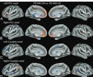
occipital areas. The PD-MCI-LD group showed decreased RSFC in the medial frontal areas and middle cingulate cortex, while increased RSFC was seen mainly in the parietal and occipital areas, compared with the de novo PD-IC group. In direct comparison between the PD-MCI-SD and MCI-LD groups, the PD-MCI-SD group showed decreased RSFC in the hippocampus, parietal areas, cu-neus, and thalamus; on the contrary, the PD-MCI-LD group showed decreased RSFC in the medial and inferior frontal ar-eas, primary motor area, cingulate cortex, inferior temporal area, and insula (Fig 2A, -B; On-line Fig 2 and On-line Table 2).
Group Comparison of RSFC by Using the Caudate Seeds
Compared with the de novo PD-IC group, the PD-MCI-SD group exhibited decreased RSFC in the putamen, tempo-ral areas, precuneus, and insula and in-creased RSFC in the primary sensory motor areas. On the other hand, the PD-MCI-LD group showed decreased RSFC in the occipital area and thalamus and increased RSFC in the frontal areas, in-cluding the primary and supplementary motor areas, hippocampus/parahip-pocampal gyrus and other temporal ar-eas, and the supramarginal gyri. In di-rect comparison between the PD-MCI-SD and PD-MCI-LD groups, the PD-PD-MCI-SD group showed decreased RSFC in the putamen, globus pallidus, frontal and temporal areas, middle cingulate cortex, and insula, while the PD-MCI-LD group showed decreased RSFC mainly in the
[image:4.594.54.541.57.215.2]pari-FIG 1. Tract-Based Spatial Statistics analysis in the PD-MCI groups. Warm colors indicate in-creased DTI values, and cool colors indicate dein-creased DTI values in the PD-MCI-SD group compared with PD-MCI-LD group (P⬍.05, family-wise error– corrected). Images are oriented according to neurological convention (right is right).
Table 1: Demographic and clinical characteristics of the patientsa
De Novo PD-IC (n= 15)
PD-MCI-SD (n= 16)
PD-MCI-LD
(n= 43) PValueb
Post Hoc Analysis P1c P2d P3e
Age (yr) 65.7⫾6.4 69.1⫾7.2 64.9⫾8.9 .221 – – –
Age at onset (yr) 64.9⫾6.5 68.5⫾7.3 61.6⫾9.0 .017 .672 .555 .016
Male (No.) (%) 6 (40.0) 8 (50.0) 17 (39.5) .604 – – –
Education duration (yr) 10.6⫾4.7 8.8⫾4.2 9.5⫾5.2 .598 – – –
Parkinsonism duration (mo) (median) (range)
11 (2–18) 5 (1–11) 25 (12–120) ⬍.001 .015 ⬍.001 ⬍.001
UPDRS III 19.1⫾8.3 25.4⫾8.8 25.6⫾11.2 .100 – – –
K-MMSE 28.6⫾1.2 26.3⫾1.5 26.9⫾2.1 .002 .003 .009 .899
Levodopa-equivalent dose (mg) (median) (range)
0.0 (0.0–0.0) 0.0 (0.0–360.0) 25.0 (0.0–1050.0) .008 .030 .006 1.000
BDI 11.7⫾7.6 14.1⫾8.7 14.8⫾10.2 .543 – – –
Interval between MRI scan and NP test (day) (median) (range)
0 (0–34) 0 (0–49) 0 (0–50) .266 – – –
Note:—BDI indicates Beck Depression Inventory; K-MMSE, the Korean version of the Mini-Mental State Examination; NP test, neuropsychological test; UPDRS III, Unified Parkinson’s Disease Rating Scale, Part III; –, not significant.
aUnless otherwise indicated, data are means.
b
Pvalues for comparison among 3 groups.
cPvalues for comparison between de novo PD-IC and PD-MCI-SD groups. d
[image:4.594.55.376.287.612.2]etal and occipital areas (Fig 2C, -D; On-line Fig 3 and On-line Table 3).
Correlation Analysis
The RSFC between the left caudate and left middle frontal gyrus (Montreal Neurological Institute coordinates [⫺36, 33, 33]) was sig-nificantly correlated with the duration of parkinsonism before MCI (r⫽0.292,P⫽.025). RSFCs between the left PCC and left hip-pocampus (Montreal Neurological Institute coordinates [⫺24,⫺36,
⫺3]) and the left PCC and left medial frontal gyrus (Montreal Neu-rological Institute coordinates [⫺6, 54, 24]) were not significantly correlated with the duration of parkinsonism before MCI (Fig 3).
DISCUSSION
The present study examined the different patterns of structural and functional changes in patients with PD-MCI according to the
duration of parkinsonism before MCI. First, the PD-MCI-SD group showed decreased frontostriatal RSFC, which was corre-lated with the duration of parkinsonism before MCI, and im-paired frontal WM integrity compared with the PD-MCI-LD group. Second, the pattern of RSFC was topographically different between the 2 groups. The PD-MCI-SD group showed decreased RSFC in the DMN, while the PD-MCI-LD group showed de-creased RSFC in the medial frontal areas with the PCC seeds. Third, no difference was found in cortical thickness among all group contrasts.
As mentioned earlier, the PD-MCI-SD group showed not only decreased frontostriatal RSFC but also decreased fractional an-isotropy values and increased mean diffusivity and radial diffusiv-ity values in the frontal WM compared with the PD-MCI-LD group. These functional and microstructural differences might be attributable to both striatal26,27and frontal28,29pathologies. Re-cently, some pathologic studies have suggested striatal-amyloid26or␣ -sy-nuclein27deposits as a primary substrate for subcortical dementia in PD. More-over, a higher degree of amyloid deposits in the striatum was found in patients with dementia with Lewy bodies than in those with PD with dementia, suggest-ing the possible role of amyloid deposi-tion in the acceleradeposi-tion of cognitive de-cline in ␣-synuclein-related cognitive disorders.30A previous imaging study also showed atrophy of the caudate nu-cleus and frontal cortex in patients with PD-MCI who subsequently converted to PD with dementia compared with those without conversion to PD with demen-tia.31Furthermore, our results showed that frontostriatal RSFC had significant correlation with the duration of parkin-sonism before MCI. In other words, pa-tients with more disrupted RSFC among these areas had MCI with a shorter inter-val after the onset of motor symptoms. Although the effort to determine which substrate has a primary role in
accelerat-FIG 2. RSFC analysis in the PD-MCI groups by using the PCC and caudate as a seed. Warm colors indicate increased connectivity, and cool colors indicate decreased connectivity in PD-MCI-SD group compared with PD-MCI-LD group. All demonstrated clusters are significant at aP⬍.05 level, with correction for multiple comparisons.
[image:5.594.56.375.247.511.2] [image:5.594.55.532.567.703.2]ing cognitive decline should be continued, functional or micro-structural alterations in the frontostriatal circuit may be a useful imaging biomarker for more rapid cognitive decline in patients with PD-MCI.
Compared with the PD-MCI-LD group, the PD-MCI-SD group also showed decreased RSFC in the DMN, including the hippocampus and inferior parietal lobule when PCC was used as a seed. Our results are in line with those in previous reports. Ac-cording to previous imaging studies, patients with a shorter du-ration of parkinsonism before cognitive impairment in PD with dementia and PD-MCI had decreased GM volume in the PCC11 and in the inferior parietal and orbitofrontal areas,12respectively. These areas substantially overlap with the DMN, which is suscep-tible to decline in patients with AD.13Moreover, a recent study reported AD pattern atrophy involving the hippocampus and temporal-parietal cortex as a predictor for 2-year future cognitive decline in PD,32suggesting that an AD imaging pattern is an im-portant predictor for more rapid cognitive decline. Decreased RSFC between the PCC and the hippocampus has been observed in early AD and MCI.13,33In AD, tauopathy involving the medial temporal lobe, including the hippocampus, is considered a possi-ble cause of the decreased connectivity.34Although this finding is less clear in PD,17,18pathologic evidence has suggested that the degree of Lewy body,35Lewy neurite,36-amyloid, or neurofibril-lary tangle deposit37in the entorhinal cortex or hippocampus is associated with cognitive dysfunction.
Most interesting, the PD-MCI-LD group had decreased RSFC primarily in the medial frontal areas and cingulate when using the PCC seeds compared with the PD-MCI-SD group. Braak et al38 reported sequential topographic extension of Lewy neurite and Lewy body deposits in PD, with these areas showing substantial overlap with the affected areas in stage 5, which is the first stage of neocortical involvement. One pathologic study39 found that among the patients who remained without dementia for a long time, the patients who had longer disease durations until death had more extensive Lewy body deposits, consistent with the PD stage of Braak et al. These patients had no or a low AD pathology burden. Therefore, decreased RSFC in these areas might be asso-ciated with cortical PD pathology accumulated for longer disease durations in the PD-MCI-LD group relative to the PD-MCI-SD group. In addition, when the caudate seeds were used, the PD-MCI-LD group showed decreased RSFC in mainly parieto-occip-ital areas compared with the PD-MCI-SD group. These areas, which are cognitive- and action-specific domains, have functional connection with the caudate tail, while the caudate head has a stronger functional connection with the prefrontal areas.40 Fur-ther pathologic study is warranted to define different topographic areas involved within the caudate between the 2 groups.
Our study has several limitations. First, our cohort was not pathologically proved to have PD. Instead, we used dopamine transporter imaging to reduce the possibility of misdiagnosis. Sec-ond, the diagnosis of PD-MCI in this study did not fully satisfy the level 2 criteria of the Movement Disorder Society Task Force guideline for some patients.24Therefore, the possibility of pa-tients with false-positive diagnoses in the PD-MCI group cannot be excluded. Third, although only the patients who underwent both MR imaging and neuropsychological tests within a 2-month
interval were included, there were 1- to 50-day intervals in 21 patients in our cohort. While the cognitive state might not change during this relatively short period, this range of intervals might have affected the results. Fourth, because we used seed-based RSFC analysis with only PCC and caudate seeds, the role of the remaining areas for cognitive decline rates could have been missed in patients with PD. Moreover, recent studies have sug-gested that dorsal attention41and frontoparietal networks41,42are associated with cognitive dysfunction in patients with PD-MCI. Therefore, future study with a data-driven approach of the whole brain should be conducted to consolidate our results and find other relevant networks influencing the cognitive decline rate in these patients. Fifth, it is still unclear what RSFC and DTI values exactly represent in the brain. Therefore, caution is needed to interpret our results until underlying pathologic evidence is elu-cidated. Finally, this study is a cross-sectional one, so our findings cannot be applied to predict the rate of development of cognitive decline. A prospective study with regular follow-up with compre-hensive neuropsychological testing is warranted. However, we compared each PD-MCI group with the de novo PD-IC group, and areas showing decreased RSFC in these comparisons were similar to those in direct comparison between the 2 PD-MCI groups. Therefore, we hope that our results can provide an a priori hypothesis for future studies to define imaging biomarkers for the cognitive decline rate in drug-naïve patients with de novo PD-IC.
CONCLUSIONS
Our results indicate that changes in RSFC and WM integrity in PD-MCI according to the duration of parkinsonism before MCI are more sensitive imaging biomarkers than cortical thickness. We also found topographically different functional and micro-structural substrates corresponding to the timing of MCI in PD-MCI.
REFERENCES
1. Emre M, Aarsland D, Brown R, et al.Clinical diagnostic criteria for dementia associated with Parkinson’s disease.Mov Disord2007;22: 1689 –707; quiz 837Medline
2. Litvan I, Aarsland D, Adler CH, et al.MDS Task Force on mild cog-nitive impairment in Parkinson’s disease: critical review of PD-MCI.Mov Disord2011;26:1814 –24CrossRef Medline
3. Adler CH, Caviness JN, Sabbagh MN, et al.Heterogeneous neuro-pathological findings in Parkinson’s disease with mild cognitive impairment.Acta Neuropathol2010;120:827–28CrossRef Medline
4. Jellinger KA.Neuropathology in Parkinson’s disease with mild cog-nitive impairment.Acta Neuropathol2010;120:829 –30; author reply 31CrossRef Medline
5. Alves G, Brønnick K, Aarsland D, et al.CSF amyloid-beta and tau proteins, and cognitive performance, in early and untreated Par-kinson’s disease: the Norwegian ParkWest study.J Neurol Neuro-surg Psychiatry2010;81:1080 – 86CrossRef Medline
6. Montine TJ, Shi M, Quinn JF, et al.CSF(42) and tau in Parkinson’s disease with cognitive impairment.Mov Disord2010;25:2682– 85
CrossRef Medline
7. Compta Y, Pereira JB, Rios J, et al.Combined dementia-risk bio-markers in Parkinson’s disease: a prospective longitudinal study.
Parkinsonism Relat Disord2013;19:717–24CrossRef Medline
9. Compta Y, Parkkinen L, O’Sullivan SS, et al.Lewy- and Alzheimer-type pathologies in Parkinson’s disease dementia: which is more important?Brain2011;134:1493–505CrossRef Medline
10. Ballard C, Ziabreva I, Perry R, et al.Differences in neuropathologic characteristics across the Lewy body dementia spectrum.Neurology 2006;67:1931–34CrossRef Medline
11. Song SK, Lee JE, Park HJ, et al.The pattern of cortical atrophy in patients with Parkinson’s disease according to cognitive status.
Mov Disord2011;26:289 –96CrossRef Medline
12. Lee JE, Cho KH, Kim M, et al.The pattern of cortical atrophy in Parkinson’s disease with mild cognitive impairment according to the timing of cognitive dysfunction. J Neurol 2012;259:469 –73
CrossRef Medline
13. Greicius MD, Srivastava G, Reiss AL, et al.Default-mode network activity distinguishes Alzheimer’s disease from healthy aging: evi-dence from functional MRI.Proc Natl Acad Sci U S A2004;101: 4637– 42CrossRef Medline
14. Katsuse O, Iseki E, Marui W, et al.Developmental stages of cortical Lewy bodies and their relation to axonal transport blockage in brains of patients with dementia with Lewy bodies.J Neurol Sci 2003;211:29 –35CrossRef Medline
15. Bellucci A, Zaltieri M, Navarria L, et al.From alpha-synuclein to synaptic dysfunctions: new insights into the pathophysiology of Parkinson’s disease.Brain Res2012;1476:183–202CrossRef Medline
16. Frisoni GB, Fox NC, Jack CR Jr, et al.The clinical use of structural MRI in Alzheimer disease.Nat Rev Neurol2010;6:67–77CrossRef Medline
17. Seibert TM, Murphy EA, Kaestner EJ, et al.Interregional correla-tions in Parkinson disease and Parkinson-related dementia with resting functional MR imaging. Radiology 2012;263:226 –34
CrossRef Medline
18. Tessitore A, Esposito F, Vitale C, et al.Default-mode network con-nectivity in cognitively unimpaired patients with Parkinson dis-ease.Neurology2012;79:2226 –32CrossRef Medline
19. Fox MD, Greicius M.Clinical applications of resting state func-tional connectivity.Front Syst Neurosci2010;4:19CrossRef Medline
20. Hughes AJ, Daniel SE, Kilford L, et al.Accuracy of clinical diagnosis of idiopathic Parkinson’s disease: a clinico-pathological study of 100 cases.J Neurol Neurosurg Psychiatry1992;55:181– 84CrossRef Medline
21. Tomlinson CL, Stowe R, Patel S, et al.Systematic review of levodopa dose equivalency reporting in Parkinson’s disease. Mov Disord 2010;25:2649 –53CrossRef Medline
22. Beck AT, Ward CH, Mendelson M, et al.An inventory for measuring depression.Arch Gen Psychiatry1961;4:561–71CrossRef Medline
23. Kang Y, Na DL.Seoul Neuropsychological Screening Battery.Incheon: Human Brain Research & Consulting Co; 2003
24. Litvan I, Goldman JG, Troster AI, et al.Diagnostic criteria for mild cognitive impairment in Parkinson’s disease: Movement Disorder Society Task Force guidelines.Mov Disord2012;27:349 –56CrossRef Medline
25. Slotnick SD, Moo LR, Segal JB, et al.Distinct prefrontal cortex ac-tivity associated with item memory and source memory for visual shapes.Brain Res Cogn Brain Res2003;17:75– 82CrossRef Medline
26. Kalaitzakis ME, Graeber MB, Gentleman SM, et al.Striatal
beta-am-yloid deposition in Parkinson disease with dementia.J Neuropathol Exp Neurol2008;67:155– 61CrossRef Medline
27. Tsuboi Y, Uchikado H, Dickson DW.Neuropathology of Parkin-son’s disease dementia and dementia with Lewy bodies with refer-ence to striatal pathology.Parkinsonism Relat Disord2007;13(suppl 3):S221–24CrossRef Medline
28. Mattila PM, Rinne JO, Helenius H, et al. Alpha-synuclein-immuno-reactive cortical Lewy bodies are associated with cognitive impair-ment in Parkinson’s disease. Acta Neuropathol2000;100:285–90
CrossRef Medline
29. Mattila PM, Ro¨ytta¨ M, Lo¨nnberg P, et al.Choline acetytransferase activity and striatal dopamine receptors in Parkinson’s disease in relation to cognitive impairment. Acta Neuropathol 2001;102: 160 – 66Medline
30. Jellinger KA, Attems J.Does striatal pathology distinguish Parkin-son disease with dementia and dementia with Lewy bodies?Acta Neuropathol2006;112:253– 60Medline
31. Lee JE, Cho KH, Song SK, et al.Exploratory analysis of neuropsy-chological and neuroanatomical correlates of progressive mild cog-nitive impairment in Parkinson’s disease.J Neurol Neurosurg Psy-chiatry2014;85:7–16CrossRef Medline
32. Weintraub D, Dietz N, Duda JE, et al.Alzheimer’s disease pattern of brain atrophy predicts cognitive decline in Parkinson’s disease.
Brain2012;135:170 – 80CrossRef Medline
33. Zhou Y, Dougherty JH Jr, Hubner KF, et al.Abnormal connectivity in the posterior cingulate and hippocampus in early Alzheimer’s disease and mild cognitive impairment.Alzheimers Dement2008;4: 265–70CrossRef Medline
34. Smith AD. Imaging the progression of Alzheimer pathology through the brain.Proc Natl Acad Sci U S A2002;99:4135–37Medline
35. Ko¨vari E, Gold G, Herrmann FR, et al.Lewy body densities in the entorhinal and anterior cingulate cortex predict cognitive deficits in Parkinson’s disease.Acta Neuropathol2003;106:83– 88Medline
36. Churchyard A, Lees AJ.The relationship between dementia and di-rect involvement of the hippocampus and amygdala in Parkinson’s disease.Neurology1997;49:1570 –76Medline
37. Kalaitzakis ME, Christian LM, Moran LB, et al.Dementia and visual hallucinations associated with limbic pathology in Parkinson’s dis-ease.Parkinsonism Relat Disord2009;15:196 –204CrossRef Medline
38. Braak H, Del Tredici K, Ru¨b U, et al.Staging of brain pathology related to sporadic Parkinson’s disease.Neurobiol Aging2003;24: 197–211Medline
39. Halliday G, Hely M, Reid W, et al.The progression of pathology in longitudinally followed patients with Parkinson’s disease. Acta Neuropathol2008;115:409 –15CrossRef Medline
40. Robinson JL, Laird AR, Glahn DC, et al.The functional connectivity of the human caudate: an application of meta-analytic connectivity modeling with behavioral filtering. Neuroimage 2012;60:117–29
CrossRef Medline
41. Baggio HC, Segura B, Sala-Llonch R, et al.Cognitive impairment and resting-state network connectivity in Parkinson’s disease.Hum Brain Mapp2015;36:199 –212CrossRef Medline