ORIGINAL RESEARCH
INTERVENTIONAL
What Is Meant by “TICI”?
J.E. Fugate, A.M. Klunder, and D.F. Kallmes
ABSTRACT
BACKGROUND: In 2003, Higashida et al proposed the Thrombolysis In Cerebral Infarction scale to evaluate angiographic intracranial flow. Our aim is to review how subsequently published studies define TICI.
MATERIALS AND METHODS: We used the ISI Web of Knowledge and SciVerse Scopus databases to search for “TICI” and “thrombolysis in cerebral infarction” and for articles that cited the original TICI paper from January 2004 through May 2012. Articles were categorized according to their definition of the TICI categories, typically grades 0 – 4, with grade 2 (partial reperfusion) subdivided into 2a and 2b, and rate of contrast entry to the perfused area. In addition, we catalogued the type of redefinitions of TICI subcategory 2 and additions of new categories.
RESULTS:Of 236 articles screened, 74 were included. Eight (11%) explicitly followed the TICI scale as originally defined. Thirty-seven (50%) cited Higashida but did not define their scale. Fifteen (21%) used and explained modified scales. Thirteen (18%) used the term TICI, but did not define the scale and did not cite Higashida. Eighteen (24%) specified a 2a subcategory. Nine defined grade 2a as⬍67% filling, 6 defined it as⬍50%, and 3 did not offer a percentage. Two studies added a 2c subcategory. Fifty-two (70%) used a cutoff level to define “successful reperfusion.” Of these, 65% used TICIⱖ2, 33% used TICIⱖ2b, and 2% used TICI⫽3.
CONCLUSIONS: There is substantial variability in the definition and/or application of the TICI scale in the literature. This variability could considerably impact our understanding of results of revascularization studies.
ABBREVIATIONS:TICI⫽Thrombolysis in Cerebral Infarction; TIMI⫽Thrombolysis in Myocardial Infarction
T
he Thrombolysis in Myocardial Infarction scale is a widely applied, graded response scale for assessment of treatment outcome in the coronary arteries.1In 2003, Higashida et al2pro-posed a seemingly simple modification of the TIMI scale to eval-uate intracranial perfusion assessed in cerebral angiography. This new scale, the Thrombolysis in Cerebral Infarction scale, was in-tended to standardize the grading of angiographic outcomes, par-ticularly for trials of endovascular treatment of acute ischemic stroke (Table 1). As originally described, TICI categories span from no perfusion (grade 0) to complete perfusion (grade 3). The “partial perfusion” category (grade 2) is defined as cases in which contrast passes the obstruction but with rates of entry and wash-out slower than normal and is subdivided into 2 subcategories, 2a
and 2b. Although the TICI scale has achieved fairly rapid accep-tance into the medical literature, the scale was somewhat arbi-trarily created and has not been validated or tested systematically. Although the TICI scale has been criticized because of confus-ing internal inconsistencies,3it is still widely used in the
litera-ture.4 Modifications of the TICI scale have been subsequently
proposed, such as a change in the definition of the 2a subcategory (to indicate⬍50% filling of the vascular territory) or by the ad-dition of a 2c subcategory.5Our current aim is to review published
studies that use the TICI scale to describe how the TICI scale is defined and utilized across studies.
MATERIALS AND METHODS
We performed a search of the medical literature by using the ISI Web of Knowledge and SciVerse Scopus databases. We searched for the terms “TICI” and “thrombolysis in cerebral infarction.” We also used these databases to search for all articles from January 2004 through May 2012 that cited the original TICI paper. In addition, we reviewed the reference list from all identified articles to identify other papers by using graded response scales for cere-Received September 3, 2012; accepted after revision December 13.
From the Division of Critical Care Neurology (J.E.F.) and Department of Radiology (D.F.K.), Mayo Clinic, Rochester, Minnesota; and Minnesota State University (A.M.K.), Mankato, Minnesota.
Please address correspondence to David F. Kallmes, MD, Department of Radiology, Mayo Clinic, 200 1st
St SW, Rochester, MN 55905; e-mail: [email protected]
bral perfusion, whether or not these papers referenced or utilized the original TICI paper.
In the initial description of the TICI scale in 2003, grade 0 indicates no perfusion as evidenced by no antegrade flow beyond the point of arterial occlusion.2Grade 1 is defined as penetration
with minimal perfusion” and applies when the “contrast material passes beyond the area of obstruction but fails to opacify the entire cerebral bed distal to the obstruction.” Grade 2 is broadly defined as partial perfusion, which occurs when the contrast material passes beyond the obstruction, opacifies the distal arterial bed, but the rate of entry of contrast and/or its rate of clearance from the vascular bed are slower than comparable areas not perfused by the previously occluded vessel. The opposite cerebral artery or the arterial bed proximal to the occlusion can be used for com-parison rates. Grade 2 is subdivided into 2a and 2b. A grade of 2a indicates partial filling (less than two-thirds) of the entire vascular territory and 2b indicates complete filling of the expected vascular territory, but with a perceptibly slower filling rate than normal. Finally, grade 3 is defined as complete perfusion and applies when “antegrade flow into the bed distal to the obstruction occurs as promptly as into the obstruction and clearance of the contrast material from the involved bed is as rapid as from an uninvolved other bed of the same vessel or the opposite cerebral artery.”2
With our literature search, we identified a total of 236 articles. We excluded articles that did not relate to the TICI scale (115 articles) and articles that were in languages other than English or that were not accessible in full length (49 articles). We qualita-tively assessed whether the definition of TICI in each article ad-hered to the original definition of the TICI scale and evaluated the articles that were cited when TICI was described. We then accord-ingly categorized the articles into 4 groups: 1) articles that explic-itly stated the scale and followed the original TICI scale com-pletely, 2) articles that did not explicitly define the scale but cited the original TICI paper, 3) articles that defined a modified scale, and 4) articles that used TICI but did not define the scale and did not cite the original TICI paper. We also catalogued the type and number of definitions of subcategory 2a and noted if a subcate-gory of TICI was used as a threshold for “successful revasculariza-tion.” This study was exempt from institutional review board review.
RESULTS
Of 74 total included articles, 8 (11%) followed the original TICI scale completely and explicitly stated the categories.6-13One
arti-cle claimed to have followed the scale completely but did not state the categories.14Thirty-seven (50%) articles did not explicitly
de-fine the scale but still cited the original paper by Higashida et al.15-51Modifications of the TICI scale were used in 15 (20%)
articles.4,5,52-65Of these, 8 cited only the original TICI paper, 4
cited the original TICI paper and other papers, and 3 cited only other papers. Thirteen (18%) articles used TICI but did not define the scale and did not cite the original TICI paper.13,30,56,66-75
These results are depicted in Fig 1.
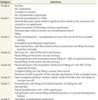
Eighteen (24%) of articles mentioned the rate of contrast fill-ing in their use of TICI. Most modifications of TICI eliminated the subcategories of 2a and 2b. Only 18 (24%) articles specified a 2a subcategory. Of these, 9 defined 2a as filling of⬍67% of the affected vascular territory (compatible with the original TICI) and 6 defined 2a as filling of⬍50% of the affected vascular terri-tory. A 2c subcategory was added in 2 articles, and a category 4 was added in 1 article. Examples of the variability in definitions of TICI categories are detailed in Table 2.
[image:2.594.54.528.54.222.2]Most articles (n⫽52, 70%) defined a threshold within the TICI scale that indicated “successful revascularization” as one of the study end points. Of these, 34 (65%) used TICIⱖ2, 17 (33%) used TICIⱖ2b (although only 1 of these studies defined a precise FIG 1. Distribution of definition and citation of the TICI scale in the literature. Articles in the English literature that use the TICI (Throm-bolysis in Cerebral Infarction) grading scale, distributed according to definition and citation of TICI.
Table 1: The original Thrombolysis in Cerebral Infarction perfusion scale2
Category Title Description
Grade 0 No Perfusion No antegrade flow beyond the point of occlusion.
Grade 1 Penetration with Minimal Perfusion
The contrast material passes beyond the area of obstruction but fails to opacify the entire cerebral bed distal to the obstruction.
Grade 2 Partial Perfusion The contrast material passes beyond the obstruction and opacifies the arterial bed distal to the obstruction. However, the rate of entry of contrast into the vessel distal to the obstruction and/or its rate of clearance from the distal bed are perceptibly slower than its entry into and/or clearance from comparable areas not perfused by the previously occluded vessel.
Grade 2a Only partial filling (less than two-thirds) of the entire vascular territory is visualized. Grade 2b Complete filling of all of the expected vascular territory is visualized but the filling
is slower than normal.
[image:2.594.301.532.235.374.2]cutoff for 2b; 67% filling of the vascular territory), and 1 used TICI⫽3. These thresholds for successful angiographic revascu-larization were prespecified in the methods in 40 (77%) of these articles.
DISCUSSION
The term “TICI” connotes a standard and widely accepted metric of revascularization, analogous to the ubiquitous TIMI outcome for coronary revascularization. In the current study, we found substan-tial variability in how the term “TICI” scale is both defined and used in the recent English literature. Far from being a consistent and uni-versal scale, we noted that only a small minority of studies, by use of the term “TICI” when reporting outcomes after revascularization, actually used the original TICI scale. Furthermore, many studies failed to provide sufficient detail to allow the reader to understand exactly what categories were used. Finally, the definition of successful revascularization varied widely among studies. These current find-ings are relevant for several reasons.
First, our understanding of the current literature has the po-tential to be greatly affected by these findings. The modification that changed the cutoff point between TICI subcategories 2a and 2b has particular relevance because a grade on the TICI scaleⱖ2b was used as an end point to define successful reperfusion in one-third of the articles that specified this end point in our study. Second, the definition of TICI will affect study design for future trials of endovascular therapy for acute ischemic stroke. The TICI
grading scale is increasingly used to define end points of revascularization success in studies. If we define success as achieving a certain grade of TICI (eg, TICI 2b) but we do not have consistent grading systems, we cannot compare or combine results of clin-ical studies. To achieve enough patients for studies to be powered adequately, it is nec-essary for investigators from different cen-ters to collaborate together. Without a stan-dardized grading scale, this will not be possible. It is essential that we communi-cate clearly with consistent terminology.
To our knowledge, our study is the first to specifically describe the varying defini-tions of the TICI scale as it is reported in the literature. Others, however, have previ-ously called attention to the confusion sur-rounding the TICI scale.3,76In 2007,
Tom-sick76acknowledged confusion about the
different revascularization scales. He noted the inconsistent descriptions and applica-tions in the literature; some focus on recan-alization, some focus on reperfusion, and others confusingly (and erroneously) use the terms interchangeably. Letters denoting acronyms for different scales are littered throughout the literature and include the TICI, TIMI, TIBI (Thrombolysis in Brain Ischemia), and AOL (Arterial Occlusive Le-sion) scales. In a previous review of the TICI scale, the inherent inconsistencies within the original TICI scale itself were identified.3For example, there is no applicable
TICI grade for a case in which greater than two-thirds but less than “complete” filling of the vascular territory is visualized. In addition, there is no applicable TICI grade for a partially revascu-larized territory with normal rate of distal opacification, a sce-nario not uncommonly encountered.3
The TIMI scale— unlike the TICI scale— has not been the sub-ject of frequent modifications. The definition of the TIMI scale throughout the abundant cardiology literature has not been sys-temically evaluated, but there is general consensus that when used for the evaluation of myocardial perfusion before and after coro-nary reperfusion therapies, it is used consistently. In the mid1990s, a quantitative assessment of coronary flow called the corrected TIMI Frame Count (CTFC) was reported in an attempt to standardize the scale,77but the original semiqualitative TIMI
scale has continued to be the standard used by interventional cardiologists. However, the TIMI scale cannot be easily applied to the more complex cerebral arteries. One review found that 7 dif-ferent operationalized versions of the TIMI scale have been used in major stroke trials, emphasizing again the need for a single, uniform, consistent scale for grading of perfusion in cerebral arteries.78
[image:3.594.55.376.57.383.2]This study has several limitations. Some articles from our lit-erature search were not reviewed because of a lack of accessibility of full-length articles or because they were written in languages Table 2: Varying definitions of TICI grades in the literature
Category Definition
Grade 0 No flow No canalization Complete occlusion
No recanalization/reperfusion Grade 1 Minimal recanalization (⬍20%)
Minimal flow (very slow) without significant flow distal to the occlusion site Limited or no reperfusion
Distal movement of thrombus without reperfusion Perfusion past initial occlusion, but limited distal branch Filling
Grade 2 Partial recanalization—recanalization of some but not all of the occluded arteries
Incomplete recanalization/reperfusion
Near-normal flow, with flow distal to the occlusion but not filling the distal branches normally
Grade 2a Perfusion of⬍50% of the MCA distribution Partial filling of the entire vascular territory
Partial perfusion with incomplete distal filling of⬍50% of expected territory Partial filling of the entire vascular territory
Grade 2b Partial perfusion with incomplete distal branch filling ofⱖ50–99% of the expected territory
Complete filling, but the filling is slower than normal
Perfusion of half or greater of the vascular distribution of the occluded artery Grade 2c Near-complete perfusion without clearly visible thrombus but with delay in
contrast run-off
Grade 3 Full perfusion with filling of all distal branches, including M3, M4 Normal flow
Partial recanalization with⬎50% reperfusion
Full perfusion with normal filling of distal branches in a normal hemodynamic fashion
other than English, creating a selection bias. However, increasing the number of studies we reviewed may have increased the ob-served variability in TICI definitions. Also, the categories into which articles were divided were subjectively chosen and were evaluated by only 2 investigators.
Further opportunities to refine our grading scales and further our understanding of brain reperfusion abound. Weaknesses in current grading scales for cerebral perfusion are not limited to confusing terminology. Vessel recanalization in the treatment of acute ischemic stroke has been shown to be associated with favor-able clinical functional outcomes, but when reperfusion is only partial, the clinical relevance of the use of different TICI grade 2 subdivisions is not known. Furthermore, there are few data re-garding the intra-observer and interobserver variability when ap-plying the TICI scale to angiography results. It also remains un-clear whether it is appropriate to apply TICI to the posterior circulation and whether the degree of collateral flow—particu-larly in cases with distal M3– 4 occlusions—modifies the effect of revascularization (as measured by TICI) on clinical outcomes.
Scales are designed to aid in the objective description of angio-graphic results, standardize data for research studies, and assist in outcome prediction.79We hope that by clarifying what we mean
by “TICI,” we will be better able to evaluate the efficacy of revas-cularization therapies for acute ischemic stroke in the future.
CONCLUSIONS
There is substantial variability in how the TICI scale is defined and applied in the cerebrovascular literature. Few studies provide suf-ficient detail for readers to understand what is meant by each TICI grade. Because TICI score is increasingly used as an outcome mea-sure in studies of revascularization therapies in acute ischemic stroke, this variability has the potential to considerably impact results and our understanding of these therapies.
Disclosures: David Kallmes—UNRELATED: Consultancy:ev3;*Grants/Grants Pend-ing:MicroVention,* Sequent Medical,* ev3,* Benvenue Medical;* Travel/Accommo-dations/Meeting Expenses Unrelated to Activities Listed:Codman* (*money paid to institution).
REFERENCES
1. TIMI Study Group.The Thrombolysis in Myocardial Infarction (TIMI) trial: phase I findings.N Engl J Med1985;312:932–36 2. Higashida RT, Furlan AJ, Roberts H, et al.Trial design and reporting
standards for intra-arterial cerebral thrombolysis for acute isch-emic stroke.Stroke2003;34:e109 –137
3. Kallmes DF.TICI: if you are not confused, then you are not paying attention.AJNR Am J Neuroradiol2012;33:975–76
4. The Interventional Management of Stroke (IMS) II Study.Stroke 2007;38:2127–35
5. Noser EA, Shaltoni HM, Hall CE, et al.Aggressive mechanical clot disruption: a safe adjunct to thrombolytic therapy in acute stroke?
Stroke2005;36:292–96
6. Kim EY, Heo JH, Lee SK, et al.Prediction of thrombolytic efficacy in acute ischemic stroke using thin-section noncontrast CT.
Neurology2006;67:1846 – 48
7. Kulcsar Z, Bonvin C, Pereira VM, et al.Penumbra system: a novel mechanical thrombectomy device for large-vessel occlusions in acute stroke.AJNR Am J Neuroradiol2010;31:628 –33
8. Lee KY, Han SW, Kim SH, et al.Early recanalization after intrave-nous administration of recombinant tissue plasminogen activator
as assessed by pre- and post-thrombolytic angiography in acute ischemic stroke patients.Stroke2007;38:192–93
9. Schumacher HC, Meyers PM, Higashida RT, et al.Reporting stan-dards for angioplasty and stent-assisted angioplasty for intracra-nial atherosclerosis.J Vasc Interv Radiol2009;20:S451–73 10. Suh DC, Kim JK, Choi CG, et al.Prognostic factors for neurologic
outcome after endovascular revascularization of acute symptom-atic occlusion of the internal carotid artery.AJNR Am J Neuroradiol 2007;28:1167–71
11. Lau AY, Wong EH, Wong A, et al.Significance of good collateral compensation in symptomatic intracranial atherosclerosis. Cere-brovasc Dis2012;33:517–24
12. Liebeskind DS, Cotsonis GA, Saver JL, et al.Collateral circulation in symptomatic intracranial atherosclerosis.J Cereb Blood Flow Metab 2011;31:1293–301
13. Roth C, Papanagiotou P, Behnke S, et al.Stent-assisted mechanical recanalization for treatment of acute intracerebral artery occlu-sions.Stroke2010;41:2559 – 67
14. Liebeskind DS, Cotsonis GA, Saver JL, et al.Collaterals dramatically alter stroke risk in intracranial atherosclerosis. Ann Neurol 2011;69:963–74
15. Almekhlafi MA, Hu WY, Hill MD, et al.Calcification and endothe-lialization of thrombi in acute stroke.Ann Neurol2008;64:344 – 48 16. Imai K, Mori T, Izumoto H, et al.MR imaging-based localized
intra-arterial thrombolysis assisted by mechanical clot disruption for acute ischemic stroke due to middle cerebral artery occlusion.
AJNR Am J Neuroradiol2011;32:748 –52
17. Kole M, Amin B, Marin H, et al.Intracranial angioplasty and stent placement for direct cerebral revascularization of nonacute intra-cranial occlusions and near occlusions.Neurosurg Focus2009;26:E3 18. Mordasini P, Frabetti N, Gralla J, et al.In vivo evaluation of the first dedicated combined flow-restoration and mechanical thrombec-tomy device in a swine model of acute vessel occlusion.AJNR Am J Neuroradiol2011;32:294 –300
19. Nogueira RG, Schwamm LH, Buonanno FS, et al.Low-pressure bal-loon angioplasty with adjuvant pharmacological therapy in pa-tients with acute ischemic stroke caused by intracranial arterial oc-clusions.Neuroradiology2008;50:331– 40
20. Raychev R, Ovbiagele B.Endovascular therapy of acute ischemic stroke.Exp Opin Pharmacother2011;12:913–30
21. Sugg RM, Noser EA, Shaltoni HM, et al.Intra-arterial reteplase com-pared to urokinase for thrombolytic recanalization in acute isch-emic stroke.AJNR Am J Neuroradiol2006;27:769 –73
22. Castano C, Dorado L, Guerrero C, et al.Mechanical thrombectomy with the Solitaire AB device in large artery occlusions of the ante-rior circulation: a pilot study.Stroke2010;41:1836 – 40
23. Deguchi I, Dembo T, Fukuoka T, et al.Usefulness of MRA-DWI mismatch in neuroendovascular therapy for acute cerebral infarc-tion.Eur J Neurol2012;19:114 –20
24. Deshmukh VR, Fiorella DJ, Albuquerque FC, et al.Intra-arterial thrombolysis for acute ischemic stroke: preliminary experience with platelet glycoprotein IIb/IIIa inhibitors as adjunctive therapy.
Neurosurgery2005;56:46 –54
25. Fesl G, Patzig M, Holtmannspoetter M, et al.Endovascular mechan-ical recanalisation after intravenous thrombolysis in acute anterior circulation stroke: the impact of a new temporary stent.Cardiovasc Intervent Radiol2012;35:1326 –31
26. Froehler MT, Tateshima S, Duckwiler G, et al.The hyperdense vessel sign on CT predicts successful recanalization with the Merci device in acute ischemic stroke.J Neurointervent Surg2013;5:289 –93 27. Hauck EF, Ogilvy CS, Siddiqui AH, et al.Direct endovascular
recan-alization of chronic carotid occlusion: should we do it? Case report.
Neurosurgery2010;67:E1152–59
28. Sandhu GS, Parikh PT, Hsu DP, et al.Outcomes of intra-arterial thrombolytic treatment in acute ischemic stroke patients with a matched defect on diffusion and perfusion MR images.J Neuroint-ervent Surg2012;4:105– 09
thrombectomy: adjunctive endovascular recanalization technique in acute stroke interventions.Stroke2012;43:1408 –11
30. Kang DH, Hwang YH, Kim YS, et al.Direct thrombus retrieval using the reperfusion catheter of the Penumbra system: forced-suction thrombectomy in acute ischemic stroke.AJNR Am J Neuroradiol 2011;32:283– 87
31. Kim SJ, Ha YS, Ryoo S, et al.Sulcal effacement on fluid attenuation inversion recovery magnetic resonance imaging in hyperacute stroke: association with collateral flow and clinical outcomes.
Stroke2012;43:386 –92
32. Kwon TH, Kim BM, Nam HS, et al.Carotid stenting in acute isch-emic stroke patients with intraluminal thrombus.Neuroradiology 2011;53:773–78
33. Liebeskind DS, Sanossian N, Yong WH, et al.CT and MRI early vessel signs reflect clot composition in acute stroke. Stroke 2011;42:1237– 43
34. Loh Y, Liebeskind DS, Shi ZS, et al.Partial recanalization of concom-itant internal carotid-middle cerebral arterial occlusions promotes distal recanalization of residual thrombus within 24 h.J Neuroint-ervent Surg2011;3:38 – 42
35. Machi P, Costalat V, Lobotesis K, et al.Solitaire FR thrombectomy system: immediate results in 56 consecutive acute ischemic stroke patients.J Neuroint Surg2012;4:62– 66
36. Machi P, Lobotesis K, Maldonado IL, et al.Endovascular treatment of tandem occlusions of the anterior cerebral circulation with Sol-itaire FR thrombectomy system: initial experience.Eur J Radiol 2012;81:3479 – 84
37. Mordasini P, Hiller M, Brekenfeld C, et al.In vivo evaluation of the Phenox CRC mechanical thrombectomy device in a swine model of acute vessel occlusion.AJNR Am J Neuroradiol2010;31:972–78 38. Park MS, Kim JT, Yoon W, et al.Intra-arterial thrombolysis after
full-dose intravenous tPA via the “Drip and Ship” approach in pa-tients with acute ischemic stroke: preliminary report.Chonnam Med J2011;47:99 –103
39. Parrilla G, Garcia-Villalba B, Espinosa de Rueda M, et al. Hemor-rhage/contrast staining areas after mechanical intra-arterial thrombectomy in acute ischemic stroke: imaging findings and clin-ical significance.AJNR Am J Neuroradiol2012;33:1791–96 40. Prothmann S, Lockau H, Dorn F, et al.The Phenox clot retriever as
part of a multimodal mechanical thrombectomy approach in acute ischemic stroke: single center experience in 56 patients.Sci World J [Epub ahead of print 24 April 2012]
41. Psychogios MN, Kreusch A, Wasser K, et al.Recanalization of large intracranial vessels using the Penumbra system: a single center ex-perience.AJNR Am J Neuroradiol2012;33:1488 –93
42. Roth C, Junk D, Papanagiotou P, et al.A comparison of 2 stroke devices: the new Aperio clot-removal device and the Solitaire AB/ FR.AJNR Am J Neuroradiol2012;33:1317–20
43. Siemonsen S, Lobel U, Sedlacik J, et al.Elevated T2-values in MRI of stroke patients shortly after symptom onset do not predict irrevers-ible tissue infarction.Brain2012;135:1981– 89
44. Sauvageau E, Levy EI.Self-expanding stent-assisted middle cerebral artery recanalization: technical note. Neuroradiology 2006;48: 405– 08
45. Stampfl S, Hartmann M, Ringleb PA, et al.Stent placement for flow restoration in acute ischemic stroke: a single-center experience with the Solitaire stent system. AJNR Am J Neuroradiol 2011;32:1245– 48
46. Tatum J, Farid H, Cooke D, et al.Mechanical embolectomy for treat-ment of large vessel acute ischemic stroke in children.J Neurointer-vent Surg2013;5:128 –34
47. Alhazzaa M, Murphy A, Lum C, et al.Angioplasty as an adjuvant therapy for the treatment of acute ischemic stroke.Can J Neurol Sci 2011;38:593–99
48. Yin NS, Benavides S, Starkman S, et al.Autopsy findings after intra-cranial thrombectomy for acute ischemic stroke: a clinicopatho-logic study of 5 patients.Stroke2010;41:938 – 47
49. Zhu L, Liebeskind DS, Jahan R, et al.Thrombus branching and vessel
curvature are important determinants of middle cerebral artery trunk recanalization with Merci thrombectomy devices. Stroke 2012;43:787–92
50. Ribo M, Molina CA, Alvarez B, et al.Intra-arterial administration of microbubbles and continuous 2-MHz ultrasound insonation to en-hance intra-arterial thrombolysis.J Neuroimaging2010;20:224 –27 51. Kulcsar Z, Bonvin C, Lovblad KO, et al.Use of the Enterprise
intra-cranial stent for revascularization of large vessel occlusions in acute stroke.Klinische Neuroradiologie[Epub ahead of print 28 Feb 2010] 52. Belisle JG, McCollom VE, Tytle TL, et al.Intraarterial therapy for
acute ischemic strokes.J Vasc Interv Radiol2009;20:327–33 53. King S, Khatri P, Carrozella J, et al.Anterior cerebral artery emboli in
combined intravenous and intra-arterial rtPA treatment of acute isch-emic stroke in the IMS I and II trials. AJNR Am J Neuroradiol 2007;28:1890 –94
54. Levy EI, Mehta R, Gupta R, et al.Self-expanding stents for recanali-zation of acute cerebrovascular occlusions.AJNR Am J Neuroradiol 2007;28:816 –22
55. Seifert M, Ahlbrecht A, Dohmen C, et al.Combined interventional stroke therapy using intracranial stent and local intraarterial thrombolysis (LIT).Neuroradiology2011;53:273– 82
56. Ribo M, Molina C, Alvarez B, et al.Buying time for recanalization in acute stroke: arterial blood infusion beyond the occluding clot as a neuroprotective strategy.J Neuroimaging2009;19:188 –90 57. Tomsick T, Broderick J, Carrozella J, et al.Revascularization results
in the Interventional Management of Stroke II trial.AJNR Am J Neuroradiol2008;29:582– 87
58. Watanabe M, Mori T, Imai K, et al.Endovascular interventions for patients with serious symptoms caused by embolic carotid T occlu-sion.Neurol Med Chir (Tokyo)2011;51:282– 88
59. Yoo AJ, Verduzco LA, Schaefer PW, et al.MRI-based selection for intra-arterial stroke therapy: value of pretreatment diffusion-weighted imaging lesion volume in selecting patients with acute stroke who will benefit from early recanalization. Stroke 2009;40:2046 –54
60. Ikushima I, Ohta H, Hirai T, et al.Balloon catheter disruption of middle cerebral artery thrombus in conjunction with thrombolysis for the treatment of acute middle cerebral artery embolism.AJNR Am J Neuroradiol2007;28:513–17
61. Khatri P, Abruzzo T, Yeatts SD, et al.Good clinical outcome after ischemic stroke with successful revascularization is time-depen-dent.Neurology2009;73:1066 –72
62. Lee JS, Hong JM, Kim EJ, et al.Comparison of the incidence of parenchymal hematoma and poor outcome in patients with carotid terminus occlusion treated with intra-arterial urokinase alone or with combined IV rtPA and intra-arterial urokinase.AJNR Am J Neuroradiol2012;33:175–79
63. Li YH, Li MH, Zhao ZG, et al.Comparison of MRI-based thrombol-ysis for patients with middle cerebral artery occlusion<orⴝ3 h and 3– 6 h.Neurology India2009;57:426 –33
64. Liu W, Yin Q, Yao L, et al.Decreased hyperintense vessels on FLAIR images after endovascular recanalization of symptomatic internal carotid artery occlusion.Eur J Radiol2012;81:1595– 600
65. Gandini R, Pampana E, Del Giudice C, et al.Acute stroke treatment using the Penumbra endovascular mechanical thrombolysis device: a single-centre experience.La Radiologia Medica2012;117:1199 –214 66. Bang JS, Oh CW, Jung C, et al.Intracranial stent placement for
recanalization of acute cerebrovascular occlusion in 32 patients.
AJNR Am J Neuroradiol2010;31:1222–25
67. Hallevi H, Barreto AD, Liebeskind DS, et al.Identifying patients at high risk for poor outcome after intra-arterial therapy for acute ischemic stroke.Stroke2009;40:1780 – 85
occlusion stroke with endovascular therapy. Neurosurg Focus 2012;32:E16
70. Khatri P, Broderick JP, Khoury JC, et al.Microcatheter contrast in-jections during intra-arterial thrombolysis may increase intracra-nial hemorrhage risk.Stroke2008;39:3283– 87
71. Kwon JH, Shin SH, Weon YC, et al.Intra-arterial adjuvant tirofiban after unsuccessful intra-arterial thrombolysis of acute ischemic stroke: preliminary experience in 16 patients.Neuroradiology2011;53:779 – 85 72. Rohde S, Haehnel S, Herweh C, et al.Mechanical thrombectomy in acute embolic stroke: preliminary results with the Revive device.
Stroke2011;42:2954 –56
73. San Roman L, Obach V, Blasco J, et al.Single-center experience of cerebral artery thrombectomy using the TREVO device in 60 pa-tients with acute ischemic stroke.Stroke2012;43:1657–59 74. Wehrschuetz M, Wehrschuetz E, Augustin M, et al.Early single center
experience with the Solitaire thrombectomy device for the treatment of acute ischemic stroke.Intervent Neuroradiol2011;17:235– 40 75. Yoo AJ, Chaudhry ZA, Nogueira RG, et al.Infarct volume is a pivotal
biomarker after intra-arterial stroke therapy. Stroke 2012;43: 1323–30
76. Tomsick T.TIMI, TIBI, TICI: I came, I saw, I got confused.AJNR Am J Neuroradiol2007;28:382– 84
77. Gibson CM, Cannon CP, Daley WL, et al.TIMI frame count: a quan-titative method of assessing coronary artery flow. Circulation 1996;93:879 – 88
78. Soares BP, Chien JD, Wintermark M.MR and CT monitoring of recanalization, reperfusion, and penumbra salvage: everything that recanalizes does not necessarily reperfuse!Stroke2009;40:S24 –27 79. Cloft HJ, Kallmes DF.Scaling back on scales with a scale of scales.