Synthesis of Ferrite Nanoparticles by Mechanochemical Processing
Using a Ball Mill
*
1Yoshikazu Todaka, Masahide Nakamura
*2, Satoshi Hattori
*3, Koichi Tsuchiya and Minoru Umemoto
Department of Production Systems Engineering, Toyohashi University of Technology, Toyohashi 441-8580, Japan
Ferrites with the spinel structure have a wide field of technological applications. In the present study, various ferrite (Fe3O4, CoFe2O4and Ni0:5Zn0:5Fe2O4) nanoparticles were synthesized by mechanochemical reaction in aqueous solution of various chlorides (FeCl3, CoCl2or NiCl2/ ZnCl2) and NaOH in a horizontal ball mill. Structures, morphologies, compositions and magnetic properties of the synthesized nanoparticles were investigated using X-ray diffraction (XRD), analytical high-resolution transmission electron microscopy (HRTEM) and vibrating sample magnetometer (VSM). It was revealed that the particle size of the ferrite nanoparticles can be controlled by milling conditions, such as the milling time and the pH value (Rvalue) of starting solution. The average size of Fe3O4particles milled for 259.2 ks and 432.0 ks atR¼1were 30 nm and 20 nm, respectively. Also, the particles milled for 259.2 ks atR¼0:5had the size of 100 nm. The formation of Fe3O4nanoparticles from the aqueous solutions with the differentRvalues proceededviatwo different processes. In the case ofR¼1,-Fe nanoparticles formed first, and then oxidized to become Fe3O4nanoparticles. Meanwhile, in the case ofR6¼1,-FeOOH phase formed, and changed to Fe3O4 nanoparticles by milling. For magnetic properties, the magnetization at 1.2 MA/m was the value of 72mWbm/kg after milling for longer than 86.4 ks withR¼1. The coercivity was the maximum of 10.8 kA/m after milling for 259.2 ks withR¼0:5. The particle sizes of CoFe2O4milled for 259.2 ks at R¼1 and Ni0:5Zn0:5Fe2O4 milled for 345.6 ks at R¼1were about 30 nm. The magnetization values of CoFe2O4 and Ni0:5Zn0:5Fe2O4were about 55mWbm/kg, with coercivity values of 43.4 kA/m and 5.3 kA/m, respectively.
(Received September 24, 2002; Accepted December 10, 2002)
Keywords: mechanochemical reaction, ball mill, ferrite, spinel structure, nanoparticle, magnetic property
1. Introduction
Recently, electronic devices, such as a personal computer and a cellular phone, have been spread by development of information technology. Because of this, there has been an increasing demand for development of various electromag-netic materials,e.g., high-density magnetic recording media and electromagnetic wave absorption materials. The spinel ferrites have been attracting attention since they exhibit wide variety of electromagnetic properties depending on the structure and composition. The spinel ferrites, known as the ferrimagnetic oxide, have the general form of MeFe2O4, whereMeis a divalent metallic ion, such as Fe2þ, Co2þ, Ni2þ
and Zn2þ. These divalent metallic ions can be distributed on
the tetrahedral position (A-site) surrounded by four oxygen ions, or the octahedral position (B-site) surrounded by six oxygen ions in the spinel structure. The composition and distribution ofMeions significantly affect the electronic and magnetic properties. Generally, various ferrites particles are prepared from aqueous solutions by precipitation method. Kiyama et al.1–4) and Ishikawa et al.5) have reported the optimum condition for the formation of ferrite particles from the aqueous solutions of ferrous salts, in which alkaline solutions were added as precipitation agents. According to their reports, the formation of ferrite particles sensitively depends on temperature, pH value in solutions and so on. In the present study, ferrite nanoparticles were synthesized by controlling the milling conditions, i.e.the milling time and the pH value in the starting solution, in mechanochemical processing using a ball mill. Mechanochemical processing makes use of enhancement of chemical reactions by
mechanical energy. Chemical reactions, which normally require high temperatures, can be thus activated during milling. In the auxiliary experiment, the solution (R¼1) consisting of FeCl3 and NaOH was left for a few weeks or stirred for a few hours at room temperature. The synthesized particles were dark reddish-brown, and the X-ray diffraction pattern from the particles were amorphous-like. Although the particles cannot be identified, the synthesized particles were not Fe3O4. It seems that Fe3O4 particles cannot be synthe-sized by only leaving or stirring the solution withR¼1 at room temperature. In the mechanochemical processing using a ball mill, however, the Fe3O4 particles were synthesized from the solution because of the chemical reactions activated by mechanical energy in the ball milling. Dinget al.6–9)have, previously, synthesized a number of metallic, oxide and sulfide nanoparticles by the mechanochemical processing using a ball mill. Suitably designed displacement reactions in solid state have been shown to generate uniform nanoparti-cles in a soluble by-product phase. Selective removal of the by-products phase results in the separated nanoparticles of the desired phase.
In the present study, the nanoparticles of various spinel ferrites, Fe3O4, CoFe2O4and Ni0:5Zn0:5Fe2O4, were
synthe-sized by mechanochemical processing, using the reaction in the solution consisting of various chlorides (FeCl3, CoCl2or NiCl2/ZnCl2) and NaOH. In the spinel ferrites, Fe3O4 is the most fundamental system. CoFe2O4 has high magnetocrys-talline anisotropy, and also Ni0:5Zn0:5Fe2O4 can be used as electromagnetic wave absorbers for high frequency. The structures, morphologies and magnetic properties for the synthesized nanoparticles were investigated.
2. Experimental Procedure
2.1 Synthesis of ferrite nanoparticles
The nanoparticles of the various ferrites, Fe3O4, CoFe2O4
*1This Paper was Originally Published in Japanese in J. Japan Inst. Metals
66(2002) 34–39.
*2Graduate Student, Toyohashi University of Technology. Present address:
TOKAI RIKA Co., Ltd.
*3Graduate Student, Toyohashi University of Technology.
and Ni0:5Zn0:5Fe2O4, were synthesized by mechanochemical
reaction. The aqueous solutions (100 g) consisting of various chlorides (FeCl3, CoCl2 or NiCl2/ZnCl2) and NaOH were milled using a horizontal ball mill (1801min1) using SUS304 balls (9:6mm, 1 kg) and SUS304 vial (500 cm3). The Fe3O4 nanoparticles were synthesized by milling for various times in the starting solution withR¼1, whereRis defined as the molar ratio of OH and Fe3þ in the starting
solution, i.e. R¼3[OH]=[Fe3þ]. Also, the R value (pH
value) in the starting solution was changed by adding various amounts of NaOH, and the nanoparticles were synthesized by milling in the starting solutions with variousRvalues. In the synthesis of CoFe2O4and Ni0:5Zn0:5Fe2O4nanoparticles, the
R value is defined as R¼4[OH]=[Fe3þ], and their nanoparticles were synthesized for various times in the starting solution withR¼1. The solution and the balls were loaded and sealed under air. The vial temperature was kept constant during the experiments by air cooling. The pH values of the colloidal solutions after milling were measured using a pH meter. The colloidal solution was diluted with deionized water, and then the synthesized nanoparticles were separated from the colloidal solution using a centrifuge. After removing the solution, the nanoparticles were dried by evacuating at room temperature.
2.2 Characterization of ferrite nanoparticles
X-ray diffraction (XRD) measurements were carried out on a Rigaku RINT-2200 diffractometer with Co–Kradiation
(¼0:1789nm) generated at 30 kV and 30 mA. The nano-particles were mounted on a glass sample holder in the glove box filled with Ar (<0:1ppm O2) and the surface was covered by a collodion/isoamyl acetate solution to prevent oxidation before exposing the sample to air. The nanopar-ticles were observed on a HITACHI H-800 transmission electron microscope (TEM) operated at 200 kV. High-resolution transmission electron microscopy (HRTEM) and composition analysis were carried out on a JEOL JEM-2010 equipped with a Voyager III energy dispersive X-ray spectroscopy (EDX) system operated at 200 kV. For the TEM samples, the nanoparticles were dispersed ultrasoni-cally in isopropyl alcohol and put onto a carbon coated copper grid. The H2gas produced during a ball milling was analyzed on a SHIMAZU GC-4BPT gas chromatograph using a carrier gas of Ar. The sample gas of 20 mm3 was collected from the milling vial, and then was fed into the gas chromatograph. The magnetic measurements of the nano-particles were carried out on a Riken Denshi BHV-55 vibrating sample magnetometer (VSM) at room temperature. The saturation magnetization was measured at a maximum field of 1.2 MA/m. The nanoparticles was loaded in a polymer vial, then was sealed and fixed by grease.
3. Results and Discussion
3.1 Fe3O4nanoparticles
3.1.1 Synthesis of Fe3O4nanoparticles
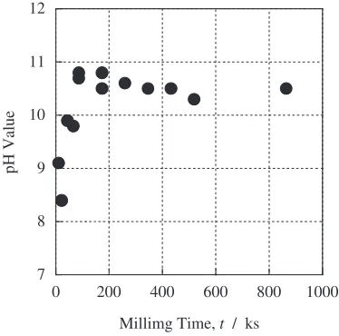
Figure 1 shows the pH values of the colloidal solutions after milling for various time with R¼1. The pH values increased rapidly with milling time for the first 86.4 ks, and started to level off at the value of about 10.5 at milling time
longer than 86.4 ks. It is suggested that the chemical reactions in the starting solution finished completely with the first 86.4 ks milling.
The XRD patterns of the synthesized nanoparticles after milling for various time withR¼1are shown in Fig.2. The peak positions agree with those of the spinel structure. Thus the synthesized nanoparticles are Fe3O4 and/or -Fe2O3
7 8 9 10 11 12
0 200 400 600 800 1000
pH Value
Millimg Time, t / ks
Fig. 1 pH values of colloidal solutions after milling for various time with R¼1.
Intensity (arb
. unit)
Milling Time 432.0 ks
43.2 ks
21.6 ks
1 1 1
2 2 0
3 1 1
4 0 0
4 2 2
5 1 1
4 4 0
6 2 0 5 3 3 4 4 4
α-Fe
86.4 ks
2 2 2
2
θ
(Co-K
α)
20
°
40
°
60
°
80
°
100
°
35
4
100
3 16 10 24 34
3 52
5 5 5 22 2 2
γ-Fe2O3 JCPDS: 39-1346
30 8
100
8 20 10 30 40
2 4104 2 Number: Intensity Fe3O4
JCPDS: 19-0629
[image:2.595.333.520.74.259.2] [image:2.595.321.530.311.645.2]phases. Although the patterns of Fe3O4and-Fe2O3 phases are similar,10,11) some peaks corresponding to -Fe2O3 phase, such as (110), (210) and (211) peaks, can not be seen in the XRD patterns. Therefore, the synthesized nanoparticles seem to be Fe3O4phase. In these diffraction patterns, no trace of the reactants and by-products,i.e.FeCl3, NaOH, NaCl and so on, can be seen. The diffraction peaks after milling for 21.6 ks are broad, that is, the nanoparticles milled for short
time seem to be amorphous-like. Those peaks become sharper with milling time, indicating that the nanoparticles are crystallized by mechanical energy during milling process. In the nanoparticles milled for short time, it can be seen the peak from -Fe phase at around 52.4 deg. The peak disappeared with further milling.
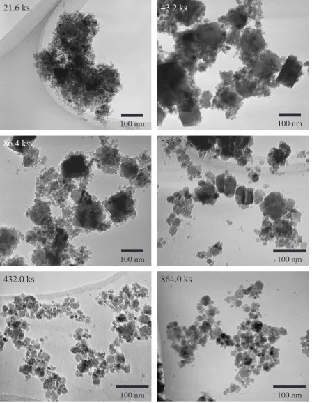
Figure 3 shows the TEM images of the Fe3O4 nanopar-ticles after milling for various time withR¼1. In the TEM
100 nm
21.6 ks
100 nm
43.2 ks
100 nm
864.0 ks
100 nm
432.0 ks
100 nm
259.2 ks
100 nm
86.4 ks
[image:3.595.76.524.73.651.2]image of the sample after milling for 21.6 ks, the aggregated particle consisting of numerous particles of a few nanometers can be seen. The particle sizes after milling for 43.2 ks and 86.4 ks had a wide distribution between a few nm and 100 nm. The images of the nanoparticles milled for more than 86.4 ks show that the particle size decreases with milling time and becomes more uniform. In the nanoparticles milled for 259.2 ks, the size was between a few nm and 50 nm, and also the nanoparticles milled for 432.0 ks and 864.0 ks were about 20 nm in diameter. The crystallite size estimated with the width of the spinel (311) peak in Fig.2 using the Scherrer equation.12) In the nanoparticles after milling for 259.2 ks, 432.0 ks and 864.0 ks, their crystallite sizes were 18 nm, 14 nm and 12 nm, respectively. Those crystallite sizes estimated using the Scherrer equation agree well with the particle sizes measured from the TEM images (Fig.3). The above results indicate the following: the chemical reactions in the starting solution with R¼1finish completely within the first 86.4 ks milling. The Fe3O4 nanoparticles form and grow in the period. Then, the synthesized nanoparticles are grinded by milling. In the ball milling, the grinding is one of the important effect in order to form the nanoparticles with uniform particle size, as well as the activation effect of chemical reaction. Size of the nanoparticles reach the final diameter of about 20 nm and become uniform after milling for more than 432.0 ks.
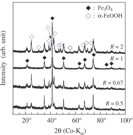
Figure 4 shows the XRD patterns of the synthesized nanoparticles after milling with variousRvalues for 259.2 ks. It can be seen that the peak positions agree with those of the spinel structure, i.e.Fe3O4 phase, in all the patterns. In the patterns of the nanoparticles milled withR6¼1, other peaks are also seen. Those peak positions coincide with those for the-FeOOH (goethite) phase.13)
The TEM images of the synthesized nanoparticles after milling with various R values for 259.2 ks are shown in Fig.5. The two different morphology, needle and irregular
shapes, can be seen in the images of the nanoparticles milled with R6¼1. From the XRD measurements (Fig.4), the needle shaped nanoparticles are the -FeOOH phase, and others are Fe3O4phase. The particle size of the Fe3O4phase increased with the smallerRvalues between 0.5 and 2.0. It was possible to separate the Fe3O4from-FeOOH
nanopar-: Fe3O4
: α-FeOOH
R = 0.67
R = 0.5
R = 1
R = 2
Intensity (arb. unit)
2
θ
(Co-K
α)
20
°
40
°
60
°
80
°
100
°
Fig. 4 XRD patterns of nanoparticles after milling with variousRvalues for 259.2 ks.
100 nm
R = 2
100 nm
R = 0.67
100 nm
R = 0.5
[image:4.595.313.540.74.672.2] [image:4.595.57.282.526.754.2]ticles by an application of magnetic field because-FeOOH phase is antiferromagnetic.
3.1.2 Formation process of Fe3O4 nanoparticles It is considered that mechanical energy accompanying collisions between balls and ball/vial during milling is necessary to synthesize Fe3O4nanoparticles. To confirm this, similar experiments were conducted without balls with the aqueous solution withR¼1. The XRD results indicated that Fe3O4nanoparticles do not form even after prolonged milling for 432.0 ks. Therefore, it is apparent that the mechanical energy supplied by ball milling is necessary for the formation of Fe3O4nanoparticles.
The XRD patterns in Fig.2 show that the nanoparticles milled for less than 86.4 ks withR¼1consisted of-Fe and Fe3O4 phases. This suggests that -Fe nanoparticles first form from the aqueous solution of FeCl3 and NaOH with R¼1 during the milling, then they are oxidized to Fe3O4. For the oxidization of -Fe nanoparticles, oxygen can be supplied from the air in the milling vial. Also, it is possible that oxygen is generated by the decomposition of H2O in the aqueous solution. Oxidation of-Fe particles with the H2O decomposition is possible since the free energy change,G, of Fe3O4from-Fe and O2at 293 K is1017:0kJ/mol and theGof the H2O decomposition to H2and O2is 238.0 kJ/ mol.14)To investigate if the oxidation of-Fe nanoparticles is due to the H2O decomposition, the milling of the aqueous solution withR¼1 for 259.2 ks was carried out in the vial atmosphere of Ar. The water for the aqueous solution was deoxygenated. The synthesized nanoparticles were the spinel structure in the XRD pattern. Also, the gas in the vial after milling for 432.0 ks, which was carried out in the vial atmosphere of air, was analyzed using a gas chromatography, and the existence of H2 gas (about 90 vol%) was confirmed. These results indicate that the oxidation of-Fe nanoparticles by the oxygen generated by decomposition is primary formation mechanism for Fe3O4nanoparticles.
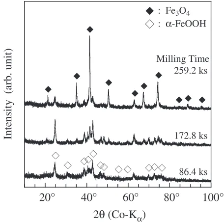
Meanwhile, the XRD patterns in Fig.4 show that the -FeOOH phase synthesized after milling withR6¼1. Figure6
shows the XRD patterns of the nanoparticles synthesized after milling for various time with R¼0:5. Although the peaks corresponding to -FeOOH phase can be seen after milling for 86.4 ks, the intensity of those peaks decreased with milling time. On the other hand, the peaks of the spinel phase appeared after milling for 172.8 ks, and the intensity of those peaks increased with milling time. This result indicates that the-FeOOH phase is synthesized first from the aqueous solutions withR6¼1, then is oxidized to become the Fe3O4 nanoparticles by mechanical energy during milling.
3.1.3 Magnetic properties of Fe3O4nanoparticles Figure 7 shows the magnetic curves for the Fe3O4 nanoparticles milled for 21.6 ks and 86.4 ks with R¼1. Similar curves were observed for the nanoparticles milled for 86.4 ks or longer. The both magnetization curves were not saturated at 1.2 MA/m. It seems that part of the synthesized nanoparticles are superparamagnetic because their particle sizes are smaller than the critical size for single domain particle for Fe3O4 (about 40 nm4)), supported by the TEM observations in Fig.3. The magnetization and coercivity of the Fe3O4 nanoparticles milled for various time withR¼1
are shown in Fig. 8. The magnetization at 1.2 MA/m
increased rapidly with milling time because of the crystal-lization of the nanoparticles, corresponding to the results of the XRD measurements in Fig.2. The magnetization started to level off at the value of about 72mWbm/kg. This value, however, is lower than the reported magnetization of Fe3O4 phase at room temperature (116mWbm/kg15)). It is because the synthesized nanoparticles are superparamagnetic, and also their structures are imperfect with the vacancies in the octahedral positions. In the XRD patterns shown in Fig. 2, the intensity of the (222) reflection was extremely weak, indicating that the octahedral positions have some vacan-cies.16) The coercivity of the nanoparticles after milling between 86.4 ks and 172.8 ks was about 6.8 kA/m, and decreased with milling time. It is well known that coercivity is sensitive to magnetic domain size, which limited by the
Milling Time 259.2 ks
86.4 ks 172.8 ks
: Fe3O4
: α-FeOOH
Intensity (arb. unit)
2
θ
(Co-K
α)
20
°
40
°
60
°
80
°
100
°
Fig. 6 XRD patterns of nanoparticles after milling for various time with R¼0:5.
-80 -60 -40 -20 0 20 40 60 80
-1.2 -0.8 -0.4 0 0.4 0.8 1.2
Magnetic Field, H / MA m-1
Magnetization,
M
/
µ
Wb
m
kg
-1
Milling Time 86.4 ks
21.6 ks
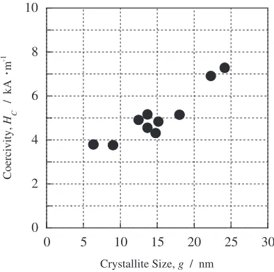
[image:5.595.312.540.72.298.2] [image:5.595.324.527.357.558.2]particle size. In the present study, the particle sizes measured from the TEM images are close to the crystallite sizes estimated from the XRD patterns. Therefore, it is considered that the coercivity of the synthesized nanoparticles depends on the crystallite size. Figure 9 shows the influence of crystallite size on the coercivity of the synthesized nanopar-ticles. Thus, the coercivity and the crystallite size exhibit a good correlation. It also indicates that the particle sizes of the synthesized nanoparticles are smaller than the single mag-netic domain size of Fe3O4 and the nanoparticles are superparamagnetic because the coercivity increased with the crystallite size.
3.2 CoFe2O4nanoparticles
Figure 10 shows the TEM image of the nanoparticles synthesized after milling for 259.2 ks. It can be seen that the nanoparticles are about 30 nm in diameter. The result of XRD measurement indicated that the crystal structure of nanopar-ticles is the spinel structure. The compositions of the individual nanoparticles, determined by an analytical HRTEM, were 21.7 at%Co and 78.3 at%Fe, indicating a deficiency of approximately 15 at%Co from the stoichio-metric composition of CoFe2O4.
Figure 11 shows the magnetic curve of the CoFe2O4 nanoparticles milled for 259.2 ks. The magnetization and coercivity were 56.5mWbm/kg and 43.4 kA/m, respectively. This magnetization value is lower than the value of CoFe2O4 at room temperature (101mWbm/kg15)). It seems that part of the synthesized nanoparticles are superparamagnetic because
2 4 6 8 10
0 200 400 600 800 1000
Coercivity,
Hc
/ kA
m
-1
Millimg Time, t / ks 20
40 60 80 100
Magnetization,
M
/
µ
Wb
m
kg
-1
Fig. 8 Magnetization at 1.2 MA/m and coercivity of Fe3O4nanoparticles after milling for various time withR¼1.
0 2 4 6 8 10
0 5 10 15 20 25 30
Coercivity,
H C
/ kA
m
-1
Crystallite Size, g / nm
Fig. 9 Influence of crystallite size on coercivity of Fe3O4nanoparticles.
-60 -40 -20 0 20 40 60
-1.2 -0.8 -0.4 0 0.4 0.8 1.2
CoFe2O4 Ni0.5Zn0.5Fe2O4
Magnetic Field, H / MA m-1
Magnetization,
M
/
µ
Wb
m
kg
[image:6.595.71.265.73.355.2]-1
Fig. 11 Magnetization curves of CoFe2O4 and Ni0:5Zn0:5Fe2O4 synthe-sized nanoparticles.
100 nm
[image:6.595.312.541.290.486.2] [image:6.595.70.267.409.605.2] [image:6.595.325.527.546.746.2]their particle sizes are smaller than the critical size for single domain particle for CoFe2O4, which is approximately 60 nm,17)supported by the TEM observations in Fig. 10. In the XRD pattern, the intensity of the (222) reflection was extremely weak, indicating that the octahedral positions have some vacancies. The above result is also supported by the composition analysis of the individual nanoparticles. Ac-cording to the composition analysis, the deficiency of the Co concentration from the stoichiometric composition of CoFe2O4 was exhibited. If the deficiency of the Co concentration is the vacancies in the octahedral positions, the octahedral positions are occupied by the Co2þof 18.5 at%
and the vacancies of 14.8 at%. On the basis of this assumption the calculated magnetization is 56mWbm/kg. Since the calculated value is closed to the magnetization of the synthesized CoFe2O4 nanoparticles in the present study, it is indicated that the octahedral positions consist of Co2þ
and some vacancies. Therefore, the magnetization of the synthesized CoFe2O4 nanoparticles is lower than the refer-ence value.
3.3 Ni0:5Zn0:5Fe2O4 nanoparticles
Figure 12 shows the TEM image of the nanoparticles synthesized after milling for 345.6 ks. It can be seen that the nanoparticles are about 30 nm in diameter. The result of XRD measurements indicated that the crystal structure of nano-particles was the spinel structure. The compositions of the individual nanoparticles, determined by an analytical HRTEM, were 7.4 at%Ni, 13.6 at%Zn and 79.0 at%Fe, indicating a deficiency of approximately 10 at%Ni from the stoichiometric composition of Ni0:5Zn0:5Fe2O4.
Figure 11 shows the magnetization curve of the Ni0:5Zn0:5Fe2O4 nanoparticles milled for 345.6 ks. The
magnetization and coercivity were 54.0mWbm/kg and 5.3 kA/m, respectively. This magnetization value is lower than the value of Ni0:5Zn0:5Fe2O4 at room temperature
(148mWbm/kg15)). This is because part of the synthesized nanoparticles are superparamagnetic, and their crystal struc-tures are imperfect with the vacancies, similarly to the case of
CoFe2O4nanoparticles. If the octahedral positions have some vacancies, the magnetization of Ni0:5Zn0:5Fe2O4 decreases. In the XRD pattern shown, the intensity of the (222) reflection is weak, indicating that the octahedral positions have some vacancies. The above result is also supported by the composition analysis of the individual nanoparticles. The deficiency in Ni concentration from the stoichiometric composition of Ni0:5Zn0:5Fe2O4 indicates that the octahedral
positions have some vacancies. Therefore, the magnetization of the synthesized Ni0:5Zn0:5Fe2O4 nanoparticles is lower
than the reference value.
4. Conclusion
The nanoparticles of the various spinel ferrites (Fe3O4, CoFe2O4 and Ni0:5Zn0:5Fe2O4) were synthesized from the
aqueous solutions consisting of various chlorides (FeCl3, CoCl2 or NiCl2/ZnCl2) and NaOH by mechanochemical processing using a ball mill. The structures, morphologies and magnetic properties in their synthesized nanoparticles were studied and can be summarized as follows:
(1) The formation of Fe3O4 nanoparticles from the aqueous solutions with the differentRvalues proceededviatwo different processes. In the case ofR¼1,-Fe nanopar-ticles were firstly synthesized, and then were oxidized to Fe3O4 nanoparticles. In the case of R6¼1, -FeOOH phase formed, and changed to the Fe3O4 nanoparticles by milling.
(2) The Fe3O4 nanoparticles with different particle sizes could be synthesized by changing the milling time and theRvalue in the starting solution. The particle size was controllable between 20 and 100 nm. In magnetic properties, the magnetization was the value of 72mWbm/kg after milling for longer than 86.4 ks with R¼1. The coercivity showed the maximum of 10.8 kA/ m after milling for 259.2 ks withR¼0:5.
(3) The CoFe2O4nanoparticles were synthesized by milling for 259.2 ks nanoparticles, were about 30 nm in diame-ter. The magnetization and coercivity were 56.5mWbm/ kg and 43.4 kA/m, respectively.
(4) The Ni0:5Zn0:5Fe2O4nanoparticles were synthesized by
milling for 345.6 ks nanoparticles, were about 30 nm in diameter. The magnetization and coercivity were 54.0mWbm/kg and 5.3 kA/m, respectively.
(5) In the synthesized nanoparticles of Fe3O4, CoFe2O4and Ni0:5Zn0:5Fe2O4, their magnetization values were lower than the reference values because part of the synthesized nanoparticles are superparamagnetic and their crystal structures are imperfect with the deficiency of cations in B-sites. It seems that the deficiency of cations can be improved by changing the composition in the starting solution.
REFERENCES
1) M. Kiyama: J. Jpn. Soc. Powder Powder Metal.42(1995) 690–694. 2) M. Kiyama and T. Takada: Bull. Inst. Chem. Res.58(1980) 193–200. 3) M. Kiyama: Bull. Chem. Soc. Japan47(1974) 1646–1650.
4) M. Kiyama: Bull. Inst. Chem. Res.60(1982) 247–253.
5) T. Ishikawa, Y. Kondo, A. Yasukawa and K. Kandori: Corros. Sci.40 (1998) 1239–1251.
100 nm
6) J. Ding, W. F. Miao, T. Tsuzuki, P. G. McCormick and R. Street: Synthesis and Processing of Nanocrystalline Powder, ed. by D. L. Bouvell, (TMS, 1996) pp. 69–79.
7) J. Ding, T. Tsuzuki, P. G. McCormick and R. Street: J. Phys. D29 (1996) 2365–2369.
8) J. Ding, T. Tsuzuki and P. G. McCormick: NanoStructured Mater.8 (1997) 739–747.
9) J. Ding, T. Tsuzuki and P. G. McCormick: J. Am. Ceram. Soc.79 (1996) 2956–2958.
10) JCPDS card, 19–0629. 11) JCPDS card, 39–1346.
12) B. D. Cullity: Elements of X-ray Diffraction, (Addison-Wesley Longman, 1978).
13) JCPDS card, 18–0639.
14) HSC Chemistry Ver. 4.1, Chemical Reaction and Equilibrium Software with Extensive Thermochemical Database, (Outokumpu Research Oy). 15) T. Hiraga, K. Okutani and T. Ojima:Ferrite, (Maruzen, 1986). 16) V. Sˇepela´k, A. Buchal, K. Tka´cˇova´ and K. D. Becker: Mater. Sci.
Forum278–281(1998) 862–867.