147
©IJRASET: All Rights are Reserved
Applying Texture Feature Extraction for Skin
Diseases using GLCM and GLRLM
Prof. Mrs. K. Chandra Prabha1, R. Abinaya 2
1
Associate Professor& Head of the Department, Department of Computer Science and Engineering, ACGCET, Karaikudi, Tamil Nadu.
2
PG Student, Department of Computer Application, ACGCET, Karaikudi, Tamil Nadu.
Abstract: The proposed system classifies different types of dermatological skin diseases. The project mainly consists of three phases training phase, testing phase, and classification phase. First, collect the five skin disease datasets. Then divide the datasets into training and testing images. The datasets are in RGB format convert the images into grayscale. For extracting texture of an image, a statistical methods GLCM and GLRLM are used. GLCM features are contrast, correlation, homogeneity, and energy. GLRLM features are SRE, LRE, RP, GLNU, RLNU, LGRE and HGRE. Compare the texture features of training images with testing images and classify skin diseases such as Eczema, Melanoma, Dermatitis, Basal cell carcinoma, and Acne by using the ANN method. Compare the accuracy levels of both GLCM and GLRLM. GLCM gives 95% accuracy and GLRLM gives 93% accuracy.
Keywords: GLCM, GLRLM, ANN, Skin Diseases, RGB, Datasets.
I. INTRODUCTION
Dermatology is the part of the drug that manages skin, hair, and nails in the amplest sense. To propose a framework which would help the patients just as specialists to analyze the skin illnesses by simply giving the picture of the influenced region of skin. proposed location framework utilizes texture feature extraction strategies GLCM and GLRLM alongside ANN strategy for classifying the skin diseases. GLCM extract the features are contrast, correlation, homogeneity, and Energy. GLRLM extract the features are SRE, LRE, RP, GLNU, RLNU, LGRE, HGRE. The Skin diseases classification system comprises of three sections where the initial section is to train the folder of images. In training, surface highlights are chosen from the training images set by utilizing GLCM and GLRLM techniques after the change of RGB images into grayscale and store the pictures into a (.mat) table. In the second part is testing the images. The testing stage goes about as approval. In the testing stage, accepts one picture as information at that point convert the RGB picture into grayscale. GLCM and GLRLM texture feature extraction strategies are utilized. After feature extractions, store the incentive in another (.mat) table. The third part is the classification part. ANN classification used to classify skin diseases such as Eczema, Melanoma, Dermatitis, Basal cell carcinoma, and Acne. Compare the accuracy levels both GLCM and GLRLM methods.
A. Literature Survey
Shuzlina Abdul-Rahman [2016]: This paper describes the skin diseases based on two feature selection methods Correlation Feature Selection (CFS) and Fast Correlation-based Filter (FCBF) help by offering a lesser number of properties with better accurateness and nearer react time.
Nibaran Das [2013]: The paper introduces an investigates on the ID of skin infections by using, Local Binary Pattern (LBP), GLCM, Discrete Cosine Transform (DCT) and Discrete Fourier Transform (DFT) have been utilized with Support Vector Machines (SVM) based classifiers.
L. G. Kabari and F. S. Bakpo [2016]: The above paper portrays the improvement of medicinal master frameworks that utilization ANN to be a promising technique for anticipating determination and conceivable treatment schedule.
II. PROPOSEDSYSTEM
148
©IJRASET: All Rights are Reserved
Low Gray Level Run Emphasis (LGRE), High Gray Level Run Emphasis (HGRE) are the features extracted by GLRLM. After extract the texture features, store the values in (.mat) table. In the testing stage, accepts one picture as info at that point convert the RGB picture into grayscale because GLCM and GLRLM texture feature extraction methods are utilized. After extract the texture features store the values in another (.mat) table. In the classification stage, the ANN classification used to classify skin diseases such as Eczema, Melanoma, Dermatitis, Basal cell carcinoma, and Acne. Accuracy levels both GLCM and GLRLM strategies. GLCM technique gives 95% and GLRLM strategy gives 93%.
[image:2.612.142.482.156.620.2]
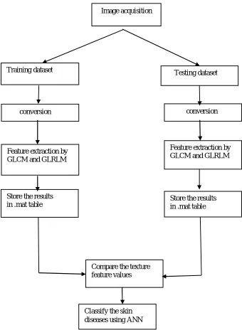
Fig. 1 Block diagram of skin diseases classification system
III. METHODOLOGIES
These methodologies are used to execute the project.
A. Texture Features Extraction
The texture is a repeated pattern of information or arrangement of the structure with regular intervals. texture refers to surface characteristics and appearance of an object given by the size, shape, density, arrangement, the proportion of its elementary parts.
Image acquisition
Training dataset Testing dataset
conversion
conversion
Feature extraction by GLCM and GLRLM
Feature extraction by GLCM and GLRLM
Store the results
in .mat table Store the results in .mat table
Compare the texture feature values
149
©IJRASET: All Rights are Reserved
1) Gray Level Co-Occurrence Matrix: A GLCM is a matrix where the number of rows and columns is equal to the number of distinct gray levels or pixel values in the image of that surface. The Texture feature calculations use the contents of the GLCM to give a measure of the variation in intensity at the pixel of interest. Typically, the co-occurrence matrix is computed based on two parameters, which are the relative distance between the pixel pair d measured in pixel number and their relative orientation. Normally, is quantized in four directions (e.g., 00,45 0, 90 0 and 135 0)
TABLE I GLCM INPUT IMAGE
TABLE III TABLE IIIII GLCM AT 00 OFFSET GLCM AT 450 OFFSET
00 pairing horizontally adjacent elements 45 0 pairing right adjacent elements 90 0 pairing vertically adjacent elements
135 0 pairing horizontally left elements
TABLE IVV
GLCM TEXTURE FEATURES
2 2 2 1
1 2 1 1
0 0 3 3
1 1 3 3
ROWS (I)
COLUMNS(J)
0 1 2 3 0 0 1 1 0 1 1 1 1 1 2 0 0 1 0 3 0 1 0 1 ROWS
(I)
COLUMNS(J)
0 1 2 3 0 1 0 0 1 1 0 2 1 0 2 0 2 2 0 3 0 0 0 1
Features Description Formula Contrast Measurement of the intensity of
pixel and its neighbor over the image
N-1
Σ i=0 N-1Σ j=0 (i −j)2 p(i, j)
Correlation how correlated a reference pixel to its neighbor over an image.
N-1
Σi=0 N-1Σj=0 Pij ______ 1+(i-j)2 Homogeneity calculate the degree of
homogeneity of grayness in an image.
N-1
Σi,j=0 Pij ((i- ) (j- )) ________ 2 Energy measurement of the extent of
pixel pair repetitions.
N-1
150
©IJRASET: All Rights are Reserved
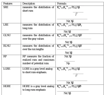
2) Gray Level Run Length Matrix: GLRLM is the number of runs with pixels of gray level i and run length j for a given direction. A set of consecutive pixels with the same gray level is called a gray level run. The number of pixels in a run is the run length. GLRLM also have four offsets like GLCM.
TABLE V GLRLM INPUT IMAGE
[image:4.612.126.482.402.728.2]
TABLE VI TABLE VIV GLRLM AT 900 OFFSET GLRLM AT 1350 OFFSET
TABLE VIIVI GLRLM texture features
0 1 2 3 0 2 3 3 2 1 1 1 3 0 3 0
GRAY LEVEL (I)
RUN LENGTH(J)
1 2 3 4
0 2 1 0 0 1 4 0 0 0 2 3 0 0 0 3 3 1 0 0
GRAY LEVEL (I)
RUN LENGTH(J)
1 2 3 4
0 4 0 0 0 1 4 0 0 0 2 0 0 1 0 3 3 1 0 0
Features Description Formula
SRE measures the distribution of
short runs
∑Ngi=1∑ Nr
j=1 P(i,j| θ)
________ j2
_______________________ Nz( θ)
LRE measures the distribution of
long runs
∑Ngi=1∑ Nr
j=1 P(i,j| θ)j 2
________________________ Nz( θ)
GLNU measures the distribution of
over the gray values
∑Ngi=1(∑ Nrj=1 P(i,j| θ))2 _______________________________________
Nz( θ)
RLNU measures the distribution of
over the run lengths
∑Nri=1(∑ Ngj=1 P(i,j| θ))2 ______________________________________
Nz( θ)
RP RP measures the fraction of
realized runs and maximum number of potential runs.
Nz( θ) ________ Np
LGRE LGRE is a gray level analog
to short runs emphasis.
∑Ngi=1∑ Nr
j=1 P(i,j| θ)
________ i2
_______________________ Nz( θ)
HGRE HGRE is a gray level analog
to long runs emphasis
∑Ngi=1∑ Nr
j=1 P(i,j| θ)i 2
151
©IJRASET: All Rights are Reserved
NG NUMBER OF INTENSITY VALUES IN THE IMAGE
Nr Number of run lengths in the image NP Number of pixels in the image
NZ (θ) Number of runs in the image along angle P(i,j | θ)
B. Classification
After texture features have been extracted which are contributes to a classifier to characterize skin diseases. In order, Artificial Neural Network weight enhancement has been utilized to characterize the skin diseases.
1) Artificial Neural Network: An ANN is prepared by changing the loads, to foresee the right class. An ANN is a computational model. It depends on the structure and elements of the neural system. ANNs have three layers that are interconnected. The primary layer comprises of info neurons. Those neurons send information on to the second layer, which thus sends the yield to neurons of the third layer. ANN classify skin diseases such as Basal cell carcinoma, Eczema, Melanoma, Dermatitis, and Acne. ANN classification additionally shows the Performance, Training state, Error Histogram, Regression, Fit of the training and Testing images.
[image:5.612.186.438.258.378.2]
Fig. 2 ANN classification
IV. PROJECTIMPLEMENTATION
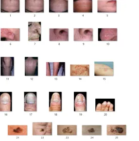
A. Image Acquisition
Five skin diseases images were collected such as Eczema, Melanoma, Dermatitis, basal cell carcinoma, Acne from
www.dermnet.com data source.
[image:5.612.166.452.427.717.2]
152
©IJRASET: All Rights are Reserved
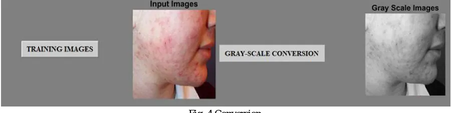
B. Conversion
The datasets are in RGB format convert the dataset into grayscale because of GLCM and GLRLM texture feature extraction methods are used. Grayscale is the accumulation or the scope of monochromic (dark) shades, extending from unadulterated white on the lightest end to unadulterated dark on the contrary end. Grayscale just contains luminance data and no shading data
[image:6.612.82.533.131.244.2]
Fig. 4 Conversion
C. Feature Extraction
The procedure of picture highlights extraction is completed with surface examination utilizing the Gray Level Co-Occurrence Matrix (GLCM) and Gray Level Run Length Matrix (GLRLM) technique. The surface is a rehashed example of data or a course of action of the structure with normal interims. A surface alludes to surface qualities and presence of an article given by size, shape thickness, game plan and extent of basic parts.
TABLE VIIX GLCM texture feature values
TABLE X
GLRLM TEXTURE FEATURE VALUES Data
sets
GLRLM TEXTURE FEATURES
SRE LRE GLN RP RLN LGRE HGRE 1 0.01 0.11 0.67 0.02 2.06 0.01 0.67 2 0.04 0.02 6.23 0.01 9.32 0.14 6.23 3 0.01 0.12 1.07 0.02 2.51 0.01 1.07 4 0.01 0.15 0.95 0.02 1.78 0.01 0.95
D. Classification
Classifies skin diseases such asBasal cell carcinoma, Eczema, Melanoma, Dermatitis, and Acne.
Fig.5 Classification of skin diseases Data
sets
GLCM TEXTURE FEATURES
[image:6.612.164.455.360.574.2]153
©IJRASET: All Rights are Reserved
V. RESULT Accuracy= TP+TN
______________ X 100% TP+FP+FN+TN
TP True positive TN True Negative FP False positive FN False Negative GLCM gives 95% accuracy while GLRLM gives 93% accuracy.
VI. CONCLUSIONS
In the proposed work classifies the five skin diseases, for example, basal cell carcinoma, melanoma, Eczema, Dermatitis, and Acne. Four texture features are extracted in GLCM, for example, Contrast, Correlation, Homogeneity, and Energy. Seven texture features are extracted in GLRLM, for example, LRE, HRE, RP, HGRE, LGRE, GLN, and RLN. Compared to GLRLM, GLCM gives better accuracy.
REFERENCES
[1] M. Shamsul Ari n, M. GolamKibria, Adnan Firoze, M. Ashraful Amin, Hong Yan, "Dermatological Disease Diagnosis Using Color-skin Images", Proceedings of the 2012 International Conference on Machine Learning and Cybernetics, Xian, 15-17 July/2015.
[2] L. G. Kabari and F. S. Bakpo, Member, IEEE, "Diagnosing Skin Diseases Using an Artificial Neural Network", 2016 IEEE.
[3] Shuzlina Abdul-Rahman, Ahmad KhairilNorhan, Marina Yuso, Azlinah Mohamed, SoanitaMutalib, "Dermatology Diagnosis with Feature Selection Methods and Artificial Neural Network", 2016 IEEE EMBS.
[4] Nibaran Das, Anabik Pal, SanjoyMazumder, SomenathSarkar, DwijendranathGangopadhyay, MitaNasipuri, "An SVM based skin disease identification using Local Binary Patterns", 2013 Third International Conference on Advances in Computing and Communications.
[5] Nesreen Abdel Wahab, Manal Abdel Wahed, Abdallah S. A. Mohamed, "Texture Features Neural Classifier of Some Skin Diseases", 2004.