Open Access
Review
Tumor suppressor gene E-cadherin and its role in normal and
malignant cells
Nives Pe
ć
ina-Šlaus*
Address: Department of Biology, School of Medicine, University of Zagreb, Šalata 3, HR-10000 Zagreb, Croatia
Email: Nives Pećina-Šlaus* - [email protected] * Corresponding author
E-cadherinCDH1cell adhesionwnt signalling
Abstract
E-cadherin tumor suppressor genes are particularly active area of research in development and tumorigenesis. The calcium-dependent interactions among E-cadherin molecules are critical for the formation and maintenance of adherent junctions in areas of epithelial cell-cell contact. Loss of E-cadherin-mediated-adhesion characterises the transition from benign lesions to invasive, metastatic cancer. Nevertheless, there is evidence that E-cadherins may also play a role in the wnt signal transduction pathway, together with other key molecules involved in it, such as beta-catenins and adenomatous poliposis coli gene products.
The structure and function of E-cadherin, gene and protein, in normal as well as in tumor cells are reviewed in this paper.
Introduction
The control of cellular adhesion and motility is one of the crucial mechanisms responsible for tumor initiation and progression. The genes involved are also contributors to malignancy along with genes responsible for cell prolifer-ation and survival. I propose to summarize the current knowledge on one very important tumor suppressor gene,
viz. E-cadherin [1,2].
E-cadherin is one of the most important molecules in cell-cell adhesion in epithelial tissues. It is localized on the surfaces of epithelial cells in regions of cell-cell contact known as adherens junctions [3]. As a member of a large family of genes coding for calcium-dependent cell adhe-sion molecules (CAMs), the cadherin glycoproteins are expressed by a variety of tissues, mediating adhesion through homotypic binding. Classical cadherins – E- and
N-cadherins being the best characterized – play important roles in the formation of tissues during gastrulation, neu-rulation and organogenesis [4].
E-cadherin has probably been studied in most detail. It is essential for the formation and maintenance of epithelia, was first identified in chicken, and was originally called L-CAM [5]. The mouse counterpart of this protein, uvo-morulin [6], has 80% identity in both nucleotide and amino acid sequences to the human counterpart [7].
Besides its role in normal cells, this highly conserved gene can play a major role in malignant cell transformation, and especially in tumor development and progression. The suppression of E-cadherin expression is regarded as one of the main molecular events responsible for dysfunc-tion in cell-cell adhesion. Most tumors have abnormal
Published: 14 October 2003
Cancer Cell International 2003, 3:17
Received: 17 September 2003 Accepted: 14 October 2003 This article is available from: http://www.cancerci.com/content/3/1/17
cellular architecture, and loss of tissue integrity can lead to local invasion. Thus, loss of function of E-cadherin tumor suppressor protein correlates with increased invasiveness and metastasis of tumors [8], resulting in it being referred to as the "suppressor of invasion" gene.
E-cadherin gene and protein structure
The human epithelial (E)-cadherin gene CDH1 maps to chromosome 16q22.1. Berx et al. [1] isolated the full-length gene by using recombinant lambda phage, cosmid and P1 phage clones. The gene they cloned encompasses 16 exons and spans a region of ~100 kb. The exons range from 115 to 2245 bp. Further analysis of the gene showed 15 introns ranging from 120 bp (intron 4) to 65 kb (intron 2). The intron-exon boundaries are highly con-served in comparison with other "classical cadherins", and in intron 1 a 5' high-density CpG island was identi-fied that may have a role in transcription regulation [1]. This island covers the region from exon 1 to exon 2 of the human E-cadherin gene, while other exons lacked such features (fig. 1), including the biggest (exon 16 of 2245 bp).
The chromosomal location of CDH1 on 16q22.1 was later confirmed by fluorescent in situ hybridization (FISH) analysis. It is interesting that the human P-cadherin gene was recently located only 32 kb upstream from E-cadherin [9], and also the M-cadherin gene was positioned on chro-mosome 16q24.1-qter [10], which further suggests clus-tering of cadherin genes originating probably from gene duplication, while possible co-evolution might be explained by gene conversion [1]. All classical cadherin genes analyzed so far have 16 exons separated by 15 introns.
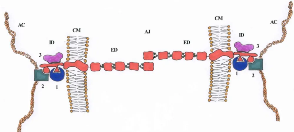
CDH1 encodes a 120 kDa glycoprotein with a large extra-cellular domain, a single transmembrane segment and a short cytoplasmic domain, which interacts with the actin cytoskeleton through linker molecules, alpha- beta- and gamma-catenins [11]. On the cytoplasmic side of the membrane, a bundle of actin filaments is linked to the
E-cadherin molecules via a protein complex. Alpha-catenin and either beta- or gamma-catenins are included in this complex. Beta- and gamma-catenins share significant homology and bind to a specific domain at the E-cadherin C-terminus. Alpha-catenin links the bound beta- or gamma-catenin to the actin cytoskeleton. (fig. 2).
Alpha-catenin has structural similarities with vinculin, one of the key components of fibroblast membrane attachment sites of microfilaments, beta-catenin shows homology to Armadillo of Drosophila melanogaster and gamma-catenin is identical to plakoglobin, a protein found in desmosomes [12].
The C-terminal cytoplasmic domain of ~150 residues is highly conserved in sequence, and has been shown exper-imentally to regulate the cell-cell binding function of the extracellular domain of E-cadherin, possibly through interaction with the cytoskeleton. The juxtamembrane region of the cadherin cytoplasmic tail has been identified as a functionally active region supporting cadherin clus-tering and adhesive strength; one of the interacting pro-teins involved in clustering and cell adhesion is p120ctn [13].
The structure of the extracellular domain of classical E-cadherin contains five tandem repeats of a 100-residue-amino-acid-motif, and the biggest part of N-terminal of these repeats contains the sites with adhesive activity. This part of the molecule also has binding sites for calcium ions situated in the pockets between the repeats. The amino acid sequences that form the Ca2+ binding pockets are highly conserved between different members of the cadherin family and between different species. Cell-cell adhesion is mediated through homotypic interactions of E-cadherin extracellular domains in a process of lateral dimerization. Parallel dimers are able to interdigitate with dimers from neighbouring cells forming the points of adhesion. Those findings introduce a cadherin-cadherin interface at the cellular surface. Until recently cadherins were thought to be involved only in homophilic Genomic organization of the human E-cadherin gene
Figure 1
interactions; however, E-cadherin has now been shown to be a ligand for two integrins, alphaEbeta7 and alpha2beta1. The first interaction might serve to retain intraepithelial lymphocytes in mucosal tissue, while the second may contribute to the organization of epithelial multilayers [14].
Cadherins have been identified in a large variety of spe-cies, including mammals, Xenopus, Drosophila, Caenorhab-ditis elegans. Many new cadherin family members have been isolated and the cadherin group of genes has grown very fast in the last decade. Proteins of several isolated molecules show a great deal of homology with the "classi-cal" cadherins [15]. Other cadherin like molecules share with the classical cadherins putative Ca2+ binding motifs in repeated extracellular domains but diverge considera-bly in various regions, particularly in the cytoplasmic domains. Even more deviations were observed for new cadherins, such as K-cadherin [16] and LI-cadherin [17], while the ret protooncogene product also shows some similarity to cadherin [18,19]. Although the extracellular domain has several similar repeats with putative Ca2+ binding motifs, this transmembrane tyrosine kinase is considered to be unrelated to the cadherins. Since the ret gene lacks matching of the splice sites to the CDH1 gene, this might be the explanation. It is possible that this
kinase has acquired its cadherin-like motifs by convergent evolution [1].
Role of E-cadherin in normal cells, wnt signalling,
adherens junctions
Expression of E-cadherin in embryonic development is very early, at the two-cell stage [12,20]. Epithelial differen-tiation and polarization (processes fundamental to cell differentiation) occur early in ontogeny in the morula stage, when the embryo compacts and each cell polarizes along its apicobasal axis to generate an epithelial-like phe-notype. E-cadherin plays an important role in the adhe-sion of the blastomeres, and early embryo's ability to compact [21]. E-cadherin is expressed in the membrane even before compaction of the morula occurs, is distrib-uted in a non-polar manner, and does not exhibit adhe-sive function [22,23]. The mechanism that renders E-cadherin functional is unknown, but it does include phos-phorylation of the protein [24]. Controlled epithelial-mesenchymal conversion is the most important exhibit of E-cadherin's function in development [25]. Loss of epithe-lial adhesion and polarity causing mesenchymal cell mor-phology occurs during mesoderm formation. Rietmacher and co-workers [12] introduced a targeted mutation in mouse embryonal stem cells and generated a mouse with-out E-cadherin sequences essential for Ca2+ binding and
Shematic illustration of E-cadherin in adherens junction Figure 2
for adhesive function. Heterozygous mutant animals were normal and fertile. In vitro, they were able to form normal blastocysts with normal blastocoels that consequently expanded. On the other hand, the homozygous E-cad-herin -/- embryos showed severe abnormalities before implantation. This included failure to maintain a polar-ized and compacted state and also failure to form a troph-ectoderm epithelium; they distort at the blastocyst stage, making the mutation lethal. The initial compaction that was observed in -/- embrios is probably due to the pres-ence of E-cadherin proteins from maternal sources [24].
Investigation of zebrafish E-cadherin expression during early embryogenesis confirmed observed expression in blastomeres, but also led to the detection of a protein expressed in the anterior mesoderm during gastrulation and developing epithelial structures [26]. In the develop-ing nervous system, CDH1 was detected at the pharyngula stage in the midbrain-hindbrain boundary and was pre-ceded by wnt 1 expression [26].
As far as normal adult epithelial tissue structure and integ-rity is concerned, E-cadherin is also involved in its main-tenance and homeostasis. As already mentioned, its function lies primarily in the formation of adherens junctions.
Cadherin mediated adhesion is a dynamic process that is regulated by several signal transduction pathways. There is also evidence that cadherins are not only targets for sig-naling pathways that regulate adhesion, but may them-selves send signals that regulate basic cellular processes, such as migration, proliferation, apoptosis and cell differ-entiation [4,27,28].
The image of individual adhesion molecule performing its function, or linear downstream signalling cascade is somewhat abandoned scheme. Instead of separating and dividing into distinct fields, the cellular mechanisms of signalling and adhesion are nowadays thought to be closely connected mechanisms where components have double (or more) functions and interconnect in a signal-ling-structural network. The clearest example is recently discovered interaction of E-cadherin with epithelial growth factor receptor (EGFR) [29].
The recently discovered wnt/wingless pathway, of mouse and Drosophila respectively is one of the most interesting kind of signal transduction, in which key components have multiple functions in adhesion and signalling. Infor-mation on Wnt signalling can be found on the wnt gene site [30] and at the connection map at Science STKE Web site [31]. In vertebrate cells, it is named after Wnt proteins, a family of highly conserved secreted signaling molecules that regulate cell-to-cell interactions during
embryogene-sis. Insights into the mechanisms of Wnt action have emerged from several systems: genetics in Drosophila and
Caenorhabditis elegans; biochemistry in cell culture; and ectopic gene expression in Xenopus embryos. Many Wnt genes in the mouse have been mutated, leading to very specific developmental defects. As currently understood, Wnt proteins bind to receptors of the Frizzled family on the cell surface. Through several cytoplasmic relay compo-nents, the signal is transduced to beta-catenin, which then enters the nucleus and forms a complex with TCF to acti-vate transcription of Wnt target genes [28].
It has been well documented that wnt genes, together with other components of wnt signalling pathway, are impli-cated in cancer [32]. Another tantalizing compartment of the wnt signalling pathway lies in downstream transcrip-tional activation. In response to WNT signalling, cytoplas-mic beta-catenin is stabilized, accumulates in the cytoplasm and enters the nucleus, where it finds a partner, a member of the DNA binding protein family LEF/TCF (T cell factor-lymphoid enhancer factor). Together they acti-vate new gene expression programs. One of the target genes for β-catenin/TCF encodes c-MYC protein [2], explaining why constitutive activation of the wnt pathway can lead to cancer.
Role of E-cadherin in malignant cells
Progressive accumulation of somatic mutations in a number of different genes characterizes the process of tumorigenesis. Many genes involved in the process of tumorigenesis are components of one of a great many sig-nal transduction pathways through which sigsig-nals traffic via molecular networks. It is now apparent that epithelial malignancy can in certain aspects be explained by altera-tions in the adhesive properties of neoplastic cells.
human cancers including breast, stomach, endometrium, ovary and thyroid [34,35]. Transgenic mouse model with loss of E-cadherin expression developed invasive carci-noma from well-differentiated adecarci-nomas [36] and finally germ-line mutations have recently been reported in early onset, diffuse-type stomach cancers [37].
Many immunohistochemical studies have examined changes in expression of the E-cadherin gene in human malignancies. In almost all non-colonic tumors exam-ined, the patterns of changes in the expression of this gene have been similar to that seen in colorectal cancer, i. e. loss of protein expression is positively correlated to loss of tumor differentiation. In oesophageal, pulmonary, squa-mous head and neck tumors, pancreatic and cervical tumors, loss of expression has been correlated with a high grade and an advanced stage of the disorder, with poor prognosis [2]. It has been reported that inactivating muta-tions of E-cadherin gene are highly frequent in infiltrating lobular breast carcinomas [38] and diffuse gastric carcino-mas [34,35]. These mutations mostly occur in combina-tion with loss of heterozygosity of the wild-type allele.
Another interesting paper [39] on E-cadherin expression demonstrates its reappearance in metastatic cells. Signifi-cant increase in re-expression of E-cadherin, together with alpha and beta-catenin, was observed in metastatic deposit of primary lobular breast cancers. This protein product was missing in the primary tumors of the same origin. Lobular breast cancers harbour E-cadherin muta-tions as well as losses of the gene locus, even in situ dis-ease. That suggests that the mutated protein has a role even before invasion process started. The findings reveal another dimension of adhesion molecules in tumor metastasis: reestablishment of cellular contact may pre-vent apoptosis. I would like to cite M. Iiyas [40] "adhesion molecule expression is the phoenix in tumor metastasis".
And finally, let us not forget yet another level of CDH1 expression regulation, i. e. E-cadherin promotor hyper-methylation. This process has been known to be an inac-tivating event in some tumors and is currently extensively researched [41].
Mutation reports
Most interesting genetic mutations and their conse-quences are listed in this section. Becker et al. [42] sug-gested that E-cadherin mutations contribute to the histopathologic appearance of stomach cancer because 13 of 26 diffuse gastric carcinomas, which had reduced homophilic cell-to-cell interactions, had abnormal gene transcripts that were not seen in non-cancerous tissue from same patients. Berx et al. [34] reported of 69 somatic mutations of the CDH1 gene. In addition to a few mis-sense mutations, those were mainly splice site and
trunca-tion mutatrunca-tions caused by insertrunca-tions, deletrunca-tions, and nonsense mutations. There was a major difference in mutation type between diffuse gastric and infiltrative lob-ular breast cancers. In diffuse gastric tumors, the predom-inant defects were exon skippings, which caused in-frame deletions. In contrast, most mutations found in infiltrat-ing lobular breast cancers were out-of-frame mutations, which yield secreted truncated E-cadherin products. In most cases these mutations occurred in combination with loss of heterozygosity.
Guilford et al. [37] reported germline mutations in the CDH1 gene in 3 familial gastric cancer kindreds of Maori origin from New Zealand. Furthermore, Richards et al. [43] analyzed 8 UK gastric cancer kindreds and identified novel germline CDH1 mutations (a nonsense and a splice site mutation) in 2 families. Both mutations were pre-dicted to truncate the E-cadherin protein in the signal pep-tide domain. Gayther et al. [44] also described germline CDH1 mutations in familial gastric cancer. In a family with a strong history of diffuse gastric carcinoma, Chun et al. [45] found the 1558insC germline mutation in the CDH1 gene. The gastric cancer was of the early onset, his-tologically diffuse type. In another family with early onset diffuse gastric cancer, Guilford et al. [37] found that the 30-year-old proband was heterozygous for A-to-T transi-tion at nucleotide 2095, which resulted in a nonsense mutation. The mutation was predicted to result in an E-cadherin peptide lacking both the transmembrane and cytoplasmic domains. Same authors [37] described a fam-ily in which multiple members with gastric cancer were heterozygous for the insertion of an additional C residue in a run of 5 cytosines at positions 2382 to 2386. The resulting frameshift led to an E-cadherin molecule lacking about half of its cytoplasmic domain. In another diffuse gastric cancer family, a heterozygous G-to-T transversion at nucleotide 70 in exon 2 of the CDH1 gene, was also identified. The mutation converted a glutamic acid (glu24) to a TAG stop codon in the signal peptide of the E-cadherin precursor protein.
Richards et al. [43] identified a splice acceptor site muta-tion, an A-to-G transition at position -2 from nucleotide 49 at the start of exon 2 of the CDH1 gene and also a germline G-to-A transition at nucleotide 59 in exon 2. The mutation, a trp20-to-ter substitution was predicted to truncate the E-cadherin gene product in the signal peptide domain, which is cleaved from the N terminus of the mature protein.
Same authors [44] found a 1-bp insertion in the CDH1 gene in the proband of a family with familial diffuse gas-tric cancer. Insertion of a G after nucleotide 1711 created a frameshift that would truncate the protein at codon 587. Another 1-bp insertion (C) at nucleotide 1588 in exon 11 was also identified in a family segregating diffuse gastric cancer.
Mutations were also found in other types of carcinoma. In an endometrial carcinoma, Risinger et al. [46] identified a GCA-to-ACA transition in codon 617 predicting a substi-tution of thr for ala in the E-cadherin molecule. Somatic loss of heterozygosity was identified in the tumor tissue. They also identified a CTG-to-GTG transversion resulting in a leu711-to-val amino acid substitution in E-cadherin. The wild type allele was not lost.
In an ovarian carcinoma, same authors [46] identified an AGC-to-GGC transition in codon 838 resulting in substi-tution of glycine for serine. The tumor tissue also showed somatic loss of heterozygosity. In an infiltrative lobular breast carcinoma, Berx et al. [1] found a GAA (glu)-to-TAA (stop) nonsense mutation in the CDH1 gene. Tumor-spe-cific loss of heterozygosity of chromosomal region 16q22.1 was demonstrated in this case. Here I have assessed only selected examples of CDH1 mutations in carcinoma tissue. The number of mutations is growing each day and can be reached at the following web site [47].
I should also mention the type of genetic changes that my group has encountered so far. Pećina-Šlaus et al. [48] indi-cated yet another type of genomic instability of CDH1 gene encountered in tumor tissue. When searching for allelic loss i. e. Loss of Heterozygosity (LOH) of the CDH1 gene in clear cell renal cell carcinoma, we came across Replication ERror (RER) positive samples, an instability problem [49] that characterizes tumor development and progression. Replication/repair machinery seems to be targeted in 10% of our clear cell renal cell carcinoma sample.
Conclusions
Reduced expression of E-cadherin is regarded as one of the main molecular events involved in dysfunction of the cell-cell adhesion system, triggering cancer invasion and metastasis. Therefore, E-cadherin is an important tumor suppressor gene. Research on E-cadherin has elucidated insights into both embryogenesis and oncogenesis. One of the most crucial exhibit of E-cadherin's function in development is the controlled epithelial-mesenchymal conversion.
The involvement of E-cadherin in wnt signaling, indicates that same molecule may have different functions and that
E-cadherin can regulate cellular response generated by external signals the cell receives. In this way it can regulate migration, proliferation, apoptosis and cell differentiation.
The method of blocking E-cadherin downregulation in tumors is one of the important future approaches in gene therapy. To target this molecule is the logical path to pre-vent metastazing potential of almost any epithelial tumor. Nevertheless, it will not be an easy enterprise since its downregulation is caused by many different mechanisms, ranging from mutations and gross deletions to repression of gene transcription, as well as signal transduction stim-ulation of E-cadherin adhesion complex formation.
Acknowledgements
I would like to thank Prof. Dr. Sc. Denys Wheatley for critically reading the manuscript and Soros Foundation for support. This work was supported by grant 0108215 from Ministry of Science and Technology, Republic of Croatia.
References
1. Berx G, Staes K, van Hengel J, Molemans F, Bussemakers MJ, van Bokhoven A and van Roy F: Cloning and characterization of the human invasion suppressor gene E-cadherin (CDH1). Genom-ics 1995, 26:281-289.
2. Guilford P: E-cadherin downregulation in cancer: fuel on the fire?Mol Med Today 1999, 5:172-177.
3. Gumbiner BM: Cell adhesion: The molecular basis of tissue architecture and morphogenesis.Cell 1996, 84:345-357. 4. Barth AI, Nathke IS and Nelson WJ: Cadherins, catenins and APC
protein: interplay between cytoskeletal complexes and sign-aling pathways.Curr Opin Cell Biol 1997, 9:683-690.
5. Gallin WJ, Sorkin BC, Edelman GM and Cunningham BA: Sequence analysis of a cDNA clone encoding the liver cell adhesion molecule, L-CAM.Proc Natl Acad Sci USA 1987, 84:2808-2812. 6. Ringwald M, Baribault H, Schmidt C and Kemler R: The structure
of the gene coding for the mouse cell adhesion molecule uvomorulin.Nucleic Acids Res 1991, 19:6533-6539.
7. Mansouri A, Spurr N, Goodfellow PN and Kemler R: Molecular cloning and chromosomal localization of the gene encoding the human cell adhesion molecule ovomorulin.Differentation
1998, 38:67-71.
8. Vleminckx K, Vakaet L Jr, Mareel M, Fiers W and van Roy F: Genetic manipulation of E-cadherin expression by epithelial tumor cells reveals an invasion supressor role.Cell 1991, 66:107-119. 9. Bussemakers MJG, van Bokhoven A, Völler M, Smit FP and Schalken
JA: The genes for the calcium-dependent cell adhesion mole-cules P- and E-cadherin are tandemly arranged in the human genome.Biochem Biophys Res Commun 1994, 203:291-1294. 10. Kaupmann K, Becker-Follmann J, Scherer G, Jockusch H and
Starzin-ski-Powitz A: The gene for the cell adhesion molecule M-cad-herin maps to mouse chromosome 8 and human chromosome 16q24.1-qter and is near the E-cadherin (uvo-morulin) locus in both species.Genomics 1992, 14:488-490. 11. Humphries MJ and Newham P: The structure of cell-adhesion
molecules.Trends Cell Biol 1998, 8:78-83.
12. Rietmacher D, Brinkmann V and Birchmeier CA: Targeted muta-tion in the mouse E-cadherin gene results in defective preim-plantation development.Proc Natl Acad Sci USA 1995, 92:855-859. 13. Yap AS, Niessen CM and Gumbiner BM: The juxtamembrane region of the chaderin cytoplasmic tail supports lateral clus-tering, adhesive strenghtening, and interaction with p120ctn.J Cell Biol 1998, 141:779-789.
14. Huber AH and Weis WI: The structure of the beta-catenin/E-cadherin complex and the molecular basis of diverse ligand recognition by beta-catenin.Cell 2001, 105:391-402.
Publish with BioMed Central and every scientist can read your work free of charge "BioMed Central will be the most significant development for disseminating the results of biomedical researc h in our lifetime."
Sir Paul Nurse, Cancer Research UK
Your research papers will be:
available free of charge to the entire biomedical community
peer reviewed and published immediately upon acceptance
cited in PubMed and archived on PubMed Central
yours — you keep the copyright
Submit your manuscript here:
http://www.biomedcentral.com/info/publishing_adv.asp
BioMedcentral
16. Xiang YY, Tanaka M, Suzuki M, Igarashi H, Kiyokawa E, Naito Y, Oth-aware Y, Shen Q, Sugimura H and Kino I: Isolation of complemen-tary DNA encoding K-cadherin, a novel rat cadherin preferentially expressed in fetal kidney and kidney carcinoma.Cancer Res 1994, 54:3034-3041.
17. Berndorff D, Gessner R, Kreft B, Schnoy N, Lajouspetter AM., Loch N, Reutter W, Hortsch M and Tauber R: Liver-intestine cadherin: Molecular cloning and characterization of a novel Ca2+-dependent cell adhesion molecule expressed in liver and intestine.J Cell Biol 1994, 125:1353-1396.
18. Schneider R: The human protooncogene ret: A communica-tive cadherin?Trends Biochem Sci 1992, 17:468-469.
19. Iwamoto T, Taniguchi M, Asai N, Ohkusu K, Nakashima I and Taka-hashi M: cDNA cloning of mouse ret proto-oncogene and its sequence similarity to the cadherin superfamily. Oncogene
1993, 8:1087-1091.
20. Larue L, Ohsugi M, Hirchenhain J and Kemler R: E-cadherin null mutant embryos fail to form a trophectoderm epithelium.
Proc Natl Acad Sci USA 1994, 91:8263-8267.
21. Fleming T, Hay M and Javed Q: Epithelial differentiation and intercellular junction formation in the mouse early embryo.
Development Suppl 1992, 17:105-113.
22. Hyafil F, Morello D, Babinet C and Jacob F: A cell surface glycopro-tein involved in the compaction of embryonal carcinoma cells and cleavage stage embryos.Cell 1980, 21:927-934. 23. Vestweber D and Kemler R: Rabbit antiserum against a purified
surface glycoprotein decompacts mouse preimplantation embryos and reacts with specific adult tissues.Exp Cell Res
1984, 152:169-178.
24. Sefton M, Johnson M. and Clayton L: Synthesis and phosphoryla-tion of uvomorulin during mouse early development. Develop-ment (Cambridge, UK) 1992, 115:313-318.
25. Thiery JP: Epithelial-mesenchymal transitions in tumour progression.Nat Rev Cancer 2002, 2:442-454.
26. Babb SG, Barnett J, Doedens AL, Cobb N, Liu Q, Sorkin BC, Yelick PC, Raymond PA and Marrs JA: Zebrafish E-cadherin: Expression during early embryogenesis and regulation during brain development.Dev Dyn 2001, 221:231-237.
27. Hulsken J, Birchmeier W and Behrens J: E-cadherin and APC com-pete for the interaction with β-catenin and the cytoskeleton.
J Cell Biol 1994, 127:2061-2069.
28. Morin PJ, Sparks AB, Korinek V, Barker N, Clevers H, Vogelstein B and Kinzler KW: Activation of β-catenin-Tcf signaling in colon cancer by mutations in β-catenin or APC. Science 1997,
275:1787-1790.
29. Pece S and Gutkind JS: Signaling from E-cadherin to the MAPK pathway by the recruitment and activation of epidermal growth factor receptors upon cell-cell contact formation.J Biol Chem 2000, 275:41227-41233.
30. The Wnt gene Homepage [http://www.stanford.edu/~rnusse/ wntwindow.html]
31. Signal Transduction Knowledge Environment [http:// www.stke.org]
32. Peifer M and Polakis P: Wnt signaling in Oncogenesis and Embriogenesis – a look outside the nucleus. Science 2000,
287:1606-1609.
33. Efstathiou JA, Liu D, Wheeler JMD, Kim HC, Beck NE, Ilyas M., Kara-yiannakis AJ, Mortensen NJMcC, Kmiot W, Playford RJ, Pignatelli M and Bodmer WF: Mutated epithelial cadherin is associated with increased tumorigenicity and los of adhesion and of responsiveness to the motogenic trefoil factor 2 in colon car-cinoma cells.PNASOnline 1999, 96:2316-2321.
34. Berx G, Becker KF, Hofler H and van Roy F: Mutations of the human E-cadherin (CDH1) gene.Hum Mutat 1998, 12:226-237. 35. Machado JC, Soares P, Carneiro F, Rocha A, Beck S, Blin N, Berx G and Sobrinho-Simoes M: E-cadherin gene mutations provide a genetic basis for the phenotypis divergence of mixed gastric carcinomas.Lab Invest 1999, 79:459-465.
36. Christofori G and Semb H: The role of the cell-adhesion mole-cule E-cadherin as a tumour-suppressor gene.Trends Biochem Sci 1999, 24:73-76.
37. Guilford P, Hopkins J, Harraway J, McLeod M, McLeod N, Harrawira P, Taite H, Scoular R, Millers A and Reeve A: E-cadherin germline mutations in familial gastric cancer.Nature 1998, 392:402-405. 38. Huiping C, Sigurgeirsdottir JR, Jonasson JG, Eiriksdottir G, Johanns-dottir JT, Egilsson V and Ingvarsson S: Chromosome alterations
and E-cadherin gene mutations in human lobular breast cancer.Br J Cancer 1999, 81:1103-1110.
39. Bukholm IK, Nesland JM and Børessen-Dale AL: Re-expression of E-cadherin, α-catenin and β-catenin, but not of γ-catenin, in metastatic tissue from breast cancer patiens.J Pathol 2000,
190:15-19.
40. Ilyas M: Adhesion molecule expression in breast cancer: the phoenix in tumor metastasis?J Pathol 2000, 190:3-5.
41. Tycko B: Epigenetic gene silencing in cancer.J Clin Invest 2000,
105:401-407.
42. Becker KF, Atkinson MJ, Reich U, Becker I, Nekarda H, Siewert JR and Hoefler H: E-Cadherin gene mutations provide clues to dif-fuse type gastric carcinomas.Cancer Res 1994, 54:3845-3852. 43. Richards FM, McKee SA, Rajpar MH, Cole TRP, Evans DGR,
Jankowski JA, McKeown C, Sanders DSA and Maher ER: Germline E-cadherin gene (CDH1) mutations predispose to familial gastric cancer and colorectal cancer. Hum Mol Genet 1999,
8:607-610.
44. Gayther SA, Gorringe KL, Ramus SJ, Huntsman D, Roviello F, Grehan N, Machado JC, Pinto E, Seruca R, Halling K and MacLeod P: Germ-line E-cadherin gene (CDH1) mutations predispose to famil-ial gastric cancer and colorectal cancer.Hum Mol Genet 1999,
8:607-610.
45. Chun YS, Lindor NM, Smyrk TC, Petersen BT, Burgart LJ, Guilford PJ and Donohue JH: Germline E-cadherin gene mutations: is pro-phylactic total gastrectomy indicated? Cancer 2001,
92:181-187.
46. Risinger JI, Berchuck A, Kohler MF and Boyd J: Mutations of the E-cadherin gene in human gynecologic cancers. Nature Genet
1994, 7:98-102.
47. Online Mendelian Inheritance in Man [http:// www.ncbi.nlm.nih.gov:80/entrez/dispomim.cgi?id=192090]
48. Pećina-Šlaus N, Pavelić K and Pavelić J: Loss of heterozygosity and protein expression of APC gene in renal cell carcinomas.J Mol Med 1999, 77:446-453.
49. Ionov Y, Peinado MA, Malkhosyan S, Shibata D and Perucho M: Ubiq-uitous somatic mutations in simple repaeted sequences reveal a new mechanism for colonic carcinogenesis.Nature