Cancer Site-Specific Isoforms of ENOX2 (tNOX),
A Cancer-Specific Cell Surface Oxidase
Brandon Hostetler&Nicole Weston&Chinpal Kim& Dorothy M. Morré&D. James Morré
Published online: 4 September 2008
#Humana Press 2008
Abstract
Introduction All neoplastic cells express one or more members of a unique family of tumor-associated cell surface ubiquinone (NADH) oxidase proteins with protein disulfide-thiol interchange activity (ENOX2 or tNOX proteins) that are characteristically blocked by quinone site inhibitors with anti-cancer activity.
Methods Analyses using two-dimensional gel electropho-resis with detection on western blots using a pan ENOX2 recombinant antibody revealed unique ENOX2 isoforms or unique combinations of isoforms of differing molecular weights and/or isoelectric points in sera of patients with cancers of different cellular or tissue origins.
Results and Discussion Isoform presence provides for broad-range cancer detection. The specific patterns and molecular weights of the isoforms present allows for identification of the cell type and/or tissue of origin of the neoplasm. ENOX2 isoform presence and relative amounts are largely independent of stage but may be proportional to tumor burden to provide indications of response to therapy and disease progression.
Keywords Cancer site-specific isoforms . tNOX . ENOX2 . ECTO-NOX . Pan ENOX2 recombinant antibody .
Two-dimensional gel electrophoresis . Proteomics
Introduction
The ENOX (ECTO-NOX) family of cell surface proteins are essential to the enlargement phase of plant and animal cell growth. In order for newly divided cells to divide again, they must first enlarge to some minimum size [1]. If enlargement is blocked, cell division will cease and the inhibited cells often will undergo apoptosis. The ENOX proteins oxidize reduced pyridine nucleotides [NAD(P)H], hence they are nicotinamide adenine dinucleotide phosphate (NAD(P)H) oxidases or NOX proteins. Since the NOX proteins participating in cell enlargement are located on the external surface of the cell, they have been designated as ECTO-NOX or EECTO-NOX proteins to distinguish them from all other NADH oxidases such as those located on the inner surface of the cell membrane [2] or in mitochondria.
While the activity of ENOX proteins is conveniently mea-sured based on their ability to oxidize NADH, they are func-tionally hydroquinone oxidases that serve as terminal oxidases in a process whereby protons and electrons are shuttled from the inside of the cell across the cell membrane to molecular oxygen at the cell surface (plasma membrane electron transport). This transport of protons and electrons involves a membrane quinone, coenzyme Q [3], and the overall process is thought to be essential to energize cell enlargement [4].
The ENOX proteins contain an active site involving copper ions, carry out a protein disulfide interchange when not oxi-dizing reduced quinones and resist proteases and other forms of degradation. Being located on the cell surface and without membrane anchoring domains, they are shed from the cell
B. Hostetler
:
N. Weston:
C. Kim NOX Technologies, Inc., 1291C Cumberland Avenue, West Lafayette, IN 47906, USA D. M. MorréDepartment of Foods and Nutrition, Purdue University, West Lafayette, IN 47907, USA
D. J. Morré (*)
Department of Medicinal Chemistry and Molecular Pharmacology, Purdue University,
surface and appear in the serum fraction of blood and in urine as relatively stable and readily accessible biofluid markers [5]. The constitutive form of the ENOX proteins (ENOX1 or CNOX) is common to all cells and organisms thus far investigated—animals, plants, bacteria, and yeasts. The human constitutive ENOX1 has been cloned (Genbank Accession No. EF432052).
A second subset of ENOX proteins is restricted to cancer. These are the tumor associated ECTO-NOX (tNOX or ENOX2) proteins [5]. Their association with the cancer cell surface helps explain the well-known uncontrolled growth of cancer cells. In contrast to the ENOX1 proteins which are activated by growth factors, the cancer-associated ENOX2 proteins are unresponsive to growth factors and constitutively activated [6]. A defining characteristic of the cancer-associated ENOX2 proteins is their ability to be inhibited by drugs such as adriamycin and cis-platinum which normally bind to DNA but also occupy quinone-binding pockets on proteins such as ENOX2 [7]. An ENOX2 or tNOX protein has been cloned (Genbank Accession No. AF20788) [8] as well. As with ENOX1 proteins, ENOX2 proteins are located on the external cell surface and are shed into the circulation. While ENOX1 proteins occur on both cancer and non-cancer cells, ENOX2 proteins are cancer specific and are absent from non-cancer cells. As such, ENOX2 proteins represent attractive diagnostic and anti-cancer therapeutic targets [5]. The presence of ENOX2 in serum is due to shedding from the patient’s cancer cells [9]. This provides a readily available non-invasive serum or blood diagnostic marker. The anti-tumor drug-inhibited ENOX2 activity is the first cell surface change reported to be associated with most, possibly all, forms of human cancer [10, 11]. However, while ENOX2 presence provides a non-invasive approach to cancer detection, until recently it offered no indication as to cancer type or location. Initially, detection utilized a pan-ENOX2 recombinant single chain variable region (scFv) of 12.1 [12] antibody carrying an S tag that cross-reacted with all known ENOX2 isoforms from solid tumors of human origin but did not differentiate among different kinds of cancer. However, analyses using this antibody, when combined with two dimensional gel electrophoretic (2DGE) separations, revealed specific ENOX2 isoforms, or patterns of isoforms, each characteristic of a particular form of cancer. The isoforms may result from alternative splicing events restricted to cancer and expressed as a family of cancer-specific mRNAs and proteins derived from the single ENOX2 gene [13].
Methods
Enriched serum proteins were concentrated for 2DGE by nickel agarose precipitation. Nickel agarose beads
(Quiagen, Hilden, Germany, Mat. No. 1018244, 50μl) were added to 0.5 ml microcentrifuge tubes with 400 μl of sera and placed on a rotary shaker overnight at 4°C. The protein enriched beads were then collected by centrifugation at 1,000 g for 30 s. The supernatants were discarded and the centrifugation step was repeated three times with pellets resuspended in distilled, deionized H2O. The samples were then resuspended in 150μl of 7 M urea, 2 M thiourea, 2% (w/v) CHAPS, 0.5% (w/v) ASB-14, 0.5% (v/v) Ampholytes pH 3–10 (Bio-Rad), 0.5% (v/v) immobilized pH gradient (IPG) buffer pH 3–10 (Amersham-Pharmacia Biotech), 65 mM dithiothreitol after which the samples were vortexed for 1 h at room temperature. The supernatants were recovered by removing the beads by centrifugation at 1,000 g for 30 s. Four to six mg of protein were loaded for analysis. The samples were electrophoresed in the first dimension by using a commercial flatbed electrophoresis system (Ettan IPGphor 3, Amersham-Pharmacia Biotech) with IPG dry strips (Amersham). A linear pH range of 3–10 on 7 cm IPG strips was used. The IPG strips were rehydrated with the samples overnight at room temperature. The strips were then focused at 50 mA per strip and at constant voltage of 300 V for 15 min, 600 V for 30 min and 1,000 V for 1 h. The samples were then electrophoresed at an increasing voltage to 4,000 V for 2 h. Finally, the strips were focused at a constant 4,000 V for 28,000 Volt-hours. After isoelectric focusing, the IPG strips were re-equilibrated for 30 min in 2.5% (w/v) SDS, 6 M urea, 30% (v/v) glycerol, 100 mM Tris-HCl (pH 8.8). The strips were placed onto linear sodium dodecyl sulfate polyacrylamide gel electrophoresis (SDS-PAGE) gels (10% (w/v) polyacrylamide) and electro-phoresed at a constant 250 V for 80 min. The samples were then transferred to nitrocellulose membranes by electro-blotting using the Bio-Rad trans-blot electrophoretic transfer cell. The membranes were blocked using milk protein (5% low fat dry milk) at room temperature for 1 h. Detec-tion was with carrying an S-tag overnight at 4°C, followed by alkaline phosphatase-linked anti-S (Novogen cat. No. 69598-3) and, after washing, detection with Western Blue nitrotetrazolium substrate (Promega, Madison, WI; Cat No. S3841) for 5–10 min at 4°C. Reactive proteins appeared reddish blue. Images were scanned using Adobe Photo-shop, and comparisons were made between cancer and non-cancer samples analyzed in parallel. For interpretative purposes, the blots were divided into quadrants I–IV with unreactive serum albumin at the center (Fig.1).
assembly of a single scFv cDNA with the light and heavy chains ligated together with an appropriate linker [14] having an S-peptide (His-Gln-Thr-Ala-Ala-Ala-Lys-Phe-Gln-Arg-Gln-His) tag linked to the C terminus was trans-fected into BL21(DE3) competent Escherichia coli cells
(Stratagene, La Jolla, CA, USA) according to Davis et al. [15] and the recombinant scFv protein was expressed. The pellet containing denatured inclusion bodies was collected from broken cell preparations and the scFv was renatured from the inclusion bodies according to Goldberg et al. [16].
Mr kDa
Fig. 1 2-D gel western blot of ENOX2 isoforms comparing pooled non-cancer (a) and pooled cancer (b) patient sera. ENOX2-enriched from pooled cancer sera (major carcinomas) fractions (b) compared to non-cancer (a). The approximate location of unreactive (at back-ground) albumin is labeled for comparison. ENOX2 isoforms are
restricted to quadrants I and IV. Detection used recombinant scFv-S (S-tag peptide: His-Gln-Thr-Ala-Ala-Ala-Lys-Phe-Gln-Arg-Gln-His) antibody visualized with alkaline phosphatase-linked S protein (S-AP). The >10 ENOX2 isoforms (b) of pooled sera are absent from non-cancer (a) and are cancer site-specific as indicated in Fig.2
Mr
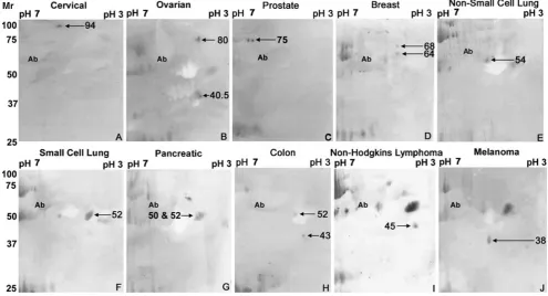
Fig. 2 Western blots of 2DGE of sera from cancer patients analyzed individually. Cancer sites are presented in the order of decreasing molecular weight of the major isoform present.a Cervical cancer, b
ovarian cancer,cprostate cancer,dbreast cancer,enon-small cell lung
cancer,fsmall cell lung cancer,gpancreatic cancer,hcolon cancer,i
The epitope of the scFv was that of the original MAB 12.1 and was to ENOX2 in the region of the Glu-Glu-Met-Thr-Glu drug (capsaicin)-binding motif [8,12].
Patient sera were from patients with clinically diagnosed and staged active disease from IRB-approved studies (See Acknowledgments). Informed consent was used. Non-cancer comparisons were with sera from healthy volunteers from the sources indicated and/or from multiple sets of pooled outpatient sera.
Results
The present report provides a method for the serum analysis of particular isoforms of the ENOX2 or tNOX (for tumor-specific NADH oxidase). The method entails 2DGE and immunoblotting using a recombinant antibody cross reac-tive with all of the various ENOX2 isoforms which charac-terize different types of cancers.
Analytical 2DGE of ECTO-NOX enriched serum proteins from a preparation of sera pooled from carcinoma patients (cervical, breast, ovarian, prostate, lung, and colon) concen-trated by nickel agarose precipitation, followed by immuno-blot analysis revealed multiple protein species in quadrants I and IV (Fig.1b) none of which were present in non-cancer sera (Fig. 1a). With separation in the first dimension by isoelectric focusing over the pH range of 3–10 and in the second dimension by 10% SDS-PAGE, a variety of acidic proteins of molecular weights between 34 and 100 kDa and absent from non-cancer sera were observed in quadrants I and IV (Fig.1b). Isoelectric points of the tNOX isoforms were in the range pH 3.9 and 6.3. Quadrants II and III of both cancer and non-cancer patients contained heavy (ca. 52 kDa) and light (ca. 25 kDa) immunoglobulin chains that were cross-reactive with the recombinant scFv at the relatively high amounts present in sera (Fig. 1) which served as convenient loading controls. Albumin and other serum proteins did not react with the antibody. It is unlikely that some of the spots with isoelectric points in the pH range 3.9–6.4, most of which have been identified as ENOX2 proteins based on capsaicin-inhibited enzymatic activities, are other proteins that cross react with the anti-body. Even albumin, a major protein of serum, the ap-proximate location of which is indicated on all of the gels to emphasize this point, is completely unreactive.
Sera from individual patients with the various forms of cancer were analyzed by 2DGE and immunoblotting to assign each of the ENOX2 isoforms to a cancer of a particular tissue of origin (Fig.2). Sera from cervical cancer patients contained the 94-kDa ENOX2 isoform (Fig. 2a), sera of ovarian cancer patients contained ENOX2 isoforms of 80 kDa and 40.5 kDa (Fig. 2b) and sera from patients with prostate cancer contained one or more 75-kDa ENOX2
isoforms resulting from small variations in isoelectric points (Fig. 2c). Sera of breast cancer patients contained 64 and 68-kDa ENOX2 isoforms (Fig.2d), sera from patients with non-small cell lung cancer contained a 54-kDa ENOX2 isoform (Fig. 2e), and sera from patients with small cell lung cancer contained a 52-kDa ENOX2 isoform together with a 40.5-kDa isoform similar to that seen for ovarian cancer (Fig. 2f). ENOX2 isoforms of Mr 50 and 52-kDa characterized sera of pancreatic cancer patients (Fig. 2g) whereas sera of colon cancer patients contain ENOX2 isoforms of 52 and 43 kDa (Fig. 2h). Figure 2i shows results from sera of a patient with non-Hodgkins lymphoma illustrating the 45-kDa ENOX2 isoform of low isoelectric point characteristic of leukemias and lymphomas. Sera of patients with malignant melanoma contain a ENOX2 isoform of Mr 38 kDa (Fig.2j).
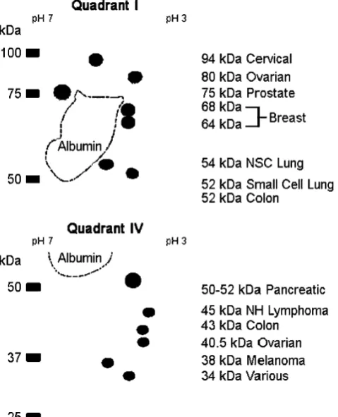
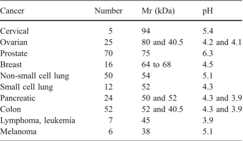
Results are depicted in a generalized analytical 2-D gel map of ENOX2 isoforms (Fig. 3) to show corresponding molecular weights. Isoelectric points together with the number of patients for which sera were analyzed are summarized in Table 1. The number of patients ranged from five (cervical cancer) to more than 70 (prostate cancer). The results were highly reproducible both with
regard to molecular weight (±1 kDa) and isoelectric point (±0.1 pH unit).
Analyses of breast, non-small cell lung, colon, prostate, and ovarian carcinoma patients where approximately equal numbers of tumor grades I through IV were available for analysis, revealed readily detectable spots even with grade I suggesting the potential for very early detection. However, between grade I and grade III, spot size was less than doubled (spot size doubles with a ca. tenfold increase in amount of immunoreactive protein based on experiments with recombinant ENOX2). As a result, the 2DGE system with detection by western blot analysis does not provide for precise quantitation of isoform amounts since spot size varies as the log of protein amount. Comparing 15 patients with stage I to IV prostate cancer and prostate specific antigen (PSA) levels between 5 and 250 ng/ml, the spot diameter and log PSA were correlated (r2=0.7) despite the log-linear relationship and the relatively low diagnostic specificity of the PSA [17].
Discussion
2DGE generates cancer-specific isoform patterns and compositions indicative of cancer presence, tumor type, disease severity, and possibly therapeutic response. The protocol resulted in the detection of more than ten cancer-specific ENOX2 isoforms which are resolved to indicate cancer presence and tissue of origin. The ENOX2 isoforms are identified on the basis of their molecular mass and isoelectric point with detection using an ENOX2-specific recombinant antibody single chain variable region scFv fragment expressed in bacteria which recognizes all ENOX2 proteins. The latter was produced from ENOX2-specific IgG rescued from ENOX2-ENOX2-specific monoclonal antibody producing hybridoma cells [12].
Each of the ENOX2 isoforms signaling cervical (94 kDa), ovarian (80 kDa), prostate (75 kDa), breast
(64–68 kDa), non-small cell lung (54 kDa), leukemia/ lymphoma (45 kDa), and melanoma (38 kDa) is repre-sented on 2DGE/western blots by ENOX2 antibody-reactive species of unique molecular weights. Colon, pancreatic, and small cell lung cancer share a common isoform of molecular weight 52 kDa. However, with colon cancer, the 52-kDa isoform is accompanied by a 40.5-kDa isoform with an isoelectric point of pH 3.9. For pancreatic cancer, the 52-kDa isoform exhibits an isoelectric point of pH 3.9 (compared to 4.3 for the 52-kDa isoform of colon and small cell lung cancer) and is accompanied by a 50-kDa isoform (Fig. 2g). The 40.5-kDa isoform which accompanies the 80-kDa isoform indicative of ovarian cancer appears to be the same or similar to the 40.5-kDa isoform characteristic of colon cancer. With small cell lung cancer, no obvious secondary ENOX2 isoform markers have been observed on any of the 2DGE western blots such that presence of the 52-kDa ENOX2 isoform and absence of the 40.5-kDa ENOX2 isoform serves to distinguish small cell lung from colon cancer.
Comparisons with known amounts of 34 kDa recombi-nant ENOX2 proteins produced in bacteria were used to demonstrate that ENOX2 proteins were absent or present at levels below the limits of detection (less than 10 pmol/ml of serum) from sera of healthy volunteers or patients with diseases other than cancer. In contrast, circulating ENOX2 has been detected in sera of more than 500 cancer patients representing all major forms of human cancer, including leukemias and lymphomas based on spectrophotometric measurements [10,11].
The presence of the ENOX2 protein was earlier dem-onstrated in a number of human tumor tissues and xeno-grafts from cytochemical studies [12]. However, serum analyses had already suggested a much broader association with cancer. ENOX2 proteins are ectoproteins reversibly bound at the outer leaflet of the plasma membrane [18]. As is characteristic of other examples of ectoproteins (sialyl and galactosyl transferases, dipeptidylamino peptidase IV, etc.), the ENOX2 proteins are shed [9,10]. They appear in soluble form in conditioned media of cultured cells [9], in patient sera [19] and in patient urine [20]. The ENOX2 isoforms from sera of cancer patients exhibit the same degree and specificity of inhibition by anti-cancer drugs (i.e., the EC50 values for inhibition of activity are the same or similar) as do the membrane-associated forms [10,11]. In contrast, no drug-responsive NOX activities were found with sera from healthy volunteers or sera from patients with diseases other than cancer. As such, the antitumor-responsive ENOX2 activity represents the first reported cell surface change undetected from non-cancer cells and potentially associated with most, if not all, forms of human cancer. ENOX2 isoform presence at all stages of cancer progression (I–IV) offer the potential for
non-Table 1 Sera from patients with different cancers exhibit patterns of ENOX2 isoforms with differing molecular weights and isoelectric points (pH)
invasive, early detection of cancer as well as the monitoring of therapeutic response and cancer recurrence and other diagnostic applications.
The ENOX2 (tNOX) gene is present in the human genome as a single copy, with no obvious homologs and a single constitutive ENOX1 (CNOX) ortholog. It is not a gene mutated in cancer but is universally present. The ENOX2 splice variants all appear to be variations resulting from an exon 4 minus splicing event that allows for down-stream initiation and expression at the cell surface of the ENOX2 protein only in cancer cells [13]. Without the exon 4 deletion, mRNA derived from the gene does not appear to be translated into protein.
Analysis of ENOX2 gene transcripts and the results from the findings with the pan-ENOX2 recombinant antibody establish that most, if not all, ENOX2 isoforms share a common region of protein structure of about 25 kDa (230 amino acids) at the carboxy terminus. This region contains the region of the protein responsible for its functional enzymatic activities, its response to anticancer drugs (drug-binding site) and the determinants in exon 5 at or near the drug-binding site recognized by the recombinant antibody. These properties are shared by all of the ENOX2 isoforms recognized on western blots. However, the remainder of the protein sequence of each of the isoforms must contain sequences which are unique to that particular isoform to account for their demonstrated differences in relative molecular weight and isoelectric point. These sequences will be available to generate monoclonal or peptide anti-bodies that recognize only these isoform-specific regions of the ENOX2 proteins.
Acknowledgments We thank Dr. Douglas Schwartzentrueber, M.D. and Dr. Kenneth Pennington, M.D. of Goshen Medical Center, Goshen, Indiana; Dr. Theodore Logan, M.D. of the Indiana University Medical Center, Indianapolis, IN and the Greater Baltimore Medical Center, Baltimore, Maryland (Dr. Paul Celano, M.D.) for providing serum samples for the studies. IRB-approved informed consent was obtained by the providing institution.
References
1. Baserga R. The biology of cell reproduction. Cambridge, MA: Harvard University Press; 1985.
2. Lambeth JD, Cheng G, Arnold RS, Edens WA. Novel homologs of gp91phox. TIBS. 2000;25:549–61.
3. Morre DJ, Pogue R, Morre DM. A multifunctional ubiquinol oxidase of the external cell surface and sera. BioFactors. 1999;9:179–87. 4. Morre DJ, Kim C, Hicks-Berger C. ATP-dependent and
drug-inhibited vesicle enlargement reconstituted using synthetic lipids and recombinant proteins. BioFactors. 2006;28:105–17.
5. Morre DJ, Morre DM. Cell surface NADH oxidases (ECTO-NOX proteins) with roles in cancer, cellular time-keeping, growth, aging and neurodegenerative disease. Free Radical Res. 2003; 37:795–808.
6. Bruno M, Brightman AO, Lawrence J, Werderitsh D, Morré DM, Morré DJ. Stimulation of NADH oxidase activity from rat liver plasma membranes by growth factors and hormones is decreased or absent with hepatoma plasma membranes. Biochem J. 1992;284:625–28.
7. Morre DJ. NADH oxidase: a multifunctional ectoprotein of the eukaryotic cell surface. In: Asard H, Berczi A, Caubergs R, editors. Plasma membrane redox systems and their role in biological stress and disease. The Netherlands: Kluwer, Dor-drecht; 1998. p. 121–56.
8. Chueh PJ, Kim C, Cho NM, Morré DM, Morré DJ. Molecular cloning and characterization of a tumor-associated, growth-related, and time-keeping hydroquinone (NADH) oxidase (tNOX) of the HeLa cell surface. Biochemistry. 2002;41:3732–41. 9. Wilkinson FE, Kim C, Cho NM, et al. Isolation and identification
of a protein with capsaicin-inhibited NADH oxidase activity from culture media conditioned by growth of HeLa cells. Arch Biochem Biophys. 1996;336:275–82.
10. Morre DJ, Caldwell S, Mayorga A, Wu L-Y, Morré DM. NADH oxidase activity from sera altered by capsaicin is widely distributed among cancer patients. Arch Biochem Biophys. 1997;342:224–30.
11. Morre DJ, Reust T. A circulating form of NADH oxidase activity responsive to the antitumor sulfonylurea N-4-(methylphenylsul-fonyl)-N’-(4-chlorophenyl)urea (LYl8l984) specific to sera of cancer patients. J Bioenerg Biomemb. 1997;29:281–9.
12. Cho NM, Chueh PJ, Kim C, Caldwell S, Morré DM, Morré DJ. Monoclonal antibody to a cancer-specific and drug-responsive hydroquinone (NADH) oxidase from sera of cancer patients. Cancer Immunol Immunother. 2002;51:121–29.
13. Tang X, Tian Z, Chueh PJ, Chen S, Morré DM, Morré DJ. Alternative splicing as the basis for specific localization of tNOX, a unique hydroquinone (NADH) oxidase, to the cancer cell surface. Biochemistry. 2007;46:12337–46.
14. Huston JS, Mudgett-Hunter M, Tai MS, McCartney J, Warren F, Haber E. Protein engineering of single-chain Fv analogs and fusion proteins. Methods Enzymol. 1991;203:46–88.
15. Davis GT, Bedzyk WD, Voss EW, Jacobs TW. Single chain antibody (SCA) encoding genes: one-step construction and expression in eukaryotic cells. Bio/Technology. 1991;9:165– 69.
16. Goldberg ME, Expert-Bezançon N, Vuillard L, Rabilloud T. Non-detergent sulphobetaines: a new class of molecules that facilitate
in vitroprotein renaturation. Structure. 1996;1:21–7.
17. Sardana G, Marshall J, Diamandis EP. Discovery of candidate tumor markers for prostate cancer via proteomic analysis of cell culture-conditioned medium. Clin Chem. 2007;53:429–37. 18. Morre DJ. NADH oxidase activity of HeLa plasma membranes
inhibited by the antitumor sulfonylurea N-(4-methylphenylsul-fonyl)-N-(4-chlorophenyl)urea (LY181984) at an external site. Biochim Biophys Acta. 1995;1240:201–08.
19. Wang S, Morre DM, Morré DJ. Sera from cancer patients contain two oscillating ECTO-NOX activities with different period lengths. Cancer Lett. 2003;190:135–41.