CODEN (USA): IAJPBB ISSN: 2349-7750
I
I
N
N
D
D
O
O
A
A
M
M
E
E
R
R
I
I
C
C
A
A
N
N
J
J
O
O
U
U
R
R
N
N
A
A
L
L
O
O
F
F
P
PH
HA
AR
RM
MA
AC
CE
EU
UT
TI
IC
CA
AL
L
S
SC
CI
IE
EN
NC
CE
ES
S
http://doi.org/10.5281/zenodo.545840
Available online at:
http://www.iajps.com
Research Article
ISOLATION, ANTIBIOGRAM AND CHARACTERIZATION OF
VANCOMYCIN-RESISTANT STAPHYLOCOCCUS AUREUS
FROM CLINICAL AND COMMUNITY ISOLATES IN
ABAKALIKI, EBONYI STATE, NIGERIA
Iroha I. R
1, Ariom T. O
1, Moses I. B *
1, Ejikeugwu P. C
1, Nwuzo A. C
1, Afiukwa F. N
1,
Nwakaeze E. A
1, Iroha C. S
2.
1
Department of Applied Microbiology, Faculty of Sciences, Ebonyi State University, Abakaliki.
2Department of Pharmacy, Federal Teaching Hospital, Abakaliki.
Received:
10 March 2017
Accepted:
25 March 2017
Published:
28 March 2017
Abstract:
This study was aimed at isolating and characterizing vancomycin-resistant Staphylococcus aureus from clinical and community samples in Abakaliki, Ebonyi State, Nigeria. Seven hundred and nine (709) clinical and community samples were obtained for this study. Antibiotic susceptibility test was done using Kirby Bauer disc diffusion method. Vancomycin resistant Staphylococcus aureus (VRSA) isolates were detected using Kirby-Bauer disc diffusion method with vancomycin antibiotic disc (30 μg). Isolates were also screened for β-lactamase enzyme production. A total of 84 (27.7 %) and 120 (29.5 %) Staphylococcus aureus isolates were obtained from the clinical and community samples respectively using standard microbiological techniques. Results showed that 55 VRSA isolates were obtained from the samples with prevalence frequency of 36.9 % and 20 % for clinical and community isolates respectively. The clinical isolates were completely resistant (100 %) to nitrofurantoin, clindamycin, ceftazidime, tetracycline and penicillin. Gentamicin was the most effective antibiotic against the S. aureus isolates obtained from clinical samples as all the isolates were completely susceptible (100 %). Ciprofloxacin was the most effective antibiotic against the S. aureus isolates obtained from community samples with a susceptibility frequency of 100 %. This was closely followed by gentamicin (75 %) and erythromycin. Exactly 38.1 % and 24.2 % of the clinical and community S. aureus isolates were positive for beta-lactamase production respectively. The HA-VRSA and CA-VRSA isolates had MARI values within the range of 0.5 to 1.0. This present discovery of multi-drug resistant VRSA with high multiple antibiotic resistance indices is in Abakaliki is a serious public health issue. Therefore, there is an urgent need to keep a strict watch on VRSA emerging from Abakaliki.
Keywords: CA-VRSA, HA-VRSA, MARI, beta-lactamase, antibiotics
Corresponding author:
Moses I. B,
Department of Applied Microbiology,
Faculty of Sciences, Ebonyi State University, Abakaliki
E-mail: [email protected]
Telephone: +2348134136233
Please cite this article in press as Iroha I. R et al, Isolation, Antibiogram and Characterization of Vancomycin-Resistant Staphylococcus Aureus from Clinical and Community Isolates In Abakaliki, Ebonyi State, Nigeria,
Indo Am. J. Pharm. Sci, 2017; 4(03).
INTRODUCTION:
Staphylococcus aureus has been implicated in a wide range of infection ranging from acute to chronic infections such as boils, bacteriuria, osteomyelitis, pneumonia, endocarditis, meningitis, septicemia and arthritis. This organism is a leading cause of human bacterial infection worldwide and is endemic in both hospital and the community (Chambers and Deleo, 2009). Multiple antibiotic resistant Staphylococcus aureus are major threats to patients’ care, owing to their stubborn intransigence to chemotherapy and disinfection (Slade et al., 2009). The resistant bacteria may spread and create broader infection control problems, both within healthcare institutions and in the community. The infections especially caused by Staphylococcus aureus have affected substantial portions of the human population, causing significant mobility and mortality (Fred, 2006). Prolonged therapy with vancomycin may lead to development of low-level resistance that compromise therapy, but that may not be detected by routine susceptibility testing methods used in hospital laboratory (Tenover, 2007). Most Staphylococcal infections are associated with serious community-acquired and nosocomial diseases which arise often in individuals with predisposing risk factors such as haemodialysis or surgery. It causes superficial, deep-skin, soft tissue infections, endocarditis and bacteremia with metastatic abscess formation and a variety of toxin–mediated diseases including gastroenteritis, staphylococcal scalded skin syndrome, toxic shock syndrome, meningitis, septicaemia and arthritis (Amghalia et al., 2009). After several attempts, scientist discovered the use of vancomycin as the frontier in the fight against
Staphylococcus aureus infection. Until recently, vancomycin was the only uniformly effective antibiotic for the treatment of Staphylococcus
infection. The euphoria was however cut short in 1997 by the detection of VRSA in Japan from clinical isolates. Based on the recognition that management of severe Staphylococcus aureus disease is challenging, research is needed to further evaluate the prevalence of vancomycin resistant Staphylococcus aureus (VRSA) in Abakaliki, Ebonyi State.
MATERIALS AND METHODS:
Sample collection
A total of 709 samples were collected for this study. Three hundred and three (303) were clinical samples
swab samples were collected using sterile swab sticks while urine, semen and sputum samples were collected using sterile specimen bottles. In the same way, the four hundred and six (406) community samples (nasal and ear swabs) were collected from apparently healthy tutors (121), artisans (62), secondary school students of Abakaliki high school (153) and petty traders from Kpirikpiri Market (70).
The Hospital patients’ group includes individuals who are at greater risk of becoming infected by this opportunistic pathogen. These individuals are generally older, have chronic underlying illnesses, and require more frequent interactions with healthcare facilities; all of which predispose them to more serious infections. The Community group includes individuals who, in general, are otherwise healthy. They are usually not predisposed by age or underlying illness to these infections, but predisposed by specific activities and community interactions that place them at an increased risk for infection contraction.The collected samples were immediately transported to the department of Applied Microbiology laboratory unit of Ebonyi State University, Abakaliki for bacteriological analysis.
Culturing, isolation, characterization and identification of the isolates
The clinical and community samples were aseptically inoculated on Mannitol Salt broth and incubated at 37
0C for 48 hours. A loopful of the inoculated mannitol
salt broth was later streaked on mannitol salt agar (MSA) and incubated at 37 0C for 24 hours. The plates were observed for creamy golden β-haemolytic colonies which are typical characteristic of
Staphylococcus aureus. These suspected S. aureus
isolates were further characterized using conventional/standard microbiology techniques such as colony morphology, Gram-staining, catalase test, motility test and other biochemical tests which include oxidase test, indole test, Simmon’s citrate
test, H2S production test, voges proskauer test,
methyl red test, sugar fermentation test, coagulase test and Staphylococcus lactase agglutination assay (Cheesbrough, 2004).
Antibiotic Susceptibility Test
The susceptibility patterns of isolated S. aureus
µg), ciprofloxacin (5 µg), ceftazidime (30 µg), sulphamethoxazole (25 µg) and clindamycin (2 µg) (Oxoid, UK) were placed on the inoculated plates using sterile forceps. The plates were incubated at 37
0C for 24 hours after which the zones of inhibition
around each disc were measured to the nearest mm with a metre rule, recorded and interpreted according to the Clinical Laboratory Standard Institute (CLSI, 2009) guidelines.
Detection of vancomycin resistant Staphylococcus aureus (VRSA)
This was done using Kirby Bauer disc diffusion method according to Clinical and Laboratory Standard Institute (CLSI, 2009) guidelines. A Mueller-Hinton agar plate was prepared according to
manufacturer’s specification. Colonies of the isolated bacteria were suspended in 5 ml of nutrient broth. The turbidity of the broth culture was adjusted to 0.5 McFarland standard, which approximately equals 1.5 108 CFU/ml.Standardized inoculum was swabbed
onto the prepared Mueller-Hinton agar plate. After at least 3 minutes, antibiotic disc impregnated with vancomycin (30 μg) was placed on Mueller-Hinton agar plate for VRSA detection. The plate was then incubated at 37 0C for 24 hours. Inhibition zone
diameter was measured to nearest millimeter and interpreted according to CLSI guidelines.
Beta-Lactamase Detection Using Nitrocefin Stick
This was done to detect the production of beta-lactamases by the S. aureus isolates. Before inoculation, the nitrocefin stick was allowed to cool to room temperature. A drop of distilled water was used to moderately moisten the tip of the nitrocefin stick. After this, the colour coded end of the stick was used to touch the colony by making sure that the reagent on the brown tip of the stick was rotated to pick mass of the cells. The colour coded tip was observed after 5 minutes for pink-red colour development to show the production of beta-lactamase.
Determination of multiple antibiotic resistance index (MARI)
Multiple antibiotic resistance indices (MARI) of the
S. aureus isolates were calculated using the technique described by Christopher et al. (2013) and Subramani
et al. (2012). This was calculated as the number of antibiotics to which the tested isolate was resistant to (a), divided by the total number of antibiotics that was tested on the isolates (b).
RESULTS:
Table 1: Clinical samples collected from Federal Teaching Hospital Abakaliki (FETHA I)
Sample
source No. of samples collected No. of isolated S. aureus VRSA isolated No. of
Wound swab 21 11 5
Urine 33 6 2
High vaginal swab 11 2 1
Sputum 7 2 0
Ear swab 17 9 2
Pus 7 2 1
Semen 21 4 0
Table 2: Clinical samples collected from Federal Teaching Hospital Abakaliki (FETHA II)
Table 3: Clinical samples collected from Mile Four General Hospital Abakaliki Sample
source
No. of samples collected
No. of S. aureus
isolated
No. of VRSA isolated
Wound swab 10 7 3
Urine 35 7 2
High vaginal swab 13 4 2
Sputum 8 2 1
Ear swab 10 5 3
Pus 9 4 2
Semen 18 2 0
TOTAL 103 31 (30.1 %) 13 (41.9 %)
Sample
source No. of samples collected No. of isolated (%) S. aureus VRSA isolated No. of
Wound swab 5 3 2
Urine 31 4 2
High vaginal swab 17 3 0
Sputum 20 3 1
Ear swab 10 4 2
Pus 0 0 0
Semen 0 0 0
Table 4: Samples collected from apparently healthy individuals in the Community
Table 5: Prevalence of VRSA in Hospital and Community isolates
Sample source
VRSA
No. of isolate tested No. positive
Hospital 84 (27.7 %) 31 (36.9 %)
Community 120 (29.6 %) 25 (20 %)
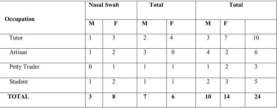
Table 6: Occupational distribution of population with Community Acquired Vancomycin Resistant
Staphylococcus aureus (CA-VRSA)
Occupation Ear Swab Nasal Swab Total No of
Samples
No of Staph.
Isolated
No of Samples
No of Staph.
Isolated
Sample
Isolate
Tutors 66 25 55 19 121 44
Artisans 32 13 30 9 62 22
Students 73 18 80 14 153 32
Petty Traders 38 12 32 10 70 22
Total 209 68 197 52 406 120 (29.6 %)
Occupation
Nasal Swab Total Total
M F M F M F
Tutor 1 3 2 4 3 7 10
Artisan 1 2 3 0 4 2 6
Petty Trader 0 1 1 1 1 2 3
Student 1 2 1 1 2 3 5
Fig 1: Percentage Susceptibility and Resistance patterns of Hospital Acquired Vancomycin Resistant
Table 7: Beta-lactamase detection in Hospital and Community isolates
Source No of isolates tested Beta-lactamase positive
Hospital 84 32
Community 120 29
Total 204 58
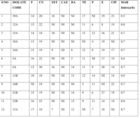
Table 8: Multiple Antibiotic Resistance Indices (MARI) of Hospital Acquired Vancomycin Resistant
Staphylococcus aureus (HA-VRSA) isolates
Key: F= Nitrofurantoin, CN= Gentamicin, SXT= Sulphamethoxazole, CAZ= ceftazidime, DA= Clindamycin, TE= Tetracycline, P= Penicillin G, E= Erythromycin, CIP= Ciprofloxacin NI= No Inhibition
S/NO ISOLATE CODE
F CN SXT CAZ DA TE P E CIP MAR Index(a/b)
1 39A 14 20 18 NI NI 17 NI 19 23 0.5
2 12A 15 22 NI NI NI 11 6 8 19 0.8
3 15A 14 18 10 NI NI 13 12 16 21 0.7
4 18A 13 19 NI NI NI NI 4 19 NI 0.7
5 34A 15 19 9 NI 8 12 8 19 17 0.7
6 5A 16 22 NI NI 5 11 NI 17 19 0.6
7 6A 12 20 16 NI 14 11 9 20 14 0.7
8 11B 10 18 NI NI 15 12 14 NI 14 0.8
9 16B NI 18 NI NI NI 5 11 NI 22 0.7
10 21B 15 19 NI NI 14 9 6 27 18 0.7
11
12 23B
13A
16
17 23
10
NI
7
NI
NI 15
12 9
NI 11
7
14
18 18
NI
0.8
Table 9: Multiple Antibiotic Resistance Indices (MARI) of CA-VRSA isolates
Key: F= Nitrofurantoin, CN= Gentamicin, SXT= Sulphamethoxazole, CAZ= ceftazidime, DA= Clindamycin, TE= Tetracycline, P= Penicillin G, E= Erythromycin, CIP= Ciprofloxacin NI= No Inhibition
DISCUSSION:
Staphylococcus aureus has long been recognized as one of the major human pathogens responsible for hospital and community acquired infections. Infections caused by vancomycin resistant S. aureus
(VRSA) have been associated with high morbidity and mortality rates. In view of the fact that
Staphylococcus aureus has the capacity to change over time, VRSA will keep on being a problem in the future, as it has been long-ago. In this study, 36 (30.8 %) S. aureus isolates were obtained from 117 clinical samples collected from FETHA I. Eleven (30.6 %) out of these isolates were identified as vancomycin resistant Staphylococcus aureus (VRSA) (Table 1). In FETHA II, 31(30.1 %) S. aureus isolates were obtained from the 103 clinical samples collected from patients. Thirteen (41.9 %) of these isolates were identified as VRSA (Table 2). In Mile Four Hospital, 17 (20.5 %) S. aureus isolates were obtained from 83 clinical samples. Seven (41.2 %) of the isolates were identified as VRSA (Table 3). One hundred and twenty (29.6 %) S. aureus isolates were obtained from the 496 community samples (nasal and ear swabs) collected from tutors, artisans, students and petty traders (Table 4). This study is in agreement with the work of Olowe et al. (2007) in Osogbo, South-western Nigeria. They reported the isolation of 42.9 % S. aureus from Staphylococci isolates. Thirty
Staphylococcus aureus isolates obtained from the community samples of nasal and ear swabs (Table 5).
The distribution of VRSA isolated from the three major hospitals in Abakaliki showed that the highest prevalence frequency of VRSA was found in FETHA II (13 (41.9 %)). This was closely followed by FETHA I (11 (35.5 %) and Mile Four General Hospital ((7 (22.6 %) being the least (Table 5). The highest number of VRSA was isolated from wound samples (10); followed by ear swabs (7), urine (6), HVS (3) and pus (3). The least prevalence of VRSA was observed in sputum samples (2). There was no VRSA isolate from semen samples (Tables 1, 2 and 3). This is similar to the report of Olowe et al. (2007) who reported that wound samples had the highest prevalence of S. aureus. Many researchers have reported an increase in the incidence of S. aureus
most of which originated from wounds (Vidhani et al., 2001). CA-VRSA was most predominant among tutors (10). The overcrowded nature of tutors’ staff
rooms and irrational use of antibiotics might possibly be the reason for the high prevalence of VRSA among them. This is because overcrowded living places have been seen as one of the risk factors for community-acquired S. aureus (Scerri et al., 2013). The least number of CA-VRSA was observed among petty traders (Table 6). CA-VRSA isolates were more prevalent in nasal swabs than ear swabs (Table
S/NO ISOLATE CODE
F CN
SXT CAZ DA TE P E CIP MAR Index(a/b)
1 3C 16 9 4 NI 15 20 4 11 24 0.7
2 30C 6 NI NI NI NI NI NI NI 19 1
3 47C 10 14 NI NI NI NI NI NI 22 0.8
4 13C 16 19 6 NI 19 15 9 23 24 0.5
(100 %) to nitrofurantoin, clindamycin, ceftazidime, tetracycline and penicillin. They also exhibited high level of resistance to sulphamethoxazole-trimethoprin (90.9 %). Gentamicin was the most effective antibiotic against the Staphylococcus aureus isolates obtained from clinical samples as all the isolates were completely susceptible (100 %) (Figure 1). This is in agreement with the work of Ibrahim et al. (2013) where antibiotic susceptibility results of S. aureus
showed absolute resistance (100 %) against ampicillin, and high resistance against cefotaxime (81 %), while resistance frequencies to ceftriaxone, erythromycin, ciprofloxacin, trimethoprim, gentamicin, levofloxacin and clindamycin were 59 %, 59 %, 41 %, 41 %, 35 %, 23 % and 18 % respectively. The susceptibility frequency of VRSA isolates to gentamicin in this study is similar to the findings of Tiwari et al. (2006) who reported a high susceptibility frequency of multidrug S. aureus
isolates to gentamicin. The CA-VRSA isolates were also multidrug resistant. All the HA-VRSA isolates were completely resistant (100 %) to ceftazidime, sulphamethoxazole and penicillin. This was closely followed by gentamicin (80 %), nitrofurantoin (80 %), clindamycin (80 %) and tetracycline (60 %). Interestingly, ciprofloxacin was the most effective antibiotic against the Staphylococcus aureus isolates obtained from community samples with a susceptibility frequency of 100 %. This was closely followed by gentamicin (75 %) and erythromycin (Figure 2). Most notable results of VRSA that was recorded by Chakraborty et al. (2011) which included eight pathogenic VRSA strains isolated from post operative pus sample in India and that is similar to the findings of our research. These resistances might result from inappropriate prescriptions due to lack of standard treatment guidelines (WHO, 2015). In developing countries, an estimated 50 % of those who need antimicrobials do not have access to them due to cost. Sometimes, these drugs are not taken by patients as prescribed and some people indulge in self-medication (WHO, 2009). Relentless exposure of
bacterial strains to a large number of β-lactams has induced active and continuous production and
mutation of β-lactamases in these bacteria, thus expanding their activity even against the newly
developed β-lactam antibiotics. Thirty-two (32) VRSA (or HA-VRSA) isolates out of the 84 clinical samples of S. aureus isolates were positive for beta-lactamase production while 29 CA-VRSA isolates were positive for beta-lactamase production (Table 7). This is in agreement with the findings of Terry et al. (2011) in South-East Nigeria who reported that 124 (64 %) of their S. aureus isolates were able to
produce β-lactamase enzyme by changing colour from yellow to red on the addition of nitrocefin
solution. Production of β-lactamase in S. aureus is reported to have been consistently high in Nigeria (Torimiro et al., 2013). They reported that 70-80 % of S. aureus isolates produced β-lactamase. Other
researchers reported such high β-lactamase prevalence (Kesah et al., 1997; Akindele et al.,
2010). The spread of β-lactamase genes had been enhanced by their integration within mobile genetic elements such as plasmids and transposons which facilitate the rapid transfer of genetic materials between microbes (Wilke et al., 2005). Multidrug resistant bacteria have created therapeutic problems especially to healthcare providers and, its consistent emergence will only cause hazard to the public health (Amalia et al., 2016). The HA-VRSA isolates had MARI values within the range of 0.5 to 0.8. These high MARI values depict the high resistances of these isolates to antibiotics (Table 8).The MARI values for CA-VRSA ranged from 0.5 to 1.0. These MARI values indicate that the CA-VRSA isolates have higher antibiotic resistance potential than the HA-VRSA isolates (Table 9). Our study is in agreement with the report of Subramani and Vignesh (2012) who revealed that all the S. aureus isolates in their study had very high MARI values of above 0.2. This signified that the bacterial isolates have been exposed to several antibiotics and the selective pressure of the antibiotics used in the management of bacterial infections could be a vital reason for resistance apart from the bacteria acquiring the resistance gene through mutation or interspecies gene transmission.
CONCLUSION:
This study has been able to show that VRSA isolates are present in some major hospitals in Abakaliki and within the community in Abakaliki, Ebonyi State, Nigeria. The report of this study has established that nitrofurantoin, clindamycin, ceftazidime, tetracycline and penicillin are now ineffective in treating VRSA infections whereas gentamycin, ciprofloxacin and erythromycin are still very effective. The high resistance frequencies exhibited by these S. aureus
serious public health problem. Therefore, it is pertinent to strictly monitor the prevalence of VRSA emerging from Abakaliki, Nigeria.
CONFLICT OF INTEREST STATEMENT
The authors declare that there are no conflicts of interest
REFERENCES:
1.Akindele, A. A., Adewuyi, I. K., Adefioye, O. A., Adedokun, S. A., Olaolu, A.O. Antibiogram and Beta-lactamase of Staphylococcus aureus isolates from different human Clinical Specimens in a Tertiary Health Institution in Ile-Ife, Nigeria. Am Eurasian Journal of Science Res., 2010; 5(4):230-233.
2.Aligholi M., Emaneini M., Jabalameli F. (2008). Emergence of high-level vancomycin-resistant
Staphylococcus aureus in the Imam Khomeini hospital in Tehran. Med Princ Pract., 17: 432– 4. 3.Amalia, A. R., Ramzi, O.S.B. and Son, R.Antibiotic resistance evolution of Methicillin Resistant Staphylococcus aureus (MRSA) and colloidal silver as the nano-weapon.International Food Research Journal,2016; 23(3): 1248-1254. 4.Amghalia, E., Nagi, A AL-Haj., Mariana, Shamsudin, N., Son Radu., RozitaRosli, Neela V. and Raha A. Rahim, A.Multiplex PCR Assay for Detection of Clinically Relevant Antibiotic Resistance Genes in Staphylococcus aureus Isolated from Malaysian Hospitals. Research Journal of Biological Sciences,2009;4 (4): 444-448.
5.Chakraborty, S. P., KarMahapatra, S. K., Bal, M. and Roy, S.(2011). Isolation and identification of vancomycin resistant Staphylococcus aureusfrom post operative pus sample.Alameen Journal of Medical Sciences, 4 (2): 52–68.
6.Chambers, H.F and Deleo, F.R. Waves of Resistant
Staphylococcus aureus in the Antibiotics Era.
Journal Review Microbiology, 2009; 7: 629-641. 7.Cheesbrough, M. District Laboratory Practice in Tropical Countries, Part Two,2ndEdition. Cambridge
University Press, UK, 2004; Pp 134-180.
8.Clinical and Laboratory Standard Institute. Performance Standard for Antimicrobial Susceptibility Testing, 17th Information Supplement
(M100-517). Wayne, Pa: Clinical and Laboratory Standard Institute; 2007.
9.Fred, C. Mechanism of Antimicrobial Resistance in Bacteria. The American Journal of Medicine, 2006;
11.Kesah C. N., Ogunsola F. T., Neemogha M. T., Odungbemi T. O. An in-vitro Study of Methicillin
and Other Antimicrobial Agent
againstStaphylococcusaureus, 1994 -1996, NigQt. J. Hosp Med., 1997; 7(3):286-88.
12.Olowe, O. A., Eniola, K. I. T., Olowe, R. A., &Olayemi, A. B.Antimicrobial Susceptibility and Beta-lactamase detection of MRSA in Osogbo, SW Nigeria. Nature and Science, 2007; 5(3), 44-48. 13.Scerri J, Monecke S, Borg M. A. Prevalence and characteristics of community carriage of methicillin-resistant Staphylococcus aureus in Malta. J Epidemiol Glob Health, 2013; 3:165–173.
14.Slade D, Lindner A. B., Paul G., Radman M. Recombination and replication in DNA repair of heavily irradiated Deinococcusradiodurans. Cell, 2009; 136: 1044–1055.
15.Subramanian P, Shanmugam N, Sivaraman U, Kumar S, Selvaraj S. Antibiotic resistance pattern of biofilm-forming uropathogens isolated from catheterised patients in Pondicherry, India. Australas Med J., 2012; 5(7):344–348.
16.Tenover, F. C., Weigel, L. M., Appelbaum, P. C. Vancomycin Resistant Staphylococcus aureus
Isolation from a Patient in Rennsylvania. Journal of Antimicrobial Agents Chemotherapy, 2004; 48: 275-280.
17.Terry Alli O.A., Ogbolu D.O., Akorede E., Onemu O.M., and Okanlawon B.M. Distribution of mecA gene amongst Staphylococcus aureus
isolates from south western Nigeria. AfricanJournal of Biomedical Research, 2011;14:9-16.
18.Tiwari H. K., Sen M. R. Emergence of vancomycin resistant Staphylococcus aureus (VRSA) from a tertiary care hospital from northern part of India. BMC Infectious Disease, 2006; 6: 156-161. 19.Torimiro, N., Moshood, A. A and Eyiolawi, S.A. Analysis of Beta-lactamase Production and Antibiotics Resistance in Staphylococcus aureus
Strains.Journal of Infectious Diseases and Immunity, 2013; 5(3) 24-28.
20.Wilke, M.S., Lovering, A .L., Strynadka, C.J.N.β -lactam Antibiotic Resistance: A Current Structural perspective. Curr. Microbiol., 2005; 8: 525-533. 21.World Health Organisation (WHO). Antimicrobial
resistance; 2015. Avaliable at