R E S E A R C H
Open Access
Pro-inflammation NF-
κ
B signaling triggers a
positive feedback via enhancing cholesterol
accumulation in liver cancer cells
Mingyan He
1†, Wenhui Zhang
2†, Yinying Dong
1†, Lishun Wang
3, Tingting Fang
1, Wenqing Tang
1, Bei Lv
1,
Guanglang Chen
3, Biwei Yang
1, Peixin Huang
1and Jinglin Xia
1,3*Abstract
Background:Hepatocellular carcinoma (HCC) develops in a complex microenvironment characterized by chronic inflammation. In recent years, cholesterol metabolic abnormalities have been implicated the importance in cancer cell physiology. This study was designed to investigate the relationship between inflammation and cholesterol accumulation in HCC cells.
Methods:Human HCC cells HepG2 and Huh7 were cultured and stimulated with lipopolysaccharide (LPS) for 24 h. The changes of HCC cells related to cholesterol metabolism including intracellular cholesterol concentrations, cholesterol uptake, and the expression of cholesterol-related genes 3-hydroxy-3-methylglutaryl-CoA reductase (HMGCR), LDL receptor (LDLR), sterol regulatory element-binding transcription factor 2 (SREBF2), and proprotein convertase subtilisin/kexin 9 (PCSK9) were comparatively analyzed. Simultaneously, the effects of nuclear factor-kappa B (NF-κB) signaling pathway on cholesterol metabolism were clarified by knocking-down of nuclear factor kappa-B kinase subunit alpha (IKKα) and TGF-beta-activated kinase 1 and MAP3K7-binding protein 3 (TAB3) via RNAi and microRNA (miR)-195. Subsequently, the roles of cholesterol accumulation in LPS induced pro-inflammatory effects were further investigated.
Results:Pro-inflammatory factor LPS significantly increased intracellular cholesterol accumulation by upregulating the expression of HMGCR, LDLR, and SREBF2, while downregulating the expression of PCSK9. These effects were revealed to depend on NF-κB signaling pathway by knocking-down and overexpression of IKKαand TAB3.
Additionally, miR-195, a regulator directly targeting IKKαand TAB3, blocked the effects of cholesterol accumulation, further supporting the critical role of pro-inflammation NF-κB signaling in regulating cholesterol accumulation. Intriguingly, the accumulation of cholesterol conversely exerted an augmented pro-inflammation effects by further activating NF-κB signaling pathway.
Conclusions:These results indicated that pro-inflammation effects of NF-κB signaling could be augmented by a positive feedback via enhancing the cholesterol accumulation in liver cancer cells.
Keywords:Lipopolysaccharide, Nuclear factor-kappa B, Cholesterol accumulation, microRNA-195, Hepatocellular carcinoma
* Correspondence:[email protected]
†Equal contributors
1Liver Cancer Institute, Zhongshan Hospital, Fudan University, Shanghai
200032, China
3Minhang Hospital, Fudan University, Shanghai 201100, China
Full list of author information is available at the end of the article
Background
Metabolic reprogramming in the uncontrolled prolifera-tion of cancer cells has been thought to play important roles in cancer biology [1]. Recently, many studies have demonstrated that cholesterol accumulate in a series of human cancers, including breast [2], colon [3], prostate [4], HCC [5], and others [6]. In addition, enzymes involved in cholesterol metabolism have been reported abnormal expression in cancer tissues [7, 8]. For instance, choles-terol acyltransferase (ACAT)2, is found to be induced and promotes esterification of excess oxysterols for secretion to avoid cytotoxicity in a subset of hepatocellular carcin-omas (HCCs) for tumor growth [9], suggestive of a spe-cific cholesterol metabolic pathway in HCCs. On the one hand, cholesterol is needed for the synthesis of mem-branes, signaling molecules, lipid raft formation, and other factors to support the rapid growth of tumor cells [8]. On the other hand, cholesterol oxidized products, namely oxysterols, exhibit inhibitory activities in cell growth and promote cell apoptosis and dampen antitumor responses [10–12]. However, mechanism and pathological signifi-cance underlying the aberrant cholesterol metabolism are still elusive.
Epidemiological studies have shown that 80% of HCCs develop in fibrotic or cirrhotic livers as a consequence of chronic liver injury [13, 14]. The NF-κB pathway, the key link of inflammatory responses, plays an important role in HCC promotion by increasing proliferation and preventing apoptosis [15]. In the inactive state, NF-κB transcription factors are complexed in the cytoplasm, either with members of the inhibitor ofκB (IκB) family (in the canonical pathway) or with the NF-κB precursor p100 (in the non-canonical pathway). Lipopolysaccharide (LPS), a component of the gram-negative bacterial wall, could activate NF-κB pathway and stimulate inflamma-tory responses. Previous studies have demonstrated that LPS/NF-κB signaling pathway promotes atherosclerotic progression and macrophage foam cell formation by in-creasing intracellular cholesterol accumulation [16, 17]. These reports inspired us to hypothesize whether there is a crosstalk between NF-κB signaling and cholesterol metabolism in cancer cells.
In the present study, we investigated the relationship between inflammation and cholesterol metabolism, and found that pro-inflammatory factor Lipopolysaccharide (LPS) increased intracellular cholesterol accumulation through NF-κB pathway in HCC cells. Cholesterol accu-mulation conversely promoted LPS/NF-κB pathway and inflammatory responses.
Methods Cell culture
Human HCC cells HepG2 and Huh7 were purchased from Cell Bank of the institute of Biochemistry and Cell
Biology, China Academy of Sciences, Shanghai, China. Cells were cultured in Dulbecco’s Modified Eagle’s Medium (DMEM) supplemented with 10% (v/v) fetal bovine serum (Gibco, BRL, USA), 10ug/mL strepto-mycin sulfate and 100ug/mL penicillin G. All cells were incubated at 37 °C in a humidified atmosphere contain-ing 5% CO2.
LDL preparation
LDL was purchased from AngYu Biotechnologies (Shanghai, China). It is isolated from blood bank pro-duced human plasma. It is purified via ultracentrifugation to homogeneity determined by agarose gel electrophoresis. Each lot is analyzed on agarose gel electrophoresis for mi-gration versus LDL.
Measurement of intracellular cholesterol
Total cellular cholesterol measurement was determined with a detection kit from Applygen (Peking, China) ac-cording to the manufacturer’s protocol. Intracellular lipids were extracted using a chloroform-methanol (2:1) mix and dried under vacuum. Total cholesterol contents were measured by an enzymatic assay. The value was normalized against total protein concentration from each sample, as determined with Bicinchoninic Acid assay (Beyotime biotechnology, China).
DIL-LDL uptake
The low density lipoprotein labeled with 1,1′ -Dioctade-cyl-3,3,3′,3′-tetramethyl- indocarbocyanideperchlorate (DIL-LDL) (Biomedical Technologies Inc., MA, USA) uptake represented the ability of cell cholesterol uptake. HCC cells were treated with different addressment, then changed to serum-free medium and incubated with 10ug/ml DIL-LDL at 37 °C for 5 h. The cells were de-tached by trypsin, washed and suspended by PBS and analyzed by flow cytometry using FL2 emission filter (FACScan, BD Biosciences, San Jose, CA, USA). Data were analyzed with FlowJo software.
Total RNA isolation and quantitative real-time quantita-tive PCR
analysis, the first-strand cDNA was synthesized using the reverse transcriptase with miRNAs-specific stem-loop pri-mer (RiboBio, Guangzhou, China). PCR amplification was conducted in ABI7500 real-time fluorescent measurement system (Applied Biosystems, USA) using the Bulge-Loop miRNA qRT-PCR Starter Kit (RiboBio, Guangzhou, China) according to the protocol provided. The relative expression level of miRNA was normalized to that of en-dogenous control U6 by using 2-△△Ctmethod.
Western blot
HCC cells were lysed in cell lysis buffer containing 1nM PMSF for 30 min at 4 °C. Lysates were collected by centri-fugation at 12,000 rpm for 30 min at 4 °C. Proteins from cell lysates were separated on the SDS-PAGE and trans-ferred onto PVDF membrane (Immobion-P Transfer Mem-brane, Millipore Corp., Billerica, MA, USA). The membrane was blocked with TBST containing 5% non-fat dry milk for 1 h and further incubated overnight at 4 °C with primary antibodies against PCSK9, LDLR, HMGCR, SREBF2 (Abcam, Cambridge, MA, USA), NF-κB p65, phospho-NF-κB p65, IKKα, TAB3 (Cell Signaling Technol-ogy, USA) and β-actin (Santa Cruz, CA, USA). After that, the membrane was incubated with horseradish peroxidase (HRP)-linked secondary antibodies (Santa Cruz Biotechnol-ogy, USA) for 2 h at room temperature. All protein bands were visualized using an electrochemiluminescence kit (Thermo, USA). Intensity of each protein band was quanti-fied by Quantity One 4.6.2 software (Bio Rad).
MiRNA inhibitor or mimic transfection
For transfections in 12-well plates, the HCC cells were seeded at a density of 1 × 10⁵ cells/well and incubated overnight and then transfected with 50 nM miR-195
mimic or 100 nM miR-195 inhibitor (GenePharma, Shanghai, China) using lipo2000 Transfection Agent (Invitrogen, USA) according to the manufacturer’s in-structions. The corresponding negative sequence of mimic or inhibitors (GenePharma, Shanghai, China) were transfected with the same concentration as con-trols. At 24 h after the transfection, cells were harvested or further incubated with LPS (1000 ng/mL) for the fol-lowing experiments.
RNA interference
Gene silencing was performed by infecting HCC cells with siRNA oligonucleotides (GenePharma, Shanghai, China). The siRNA sequences were shown in Table 2. For trans-fections in 12-well plates, 1.0 × 10⁵cells were seeded per well and incubated overnight, then transfected with 80nM TAB3-siRNA, IKKα-siRNA or negative sequence using lipo2000 Transfection Agent (Invitrogen, USA) according to the manufacturer’s protocol. At 24 h after the transfec-tion, cells were harvested or further incubated with LPS (1000 ng/mL) for the following experiments.
ELISA
HCC cells were stimulated with LPS (1000 ng/ml) for 24 h in serum-free medium in the presence or absence of LDL (200ug/ml). The concentrations of IL-8 and TNF-α in culture supernatants were measured with ELISA kits (R&D Systems, USA) according to the manu-facturer’s instruction. Absorbance was recorded at 450 nm using a microplate reader.
Statistical analysis
Data analysis was performed using SPSS16.0 statistical software (SPSS Inc, Chicago, IL). Student’s t-tests were
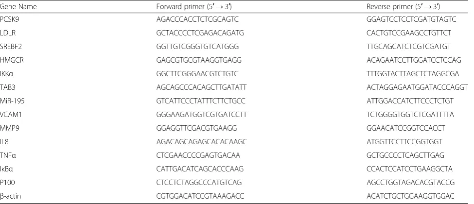
Table 1Primers for PCR used in this study
Gene Name Forward primer (5′→3′) Reverse primer (5′→3′)
PCSK9 AGACCCACCTCTCGCAGTC GGAGTCCTCCTCGATGTAGTC
LDLR GCTACCCCTCGAGACAGATG CACTGTCCGAAGCCTGTTCT
SREBF2 GGTTGTCGGGTGTCATGGG TTGCAGCATCTCGTCGATGT
HMGCR GAGCGTGCGTAAGGTGAGG ACAGAATCCTTGGATCCTCCAG
IKKα GGCTTCGGGAACGTCTGTC TTTGGTACTTAGCTCTAGGCGA
TAB3 AGCAGCCCACAGCTTGATATT ACTAGGAGAATGGATACCCAGGT
MiR-195 GTCATTCCCTATTTCTTCTGCC ATTGGACCATCTTCCCTCTGT
VCAM1 GGGAAGATGGTCGTGATCCTT TCTGGGGTGGTCTCGATTTTA
MMP9 GGAGGTTCGACGTGAAGG GGAACATCCGGTCCACCT
IL8 AGACAGCAGAGCACACAAGC ATGGTTCCTTCCGGTGGT
TNFα CTCGAACCCCGAGTGACAA GCTGCCCCTCAGCTTGAG
IκBα CATTGACATCAGCACCCAAG CCACTCCATCCTGAAGGCTA
P100 CTCCTCTAGGCCCATGTCAG AGCCTGGTAGACACGTACCG
used to assess significant differences among study groups. P< 0.05 was considered statistically signifi-cant. All experiments have been performed at least three times.
Results
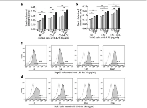
Role of NF-κB signaling in cholesterol accumulation To assess whether cholesterol metabolism could be affected by NF-κB pathway in HCC cells, we moni-tored intracellular cholesterol levels in response to LPS at dosages ranging from 0 to 1000 ng/ml in serum free (SF) medium or complete medium (CM)
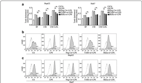
or CM with LDL loading (200 μg/ml). As shown in Fig. 1a and b, LPS significantly increased the intracel-lular cholesterol concentration in a dose-dependent manner in HepG2 and Huh7 HCC cells. And the ef-fects were not affected by constitutive cholesterol levels. Furthermore, we found that uptake of 3,3′ -dioctadecylindocarbocyanine-low density lipoprotein (DIL-LDL) in HCC cells was stimulated by LPS, sug-gesting that it may increase native LDL cholesterol uptake via LDLR (Fig. 1c and d).
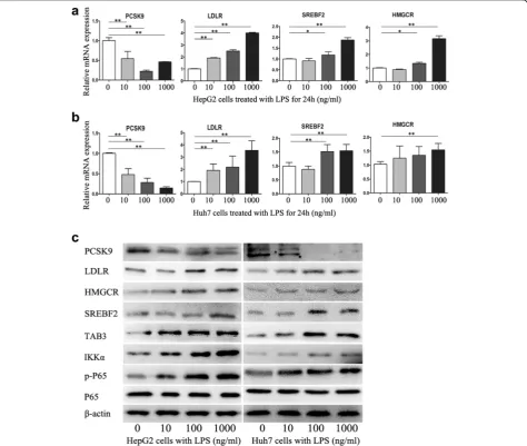
Next, we examined the effect of LPS on the expression of proprotein convertase subtilisin/kexin 9 (PCSK9),
Table 2The sequences of siRNA used in this study
Identifier Sense Primer Sequence (5′→3′) Anti-sense Primer Sequence (3′→5′)
NC siRNA CCACCUCUGAUCGAUUUAUdTdT dTdTAUAAAUCGAUCAGAGGUGG
IKKαsiRNA GCAGGCUCUUUCAGGGACAdTdT dTdTCGUCCGAGAAAGUCCCUGU
TAB3 siRNA CCAAAGGUUCCAUGAAGAAdTdT dTdTGGUUUCCAAGGUACUUCUU
Fig. 1Effects of LPS on cholesterol metabolism in HCC cells.a,bCells treated with 0, 10, 100, 1000 ng/ml LPS in serum free (SF) medium or complete medium (CM) in the absence or presence of LDL (200 ug/ml) for 24 h, the total cholesterol ester concentration was measured;c,d
LDLR, HMGCR and sterol regulatory element-binding transcription factor 2 (SREBF2) in HCC cells (Fig. 2a-c). After treatment with various concentrations of LPS for 24 h, PCSK9 mRNA and protein levels in HCC cells were significantly downregulated, and the expression of LDLR, HMGCR and SREBF2 were upregulated in a dose-dependent manner. In addition, LPS promotes the expression of IKKα, TAB3 and phosphorylated P65 (Fig. 2c), demonstrating the role of NF-κB pathway in HCC intracellular cholesterol accumulation.
IKKαand TAB3 regulates LPS-induced cholesterol accumulation
To further examine whether LPS mediated cholesterol accumulation by NF-κB pathway, we synthesized siR-NAs specifically against nuclear factor kappa-B kinase
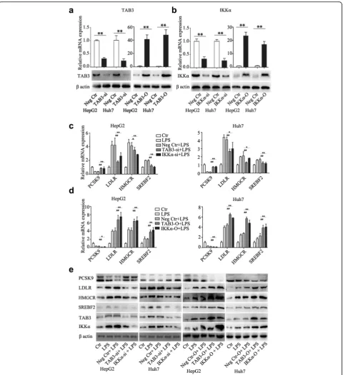
subunit alpha (IKKα) and TGF-beta-activated kinase 1 and MAP3K7-binding protein 3 (TAB3) and overex-pressed IKKα and TAB3 in HepG2 and Huh7 cells. The down- and up-regulated efficacy of TAB3 and IKKα was confirmed by quantitative real-time PCR and western blotting (Fig. 3a and b). Twenty-four hours after transfection, HCC cells were treated with LPS (1000 ng/ml) for 24 h, and PCSK9, LDLR, HMGCR, and SREBF2 expression, total cellular chol-esterol levels, and uptake of DIL-LDL were measured at the endpoints. As expected, down-regulation of TAB3 and IKKα increased PCSK9 expression and de-creased LDLR, HMGCR, and SREBF2 expression, while up-regulation of TAB3 and IKKα decreased PCSK9 expression and increased LDLR, HMGCR, and SREBF2 expression (Fig. 3c-e). Additionally,
regulation of TAB3 and IKKα abrogated the effects of LPS on cholesterol accumulation (Fig. 4a–c). These findings indicated that TAB3 and IKKα were indeed functional in LPS-induced cholesterol accumulation.
MiR-195 regulates cholesterol accumulation and cholesterol-related gene expression induced by LPS Previous findings have shown that miR-195 decreases the activity of NF-κB signaling and the expression of Fig. 3IKKαand TAB3 regulates cholesterol-related gene expression induced by LPS.a,bqRT-PCR and western blotting confirm the down- and up-regulation of IKKαor TAB3 expression in HCC cells.c,d,eTwenty-four hours after transfection, HCC cells were treated with LPS (1000 ng/ml) for 24 h, and then PCSK9, LDLR, HMGCR and SREBF2 expression were detected. Data represent the mean ± SD of triplicate samples;
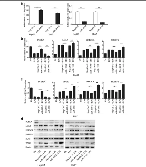
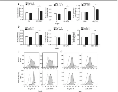
NF-κB downstream effectors by directly targeting TAB3 and IKKα[19]. To further investigate the involvement of NF-κB signaling in cholesterol metabolism, the cells overexpressing or knocking down miR-195 were estab-lished by transfecting with miR-195 mimic, mir-195 in-hibitor. As shown in Fig. 5a, these cells achieved more than a 100-fold upregulation and 80% downregulation of mRNA expression, respectively (Fig. 5a). Twenty-four hours after transfection with miR-195 mimic, miR-195 inhibitor, or negative control, HCC cells were treated with 1000 ng/mL LPS for another 24 h and then ana-lyzed for intracellular cholesterol levels and related gene expression. Relative to the controls, mRNA and protein expression levels of PCSK9 were downregulated by LPS, and LDLR, HMGCR, and SREBF2 expression was upreg-ulated. Overexpression of miR-195 increased PCSK9 ex-pression and decreased LDLR, HMGCR, and SREBF2 expression, while knockdown of miR-195 decreased PCSK9 expression and increased LDLR, HMGCR, and SREBF2 expression (Figs. 5b–d). In addition, overexpres-sion of miR-195 inhibited the total intracellular choles-terol level (Fig. 6a and b) and DIL-LDL uptake (Fig. 6c). These data indicate that miR-195 negatively regulates LPS-induced cholesterol accumulation and expression of cholesterol-related genes.
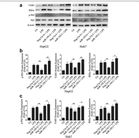
Next, we investigated the phosphorylation status of p65 and the expression levels of TAB3 and IKKα, which were the direct gene targets of miR-195. We observed that overexpression of miR-195 significantly inhibited the phosphorylation status of p65 and the expression levels of TAB3 and IKKα, while knockdown of miR-195 increased the phosphorylation status of p65 and the ex-pression levels of TAB3 and IKKα (Fig. 7), supporting our findings that NF-κB pathway played an important role in HCC intracellular cholesterol accumulation.
Cholesterol increases LPS induced pro-inflammatory effects
by LDL at 200ug/ml in both HepG2 and Huh7 cells. The data suggest that accumulated cholesterol increases LPS/NF-κB-induced inflammatory effects by increasing p-IKKαin HCC cells.
Discussion
HCC, among the most malignant of human cancers [20], develops in a complex microenvironment charac-terized by chronic inflammation [13, 14]. Emerging evi-dence has demonstrated that perpetuating hepatic inflammation promotes HCC initiation and progression [13, 21, 22]. Meanwhile, cholesterol metabolic abnormal-ities have been implicated the importance in cancer cell physiology in recent years [2–4, 23]. However, the rela-tionship between the aberrant cholesterol metabolism and inflammation in HCCs are not completely under-stood. In the present study, we demonstrated that LPS/
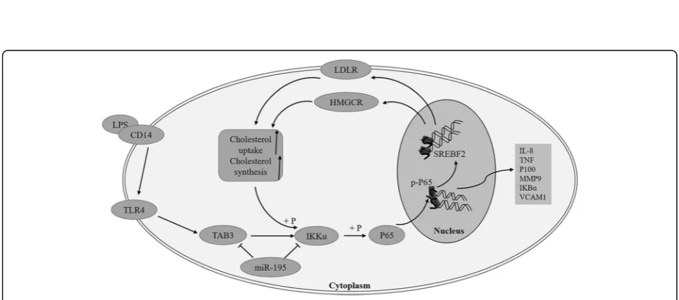
NF-κB pathway induced a significant increase in the intra-cellular cholesterol concentration and in DIL-LDL uptake in HCC cells, suggesting a key role of NF-κB pathway in promoting cholesterol accumulation. Additionally, the cholesterol accumulation conversely promoted
LPS/NF-κB-induced inflammatory effects, indicating a positive feedback of inflammation and cholesterol metabolism (Fig. 9).
NF-κB activity by directly targeting the expression of IKKα TAB3 in HCCs [19]. Here, we demonstrated that not only miR-195 but also its targeted genes TAB3 and IKKα mediate LPS/NF-κB-induced cholesterol accumu-lation and cholesterol-related gene expression, providing further evidence that NF-κB pathway play an important role in HCC intracellular cholesterol accumulation.
PCSK9, the ninth member of the proteinase K subfamily of subtilases, plays an important role in post-transcriptional degradation of LDLR, which decreases intracellular
Fig. 8LDL increases LPS induced pro-inflammatory effects. LDL increases intracellular cholesterol contents (a), LDL significantly activated the phosphorylation of p65 and IKKα(b), increased the expression of NF-κB target genes (c,d), promoted the production of IL-8 and TNF-αin HCC cells with administration of LPS for 24 h (e,f). Data represent the mean ± SD of triplicate samples; *P< 0.05, **P< 0.01
has been demonstrated that PCSK9, LDLR, and HMGCR expression are co-regulated by SREBF2 [36–38]. When cholesterol levels fall, SREBF2 is activated to up-regulate the expression of genes responsible for cholesterol synthe-sis, such as HMGCR, and for cholesterol uptake, such as LDLR. In this study, the expression of PCSK9, LDLR, HMGCR, and SREBF2 were investigated in HCC cells after stimulation with LPS. We found that LPS significantly inhibited the expression of PCSK9 and increased LDLR, HMGCR, and SREBF2 expression, suggesting that LPS may increase native LDL cholesterol uptake via LDLR and
pro-motede novocholesterol synthesis via HMGCR.
There are growing evidences that cholesterol, as an important molecule, impacts upon cancer cell physi-ology, however, the concrete role of cholesterol in cancer progression remains elusive and controversial. Analyses of the cancer Genome Atlas (TCGA) database revealed a correlation between increased activity of the cholesterol synthesis pathway and decreased survival in patients with sarcoma, acute myeloid leukemia and melanoma [39, 40], supporting the concept that cholesterol pro-motes carcinogenesis. However, some epidemiological studies have reported objective observation that poor prognosis in HCC patients were linked to decreased serum cholesterol [41, 42]. In this study, we have sug-gested that cholesterol further activated the NF-κB sig-naling pathway and promotes the expression of NF-κB target genes, indicating the pro-inflammatory effects of cholesterol in HCC cells.
Conclusions
In summary, we have experimentally demonstrated that LPS/NF-κB signaling pathway triggers an increase in intracellular cholesterol levels by promoting the expres-sion of HMGCR and LDLR in HCC cells. Cholesterol accumulation conversely promotes LPS/NF-κB induced pro-inflammatory effects.MiR-195, as a regulator of
NF-κB pathway, inhibited cholesterol accumulation by de-creasing the expression of TAB3 and IKKα. These data provide us with a better understanding of the relation-ship between LPS/NF-κB pathway and cholesterol ab-normalities in cancer cells.
Abbreviations
DIL-LDL:Low density lipoprotein labeled with 1,1′-Dioctadecyl-3,3,3′,3′ -tetramethyl-indocarbocyanideperchlorate; HCC: Hepatocellular carcinoma; HMGCR: 3-hydroxy-3-methylglutaryl-CoA reductase; IKKα: Nuclear factor kappa-B kinase subunit alpha; LDLR: LDL receptor; LPS: Lipopolysaccharide; miR-195: microRNA-195; NF-κB: Nuclear factor-kappa B; PCSK9: Proprotein convertase subtilisin/kexin 9; SREBF2: Sterol regulatory element-binding tran-scription factor 2; TAB3: TGF-beta-activated kinase 1 and MAP3K7-binding protein 3
Acknowledgements
Not applicable.
Funding
This study was sponsored by grants from the National Natural Science Foundation of China (Nos. 81272732 and 81572395), the Shanghai Leading Talent Projects (No. 048, 2013), the Shanghai Leading Academic Discipline Project (Project Number: B115), and the Shanghai Science and Technology Commission (Project Number: 14XD1401100).
Availability of data and materials
Not applicable.
Authors’contributions
JLX and LSW conceived and designed the study. MYH, WHZ, YYD performed the experiments. TTF, WQT, BL, GLC, BWY, PXH analyzed the data. All authors read and approved the final manuscript.
Competing interests
The authors declare that they have no competing interests.
Consent for publication
Not applicable.
Ethics approval and consent to participate
Not applicable.
Author details
1Liver Cancer Institute, Zhongshan Hospital, Fudan University, Shanghai
200032, China.2Cancer Hospital of Baotou City, Inner Monggolia 014030, China.3Minhang Hospital, Fudan University, Shanghai 201100, China.
Received: 7 October 2016 Accepted: 4 January 2017
References
1. Warburg O. On the origin of cancer cells. Science. 1956;123(3191):309–14. 2. de Gonzalo-Calvo D, López-Vilaró L, Nasarre L, Perez-Olabarria M, Vázquez T,
Escuin D, Badimon L, Barnadas A, Lerma E, Llorente-Cortés V. Intratumor cholesteryl ester accumulation is associated with human breast cancer proliferation and aggressive potential: a molecular and clinicopathological study. BMC Cancer. 2015;15(1):460.
3. Accioly MT, Pacheco P, Maya-Monteiro CM, Carrossini N, Robbs BK, Oliveira SS, Kaufmann C, Morgado-Diaz JA, Bozza PT, Viola JPB. Lipid bodies are reservoirs of cyclooxygenase-2 and sites of prostaglandin-E2 synthesis in colon cancer cells. Cancer Res. 2008;68(6):1732–40.
4. Yue S, Li J, Lee S, Lee HJ, Shao T, Song B, Cheng L, Masterson TA, Liu X, Ratliff TL, Cheng J. Cholesteryl ester accumulation induced by PTEN loss and PI3K/AKT activation underlies human prostate cancer aggressiveness. Cell Metab. 2014;19(3):393–406.
5. Jiang J, Xu N, Zhang X, Wu C. Lipids changes in liver cancer. J Zhejiang Univ Sci B. 2007;8(6):398–409.
6. White C. The occurence of crystals in tumours. J Pathol Bacteriol. 1909; 13(1):3–10.
7. Stine JE, Guo H, Sheng X, Han X, Schointuch MN, Gilliam TP, Gehrig PA, Zhou C, Bae-Jump VL. The HMG-CoA reductase inhibitor, simvastatin, exhibits anti-metastatic and anti-tumorigenic effects in ovarian cancer. Oncotarget. 2016;7(1):946–60.
8. Abramson HN. The lipogenesis pathway as a cancer target. J Med Chem. 2011;54(16):5615–38.
9. Lu M, Hu XH, Li Q, Xiong Y, Hu GJ. A specific cholesterol metabolic pathway is established in a subset of HCCs for tumor growth. J Mol Cell Biol. 2013; 5(6):404–15.
10. Gill S, Chow R, Brown AJ. Sterol regulators of cholesterol homeostasis and beyond: the oxysterol hypothesis revisited and revised. Prog Lipid Res. 2008; 47(6):391–404.
11. Lanterna C, Musumeci A, Raccosta L, Corna G, Moresco M, Maggioni D, Fontana R, Doglioni C, Bordignon C, Traversari C, Russo V. The
administration of drugs inhibiting cholesterol/oxysterol synthesis is safe and increases the efficacy of immunotherapeutic regimens in tumor-bearing mice. Cancer Immunol Immunother. 2016;65(11):1303–15.
13. Luedde T, Schwabe RF. NF-κB in the liver—linking injury, fibrosis and hepatocellular carcinoma. Nat Rev Gastro Hepat. 2011;8(2):108–18. 14. Fattovich G, Stroffolini T, Zagni I, Donato F. Hepatocellular carcinoma
in cirrhosis: incidence and risk factors. Gastroenterology. 2004;127(5 Suppl 1):S35–50.
15. Dapito DH, Mencin A, Gwak G, Pradere J, Jang M, Mederacke I, Caviglia JM, Khiabanian H, Adeyemi A, Bataller R, Lefkowitch JH, Bower M, Friedman R, Sartor RB, Rabadan R, Schwabe RF. Promotion of hepatocellular carcinoma by the intestinal microbiota and TLR4. Cancer Cell. 2012;21(4):504–16. 16. Li LC, Varghese Z, Moorhead JF, Lee CT, Chen JB, Ruan XZ. Cross-talk between TLR4-MyD88-NF- B and SCAP-SREBP2 pathways mediates macrophage foam cell formation. Am J Physiol Heart Circ Physiol. 2013; 304(6):H874–84.
17. Lehr HA, Sagban TA, Ihling C, Zahringer U, Hungerer KD, Blumrich M, Reifenberg K, Bhakdi S. Immunopathogenesis of atherosclerosis: endotoxin accelerates atherosclerosis in rabbits on hypercholesterolemic diet. Circulation. 2001;104(8):914–20.
18. Livak KJ, Schmittgen TD. Analysis of relative gene expression data using real-time quantitative PCR and the 2(−delta delta C(T)) method. Methods. 2001;25(4):402–8.
19. Ding J, Huang S, Wang Y, Tian Q, Zha R, Shi H, Wang Q, Ge C, Chen T, Zhao Y, Liang L, Li J, He X. Genome-wide screening reveals that miR-195 targets the TNF-alpha/NF-kappaB pathway by down-regulating IkappaB kinase alpha and TAB3 in hepatocellular carcinoma. Hepatology. 2013;58(2):654–66. 20. Venook AP, Papandreou C, Furuse J, de Guevara LL. The incidence and
epidemiology of hepatocellular carcinoma: a global and regional perspective. Oncologist. 2010;15 Suppl 4:5–13.
21. Wilson CL, Jurk D, Fullard N, Banks P, Page A, Luli S, Elsharkawy AM, Gieling RG, Chakraborty JB, Fox C, Richardson C, Callaghan K, Blair GE, Fox N, Lagnado A, Passos JF, et al. NFkappaB1 is a suppressor of neutrophil-driven hepatocellular carcinoma. Nat Commun. 2015;6:6818.
22. Sanz-Cameno P, Trapero-Marugan M, Chaparro M, Jones EA, Moreno-Otero R. Angiogenesis: from chronic liver inflammation to hepatocellular carcinoma. J Oncol. 2010;2010:272170.
23. Ribas V, García-Ruiz C, Fernández-Checa JC. Mitochondria, cholesterol and cancer cell metabolism. Clin Transl Med. 2016;5(1):22.
24. Griffiths-Jones S, Saini HK, van Dongen S, Enright AJ. miRBase: tools for microRNA genomics. Nucleic Acids Res. 2008;36(Database issue):D154–8. 25. Guo J, Wang M, Liu X. MicroRNA-195 suppresses tumor cell proliferation and metastasis by directly targeting BCOX1 in prostate carcinoma. J Exp Clin Cancer Res. 2015;34:91.
26. Cai C, Chen QB, Han ZD, Zhang YQ, He HC, Chen JH, Chen YR, Yang SB, Wu YD, Zeng YR, Qin GQ, Liang YX, Dai QS, Jiang FN, Wu SL, Zeng GH, et al. miR-195 inhibits tumor progression by targeting RPS6KB1 in human prostate cancer. Clin Cancer Res. 2015;21(21):4922–34.
27. Liu B, Qu J, Xu F, Guo Y, Wang Y, Yu H, Qian B. MiR-195 suppresses non-small cell lung cancer by targeting CHEK1. Oncotarget. 2015;6(11):9445–56. 28. Han K, Chen X, Bian N, Ma B, Yang T, Cai C, Fan Q, Zhou Y, Zhao TB.
MicroRNA profiling identifies MiR-195 suppresses osteosarcoma cell metastasis by targeting CCND1. Oncotarget. 2015;6(11):8875–89. 29. Xu T, Zhu Y, Xiong Y, Ge YY, Yun JP, Zhuang SM. MicroRNA-195 suppresses
tumorigenicity and regulates G1/S transition of human hepatocellular carcinoma cells. Hepatology. 2009;50(1):113–21.
30. Maxwell KN, Breslow JL. Adenoviral-mediated expression of Pcsk9 in mice results in a low-density lipoprotein receptor knockout phenotype. Proc Natl Acad Sci U S A. 2004;101(18):7100–5.
31. Nassoury N, Blasiole DA, Tebon OA, Benjannet S, Hamelin J, Poupon V, McPherson PS, Attie AD, Prat A, Seidah NG. The cellular trafficking of the secretory proprotein convertase PCSK9 and its dependence on the LDLR. Traffic. 2007;8(6):718–32.
32. Lagace TA, Curtis DE, Garuti R, McNutt MC, Park SW, Prather HB, Anderson NN, Ho YK, Hammer RE, Horton JD. Secreted PCSK9 decreases the number of LDL receptors in hepatocytes and in livers of parabiotic mice. J Clin Invest. 2006;116(11):2995–3005.
33. Bhat M, Skill N, Marcus V, Deschenes M, Tan X, Bouteaud J, Negi S, Awan Z, Aikin R, Kwan J, Amre R, Tabaries S, Hassanain M, Seidah NG, Maluccio M, Siegel P, et al. Decreased PCSK9 expression in human hepatocellular carcinoma. BMC Gastroenterol. 2015;15:176.
34. Chushi L, Wei W, Kangkang X, Yongzeng F, Ning X, Xiaolei C. HMGCR is up-regulated in gastric cancer and promotes the growth and migration of the cancer cells. Gene. 2016;587(1):42–7.
35. Singh R, Yadav V, Kumar S, Saini N. MicroRNA-195 inhibits proliferation, invasion and metastasis in breast cancer cells by targeting FASN, HMGCR, ACACA and CYP27B1. Sci Rep. 2015;5:17454.
36. Jeong HJ, Lee HS, Kim KS, Kim YK, Yoon D, Park SW. Sterol-dependent regulation of proprotein convertase subtilisin/kexin type 9 expression by sterol-regulatory element binding protein-2. J Lipid Res. 2008;49(2):399–409. 37. Dubuc G, Chamberland A, Wassef H, Davignon J, Seidah NG, Bernier L, Prat A. Statins upregulate PCSK9, the gene encoding the proprotein convertase neural apoptosis-regulated convertase-1 implicated in familial
hypercholesterolemia. Arterioscler Thromb Vasc Biol. 2004;24(8):1454–9. 38. Horton JD, Goldstein JL, Brown MS. SREBPs: activators of the complete
program of cholesterol and fatty acid synthesis in the liver. J Clin Invest. 2002;109(9):1125–31.
39. Kuzu OF, Noory MA, Robertson GP. The role of cholesterol in cancer. Cancer Res. 2016;76(8):2063–70.
40. Network CGAR, Weinstein JN, Collisson EA, Mills GB, Shaw KR, Ozenberger BA, Ellrott K, Shmulevich I, Sander C, Stuart JM. The Cancer Genome Atlas Pan-Cancer analysis project. Nat Genet. 2013;45:1113–20.
41. Chen Z, Keech A, Collins R, Slavin B, Chen J, Campbell TC, Peto R. Prolonged infection with hepatitis B virus and association between low blood cholesterol concentration and liver cancer. BMJ. 1993;306(6882):890–4. 42. Li WX. Serum cholesterol and cancer mortality: eleven-year prospective
cohort study on more than nine thousand persons. Zhonghua Liu Xing Bing Xue Za Zhi. 1993;14(1):6–9.
• We accept pre-submission inquiries
• Our selector tool helps you to find the most relevant journal
• We provide round the clock customer support
• Convenient online submission
• Thorough peer review
• Inclusion in PubMed and all major indexing services
• Maximum visibility for your research
Submit your manuscript at www.biomedcentral.com/submit