R E S E A R C H
Open Access
A novel antagonist of p75NTR reduces
peripheral expansion and CNS trafficking of
pro-inflammatory monocytes and spares
function after traumatic brain injury
Sangmi Lee
1, Aaron Mattingly
2, Amity Lin
1, Jeffrey Sacramento
1, Leda Mannent
3, Marie-Noelle Castel
3,
Benoit Canolle
3, Sandrine Delbary-Gossart
4, Badia Ferzaz
3, Josh M. Morganti
1, Susanna Rosi
1,2, Adam R. Ferguson
1,2,
Geoffrey T. Manley
1, Jacqueline C. Bresnahan
1and Michael S. Beattie
1,2*Abstract
Background:Traumatic brain injury (TBI) results in long-term neurological deficits, which may be mediated in part by pro-inflammatory responses in both the injured brain and the circulation. Inflammation may be involved in the subsequent development of neurodegenerative diseases and post-injury seizures. The p75 neurotrophin receptor (p75NTR) has multiple biological functions, affecting cell survival, apoptotic cell death, axonal growth, and degeneration in pathological conditions. We recently found that EVT901, a novel piperazine derivative that inhibits p75NTR oligomerization, is neuroprotective, reduces microglial activation, and improves outcomes in two models of TBI in rats. Since TBI elicits both CNS and peripheral inflammation, we used a mouse model of TBI to examine whether EVT901 would affect peripheral immune responses and trafficking to the injured brain. Methods:Cortical contusion injury (CCI)-TBI of the sensory/motor cortex was induced in C57Bl/6 wild-type mice and CCR2+/RFPheterozygote transgenic mice, followed by treatment with EVT901, a selective antagonist of p75NTR, or vehicle by i.p. injection at 4 h after injury and then daily for 7 days. Brain and blood were collected at 1 and 6 weeks after injury. Flow cytometry and histological analysis were used to determine peripheral immune responses and trafficking of peripheral immune cells into the lesion site at 1 and 6 weeks after TBI. A battery of behavioral tests administered over 6 weeks was used to evaluate neurological outcome, and stereological estimation of brain tissue volume at 6 weeks was used to assess tissue damage. Finally, multivariate principal components analysis (PCA) was used to evaluate the relationships between inflammatory events, EVT901 treatment, and neurological outcomes.
(Continued on next page)
* Correspondence:[email protected]
1Department of Neurological Surgery, Brain and Spinal Injury Center, University of California at San Francisco, Box 08991001 Potrero Ave, Bldg 1, Rm 101, San Francisco, CA 94143-0899, USA
2Biomedical Science Graduate Program, University of California at San Francisco, San Francisco, CA 94143-0899, USA
Full list of author information is available at the end of the article
(Continued from previous page)
Results:EVT901 is neuroprotective in mouse CCI-TBI and dramatically reduced the early trafficking of CCR2+ and pro-inflammatory monocytes into the lesion site. EVT901 reduced the number of CD45highCD11b+ and CD45highF4/80 + cells in the injured brain at 6 weeks. TBI produced a significant increase in peripheral pro-inflammatory monocytes (Ly6Cint-highpro-inflammatory monocytes), and this peripheral effect was also blocked by EVT901 treatment. Further, we found that blocking p75NTR with EVT901 reduces the expansion of pro-inflammatory monocytes, and their response to LPS in vitro, supporting the idea that there is a peripheral EVT901 effect that blunts inflammation. Further, 1 week of EVT901 blocks the expansion of pro-inflammatory monocytes in the circulation after TBI, reduces the number of multiple subsets of pro-inflammatory monocytes that enter the injury site at 1 and 6 weeks post-injury, and is neuroprotective, as it was in the rat.
Conclusions:Together, these findings suggest that p75NTR signaling participates in the production of the peripheral pro-inflammatory response to CNS injury and implicates p75NTR as a part of the pro-inflammatory cascade. Thus, the neuroprotective effects of p75NTR antagonists might be due to a combination of central and peripheral effects, and p75NTR may play a role in the production of peripheral inflammation in addition to its many other biological roles. Thus, p75NTR may be a therapeutic target in human TBI.
Keywords:Traumatic brain injury (TBI), p75 neurotrophin receptor (p75NTR), Inflammatory responses, Pro-inflammatory monocytes, Neuroprotection, Therapeutic target
Background
Traumatic brain injury (TBI) is a huge public health prob-lem; 1.7 million people sustain a brain injury annually, and TBI is a contributing factor to a third of all injury-related deaths (CDC statistics). Mild to severe TBI patients often suffer long-term disabilities, including cognitive, sensory and motor dysfunction, dementia, fatigue, epilepsy,
head-aches, depression, and regression of social behavior [1–6].
Despite the large numbers of TBI patients, there is no effective pharmacological treatment available. Under-standing the pathogenesis of TBI is critical; TBI triggers complex and heterogenous pathologic events initiated by the mechanical injury and hemorrhage and the sub-sequent progressive secondary injury processes [3]. In addition to necrotic and apoptotic cell death cascades [7, 8], these include robust inflammatory responses in the injured brain including activation of resident micro-glia and subsequent infiltration of peripheral leuko-cytes. Chronic TBI patients often display long-lasting inflammatory responses continuing for months to years [9, 10]. Such inflammation may contribute to structural brain abnormalities, cognitive dysfunction [10], and white matter degeneration (e.g., corpus callosum shrinkage) [11]. Thus, neuroinflammation likely plays a critical role in the pathology of chronic TBI.
TBI also increases pro-inflammatory responses in the peripheral circulation. A recent clinical study shows that trauma increased the level of MCP-1 (monocyte chemo-tactic protein-1; also known as CCL2) in the peripheral blood suggesting that peripheral immune system activa-tion may provide a surrogate biological index for TBI impairment [12]. Traumatic spinal cord injury (SCI) in-creases the number of myeloid cells in the circulation, a
pressage to the infiltration of myeloid cells into the in-jured spinal cord [13]. Ischemic brain injury releases soluble damage-associated molecular pattern (DAMP) molecules that induce peripheral inflammation via toll-like receptors (TLRs) on peripheral leukocytes [13, 14]. These data suggest that monitoring peripheral myeloid cells may predict not only the pro-inflammatory re-sponses of peripheral blood cells but also the trafficking of myeloid cells into the CNS. Indeed, some studies report that peripheral blood cells increase pro-inflammatory
genes in response to brain injury [15–17]. Furthermore,
peripheral blood mononuclear cells (PBMCs) isolated from post-traumatic stress disorder (PTSD) patients have elevated spontaneous pro-inflammatory cytokine
expres-sion, e.g., interleukin-1 beta (IL-1β), interleukin-6 (IL-6),
and tumor necrosis factor-alpha (TNF-α), providing
evi-dence that PBMCs can be a circulating biological indicator or biomarker of neuroinflammation after injury [18]. Interestingly, several preclinical studies of SCI and TBI show that attenuating leukocyte infiltration into the in-jured CNS in the acute period after trauma often results in not only inhibition of inflammatory responses in the in-jured tissue but also better neurological outcomes in the
long term [13, 19–24]. Thus, peripheral inflammation
ini-tiated in response to CNS trauma can feed forward and exacerbate dysfunction. Further, chronic peripheral in-flammation can lead to immune suppression, which is a common feature of brain and spinal cord injuries, adding significantly to post-injury morbidities [25].
We and others have previously targeted secondary in-jury processes associated with activation of the p75 neurotrophin receptor (p75NTR) as a neuroprotective
p75NTR, a low-affinity neurotrophin receptor, is a transmembrane protein in the TNF receptor superfam-ily (TNFRS-16) [30, 31]. p75NTR binds to all mature neurotrophins (NTs) with a low affinity; however, the dimerization with its multiple co-receptors conveys high-affinity for different ligands and provides ligand selectivity [32]. For example, p75NTR-Trks (A, B, and C) dimers have much greater affinity for mature NTs,
mediating cell survival and neurite outgrowth [32–34].
However, when p75NTR forms complexes with sortilin, it has a high affinity for the pro-neurotrophins (pro-nerve growth factor (NGF) and pro-brain-derived neurotrophic factor (BDNF)) and mediates apoptotic cell death [32, 35], while p75NTR-Nogo/Lingo 1 com-plexes prefer to bind with myelin-derived ligands (oligodendrocyte myelin glycoprotein (OMgp) and myelin-associated glycoprotein (MAG)), resulting in the inhibition of axonal regeneration [36, 37].
p75NTR is widely expressed in the developing brain, but its expression is restricted to few regions in the adult
brain [38–40]. However, it is up-regulated, along with its
ligand pro-NGF [26, 41] in various CNS pathologies and after injury [32]. p75NTR null mice show reduced cor-tical neuronal cell death after close axotomy [27], re-duced tissue damage and motor deficits after TBI [42], and reduced oligodendrocyte apoptosis after SCI [26], and treatment with a p75NTR antagonist results in oligodendrocyte protection and improvement in func-tional recovery after SCI [29]. A recent study has also
shown protection and improved recovery in p75NTR−/−
mice [42]. We have recently studied the effects of a novel p75NTR antagonist, EVT901, on two models of TBI in rats [43]. EVT901 is a novel piperazine-derived compound, which interacts with the first cysteine-rich domain of the extracellular region, interfering with p75NTR oligomerization. In vitro binding studies showed that EVT901 targets p75NTR, but not other members of the TNFR superfamily tested (TNFR1, TNFR2, HVEM, 4-1BB, LTR, DR5, BCMA, Fas) [43]. Further, treatment of both controlled cortical impact (CCI) and lateral fluid per-cussion (LFP) TBI in rats resulted in a dramatic preserva-tion of brain tissue volume, inhibipreserva-tion of cell death in both oligodendrocytes and cortical neurons, and promotion of neurological recovery [43]. Although p75NTR has rarely been observed on microglia, treatment with EVT901 dra-matically reduced microglial activation in the lesion area, suggesting that p75NTR blockade could reduce central neuroinflammation. There is evidence for expression of
p75NTR in leukocytes [44–46] and mesenchymal stem
cells [47, 48], and in light of the evidence linking CNS and peripheral inflammation, we wondered whether the dra-matic effects of EVT901 might reflect an additional action on peripheral immune responses to TBI. We chose a CCI-TBI since cortical contusions are common in human CCI-TBI,
and this type of injury is characterized by a“blooming”of
blood-brain barrier breakdown and hemorrhage, with many peripheral cells entering the brain over the first few days after injury [19, 49]. We found that mouse mono-cytes express p75NTR and that TBI increases their num-ber at a week following injury, while sham injuries do not. One week of treatment with EVT901 blocks this effect of TBI, suggesting a CNS injury-specific effect. We found that blocking p75NTR with EVT901 reduces the expan-sion of pro-inflammatory monocytes, and their response to lipopolysaccharide (LPS) in vitro, supporting the idea that there is a peripheral EVT901 effect that blunts in-flammation. Further, one week of EVT901 blocks the ex-pansion of pro-inflammatory monocytes in the circulation after TBI, reduces the number of multiple subsets of pro-inflammatory monocytes that enter the injury site at 1 and 6 weeks post-injury, and is neuroprotective, as it was in the rat. Together, these results support the idea that there is a central-to-peripheral-to-central loop of inflammatory responses that contribute to CNS dysfunction after injury and identify p75NTR as a target in both central and per-ipheral inflammation. Thus, p75NTR may be a unique therapeutic target for immune modulation in human TBI.
Methods
Controlled cortical impact TBI
Adult C57BL/6 wild-type (WT) mice (3–5 months old,
Jackson Laboratory) and CCR2+/RFPtransgenic
heterozy-gous mice (backcrossed with C57Bl/6 mice; [50]) were used for this study. Mice were housed individually and maintained on a 12-h light/dark cycle with food and water. All animal experiments were approved by the In-stitutional Laboratory Animal Care and Use Committee of University of California San Francisco and performed in compliance with NIH guidelines. Surgical procedures were carried out aseptically under isoflurane anesthesia. The toe pinch-reflex test was used to determine the ef-fectiveness of the anesthetic prior to surgery. Lacrilube ophthalmic ointment (Allergan Pharmaceuticals, Irvine, CA) was applied to the eyes prior to surgery. Body temperature was monitored using a rectal thermal probe and maintained at 37.5 ± 0.5 °C using a heating pad.
To generate the TBI, a controlled cortical impact (CCI) device (Custom Design & Fabrication, Inc., Sandston, VA) was used as described previously [43, 51]. Briefly, mice were mounted in a Kopf stereotaxic frame under isoflurane
anesthesia (5 % induction, 1.5–2 % maintenance). A
unilat-eral craniectomy (4.0-mm diameter) was produced in the
skull between bregma (+2 to−2 mm) and between 1.0 and
time of 150 ms was used. The injury sites were closed, and the animals maintained at 37.5 °C in a Thermocare®, Inten-sive Care Unit with Dome Cover (Thermocare, Inclined Village, NV). Sham-injury controls had all procedures, including the craniectomy. Animals were administered 50 mg/kg of cefazolin (Ancef, Novation, LCC, Irving, TX) perioperatively and 1 day post-operatively and 1.2 mg/kg of buprenorphine HCl CIII SR (Zoopharm, Windsor, CO, USA) once perioperatively.
Drug treatment
One milligram per kilogram of EVT901 (Evotec, France) or vehicle (PBS) was injected i.p. once per day for 7 days starting at 4 h post-injury. This dose was chosen based on our previous study [43].
Measurement of RFP+ monocyte signals from CCR2+/RFP Tg mice
CCR2+/RFP Tg mice were euthanized at 7 days after TBI and transcardially perfused with PBS, followed by 4 % paraformaldehyde. Brains were removed, post-fixed, and cryoprotected in 30 % sucrose. Thirty-micrometer sections were cut on a cryostat (Microm HM550, Thermo Fisher Scientific, Cambridge, MA, USA). To quantitate the infiltrating RFP+ monocytes, a total of five brain sections from every 10th section from
the region of interest (ROI; bregma 1.5 to −1.5 mm)
were used. After washing with PBS, brain tissue sec-tions were mounted with mounting media (Prolong Gold with DAPI, Invitrogen, Grand Island, NY, USA). Brain tissue sections were photographed with ×20 ob-jective on a BIOREVO all-in-one fluorescence micro-scope with a BZ-9000 Generation II analyzer (Keyence microscopes, Itasca, IL, USA), and the stitched image from a whole brain section was obtained using the
Image Stitching Function, which links XY coordinate
positions and is able to create a wide-view image. To measure the infiltrating RFP+ monocytes in the injured
brain in CCR2+/RFP Tg mice, we used proportional area
(P.A.) measurements based on previous studies with minor modifications [21, 52]. Briefly, a stitched image was opened using the BZ-9000 Generation II analyzer
(BZ-9000 Generation II, Keyence microscope). The“hybrid cell
count”function for fluorescence image types was used. To
determine the specific target area, the ipsilateral or
contra-lateral hemisphere was contoured using the“target area”
function. After setting the threshold on the RFP-positive signal on the image, the RFP-positive area and the thre-sholded area were derived. The total areas from the ipsi-lateral and contraipsi-lateral hemispheres were measured by
the“area measurement”function. P.A. was then calculated
as a ratio: (the area of the RFP signal/total area of the
ipsi-lateral or contraipsi-lateral tissue)*100. A total of 16 CCR+/RFP
Tg mice were used (n= 5 per TBI groups, vehicle vs
EVT901; andn= 3 per sham groups, vehicle vs EVT901).
Histology and immunocytochemistry for the CCI-TBI subjects
C57Bl/6 mice were euthanized at 7 days post-injury and transcardially perfused with PBS, followed by 4 % parafor-maldehyde. Brains were removed, post-fixed, and cryopro-tected in 30 % sucrose. Thirty-micrometer sections were cut on a cryostat (Microm HM550, Thermo Fisher Scien-tific). Brain sections were treated with blocking buffer (10 % goat serum/0.1 % BSA/0.01 % Triton X-100) for 1 h at RT and stained O/N at 4 °C with antibodies against CD45-FITC (1:200, eBioscience, San Diego, CA, USA), CCR2 (1:100, Abcam, Cambridge, MA, USA), CD11b (1:200, Millipore, Darmstadt, Germany), and F4/80 (1:200, AbD Serotec, Raleigh, NC). After washing with PBS, three times, secondary Abs (1:200; anti-Rabbit-Cys3; Jackson ImmunoLaboratories, West Grove, PA, USA, anti-Hamster rat-Cys3; Jackson ImmunoLabora-tories) were added and incubated for 1 h at RT. Slides were washed with PBS for 5 min, three times, and then coverslipped with mounting media with DAPI (Pro-long Gold with DAPI, Invitrogen). The stained brain tissue sections were photographed with a ×20 object-ive using the BIOREVO all-in-one fluorescence micro-scope (BZ-9000 Generation II, Keyence micromicro-scope), and positive signal was measured using BZ-9000 Gen-eration II analyzer (Keyence).
Measurement of P.A. for double-positive signals from immuno-stained tissue sections
To quantify the double-positive cells (CD45+ with CCR2+, or F4/80+) in the injured brain tissues, we
ap-plied P.A. measurements using the “single extraction”
function using the BZ-9000 Generation II analyzer (BZ-9000 Generation II, Keyence microscope). Briefly, a total of five brain tissue sections from every 10th sec-tion from the regions of interest (ROI; from bregma
+1.5 to−1.5 mm) were selected for measuring the P.A.
of immuno-stained brain sections from the ROI. Using the hybrid cell count function, for fluorescence images, the target area was specified, and the ipsilateral or contra-lateral hemisphere was contoured and the threshold set for CD45-positive signal, CCR2-positive signal, or F4/80-positive signal on the image. The total area of the ipsilat-eral and contralatipsilat-eral hemisphere was then measured. The P.A. was calculated for each hemisphere as a ratio: (the area of double-positive signal/total area of the ipsilat-eral tissue)*100. A total 16 C57Bl/6 WT mice were used:
n= 5 per TBI group (vehicle vs EVT901) and n= 3 for
Determination of brain tissue damage
To determine if EVT901 protects the brain from damage after TBI, we measured the areas of the injured brain at 7 days after TBI with or without EVT901. A total of five sections from every 10th brain sections through the ROI
(bregma 1.5 to −1.5 mm) were selected. The 30-μ
m-thick brain tissue sections were stained with cresyl violet and mounted with DPX mounting media (Sigma). The total area of the ipsilateral and contralateral hemispheres
was measured using the “area measurement” function
using BZ-9000 Generation II analyzer (BZ-9000 Gener-ation II, Keyence microscope). Brain damage was deter-mined as (area of the ipsilateral hemisphere/area of the contralateral hemisphere)*100. A total of 32 mice (16 CCR+/RFPTg mice and 16 C57Bl/6 WT mice) were used:
n= 5 per TBI group (vehicle vs EVT901) and n= 3 for
sham groups (vehicle vs EVT901).
Flow cytometric analysis
Determination of RFP-positive cells in the circulation
To examine if TBI affected RFP-positive monocytes in the circulation, we used flow cytometry with leukocytes
isolated from CCR+/RFP Tg mice. Blood was obtained by
cardiac puncture from CCR+/RFPTg mice at 7 days after
TBI with or without EVT901 treatment. Leukocytes
were isolated as described previously [13]. Briefly, 200μl
of blood was treated with 2 ml of ×1 RBC lysis buffer
(eBioscience) for 5 min at RT and 20 ml of Dulbecco’s
phosphate-buffered saline (DPBS) (cell culture facility at UCSF) were added. Leukocytes were obtained by
centri-fuging at 350×g for 5 min. The cell pellet was
re-suspended with Fluorescence-activated cell sorter
(FACS) buffer (0.02 % NaN3/1 % FBS/DPBS, pH 7.4).
The isolated leukocytes were run with flow cytometry (LSRII, BD Bioscience, San Jose, CA, USA), and RFP+ cells were determined. For negative control, leukocytes were isolated from WT mice and run with flow cytome-try. To determine cell viability, aqua amine reactive dye (AARD) (LIVE/DEAD® Fixable-Aqua Dead Cell Stain
Kit, Invitrogen) was used. A total of 16 CCR+/RFPTg mice
were used:n= 5 per TBI group (vehicle vs EVT901) and
n= 2–3 for sham group (vehicle vs EVT901).
Determination of p75NTR expression in the circulation after TBI
To examine if TBI up-regulates p75NTR expression in the circulation, we used flow cytometry with leukocytes isolated from C57Bl/6 mice at 1 and 6 weeks after TBI with or without EVT901. The leukocytes were isolated as described earlier. The isolated cells were incubated
with AARD according to the manufacturer’s protocol.
For intracellular staining, the isolated leukocytes were fixed with IC fixation buffer (eBioscience) and treated with permeabilization buffer (eBioscience). The fixed
leukocytes were stained with rabbit anti-p75NTR (Abcam) for 30 min at 4 °C and then stained with anti-rabbit-PE Ab (Invitrogen) for 30 min at 4 °C. The p75NTR-expressing cells were identified by flow cy-tometry (LSRII, BD Bioscience). Non-specific signal was determined using isotype control Ab. A total of 34
C57Bl/6 WT mice were used: n= 4–5 per TBI group
(vehicle vs EVT901) and n= 3–5 for sham groups
(ve-hicle vs EVT901).
Determination of Ly6Chighmonocytes in the circulation
To examine if TBI affects Ly6Chigh inflammatory
monocytes in the circulation, we used flow cytometry with leukocytes isolated from C57Bl/6 mice at 1 and 6 weeks after TBI with or without EVT901 treatment. The isolated cells were incubated with AARD according
to the manufacturer’s protocol. The leukocytes were
stained with anti-mouse CD16/32 Fc blocking Ab (1:10, eBioscience) at 4 °C for 10 min. After adding anti-Ly6C-PE/Cy7 Ab (BioLegend) and anti-CD11b-PE/Cy5 Ab (eBioscience), the leukocytes were incubated at 4 °C for 30 min. After washing with PBS, the cells were re-suspended in fixative (1 % platelet function assay (PFA)). The stained leukocytes were identified by flow cytometry (LSRII, BD Bioscience). Non-specific signal was determined using isotype control Ab. A total of 33
C57Bl/6 WT mice were used: n= 4–7 per TBI group
(vehicle vs EVT901) andn= 3 for sham groups (vehicle
vs EVT901).
Determination of the recruitment of monocytes into the injured brain
To determine if TBI induces recruitment of Ly6Chigh
in-flammatory monocytes into the injured brain, we used flow cytometric analysis. Brain cells were obtained as de-scribed previously [53, 54]. Briefly, mice were perfused with PBS, and brains were obtained from C57Bl/6 WT mice at 1 and 6 weeks after TBI with or without EVT901 treatment. The ipsilateral hemisphere was dis-sected in the HBSS media and mechanically dissociated with the syringe followed by pipetting with 1-ml tips. To the cell suspensions, DNase I (0.025 U/ml of final concen-tration) and collagenases (0.05 % of final concenconcen-tration) were added and incubated for 15 min at 37 °C. The cell suspensions were centrifuged, and the cell pellets were re-suspended with 1 % FBS/HBSS. The cell suspensions were
strained in a 40-μm nylon cell strainer (Becton Dickinson)
and centrifuged at 1500 rpm for 5 min. The cells were re-suspended with 30 % Isotonic Percoll solution (GE Healthcare Bio-Science AB, Uppsala, Sweden) and over-lay in the 70 % Percoll solution followed by centrifuging
at 500×gfor 25 min at RT without braking. The cells in
the interphase were obtained and re-suspended with
isolated cells were incubated with AARD according to
the manufacturer’s protocol. The cells were pre-stained
with anti-mouse CD16/32 Fc blocking Ab (1:10, eBioscience) at 4 °C for 10 min followed by adding Abs: anti-CD45-BV605 (BioLegend, San Diego, CA, USA), anti-Ly6C-PE/Cy7 (BioLegend), anti-CD11b-PE/Cy5 (eBioscience), and F4/80-BV421 (Biolegend). After in-cubating at 4 °C for 30 min, the cells were washed and re-suspended with a fixative (1 % PFA). The stained leukocytes were identified by flow cytometry (LSRII, BD Bioscience). To remove false-positive signals from multiple fluorochromes, each fluorescence compensa-tion control was run by flow cytometry (LSRII, BD Bioscience). Non-specific signal was determined using isotype control Ab. A total of 36 C57Bl/6 WT mice
were used:n= 5–7 per TBI group (vehicle vs EVT901)
andn= 3–4 for sham groups (vehicle vs EVT901).
Data analysis
Flowjo software (Tree Star, Ashland, OR) was used for analyzing data from flow cytometry. Firstly, a fluores-cence compensation was processed with Flowjo software with each fluorochrome control. For blood cells, at least 100,000 events were counted. Leukocytes were initially gated by their characteristic forward and side scatter pro-files, which represent size and granularity, respectively. Vi-able cells were determined by AARD non-labeled cells. For brain cells, 500,000 events were counted. The brain cells were initially gated by live cells. Gated cells were then analyzed for fluorescent intensity.
To determine absolute number of total cells, the cell concentration was calculated with counting beads
(CountBright™Absolute Counting Beads for flow
cytom-etry, Molecular Probes) according to the manufacturer’s
protocol.
EVT901 effects on p75NTR expression by LPS in vitro To determine if p75NTR is involved in the pro-inflammatory responses of leukocytes, we used an in vitro culture system. Blood was obtained from C57Bl/6 mice. Leukocytes were isolated as described previously [13]. Briefly, blood was obtained by cardiac puncture and was treated with ×1 RBC lysis buffer (eBioscience) for 5 min at RT and added DPBS (cell culture facility in UCSF).
Leuko-cytes were obtained by centrifuging at 350×g for 5 min.
The cell pellet was re-suspended with RPMI media with
1 % FBS. 1 × 10⁶cells were treated with LPS (10 ng/ml)
and EVT901 (30 nM), EVT901 (30 nM) alone, or vehicle alone. After 24-h incubation at 37 °C, cells were isolated by centrifugation and used for flow cytometric analysis.
The supernatant was used for TNFαELISA.
The isolated cells were stained with CD11b-PE/Cy5 (eBioscience), a pan monocyte/macrophage marker, for
30 min at 4 °C. After washing with PBS, the leukocytes were fixed with IC fixation buffer (eBioscience). And then, the cells were placed in permeabilization buffer (eBioscience). The fixed leukocytes were stained with rabbit anti-p75NTR (Abcam) for 30 min at 4 °C and then stained with anti-rabbit-PE Ab (Invitrogen) for 30 min at 4 °C. The p75NTR expressing CD11b+ cells were identified by flow cytometry (LSRII, BD Bioscience).
The cell supernatant was used for determining TNFα
pro-duction using mTNFαELISA kit (Invitrogen) according to
a manufacturer’s manual. Briefly, 50μl of standard diluent
buffer was added in the TNFαpre-coated eight-well strips
followed by adding 50 μl of cell supernatant. And then,
50μl of biotinylated mouse TNFαBiotin Conjugate solution
was added and incubated for 90 min at RT. After washing
with buffer, 100 μl of streptavidin-HRP working solution
was added in the wells and incubated for 30 min at RT.
After washing, the wells were incubated with 100μl of
Sta-bilized Chromogen for 30 min at RT, and then, the reaction
stopped by adding 100μl Stop solution. The wells were read
on a 450-nm plate reader using Magellan software (GENios plate reader, TECAN, Switzerland). A total of six mice were used for the in vitro experiments. Data were obtained from three independent in vitro experiments.
Volumetric analysis: the Cavalieri probe method
To determine if EVT901 affected tissue damage after TBI, the estimated volume of the injured brain was mea-sured by systematic volumetric analysis. Briefly, at 6 weeks after TBI, the brains of PFA-perfused animals
were collected and cut at 30μm as described above. For
each mouse, eight brain tissue sections (every 10th
sec-tion between +1.5 and −1.5 mm from Bregma) were
chosen for stereological analysis and stained with cresyl violet. The Cavalieri principle was used to generate un-biased estimates of volume using Stereo Investigator software (MBF Bioscience, Williston, VT). As a Cavalieri
probe, a 150-μm square grid was placed over the brain
tissue section and all the grid points that overlay the areas of interest were marked. The area of the cerebral cortex was indicated as in Fig. 8b (red for ipsilateral to the TBI, blue for contralateral). All eight brain tissue sections were evaluated by marking the grid points, which generated unbiased estimations of area. The vol-ume was estimated by summing the areas and multiply-ing by the tissue thickness usmultiply-ing Stereo investigator software (MBF Bioscience). Gunderson Coefficient of
Error,m= 1, was evaluated to determine the accuracy of
the stereological estimation and was under 0.05 (not
shown) in this study (n= 4–5 per group).
Behavioral outcome measures
A battery of behavioral tests was performed to assess
group were used for the behavioral testing (groups: TBI with and without EVT901 and sham with and without EVT901).
Paw placement test
Paw placement assessments were performed as previ-ously described [43, 51]. Briefly, mice were placed in a clear plastic cylinder with two mirrors placed at angles such that both sides of the mice were clearly visible. Mouse behaviors were recorded with a digital camera for 3 min, and the number of times the mice placed its left, right, or both forepaws against the cylinder during weight supported movements was recorded. Individual
placements were scored as either “left” or “right” when
0.5 s or more passed without the other limb contacting the cylinder. If both forepaws were used for weight-supported movements within 0.5 s of each other, a score
of“both” was given. Scoring was performed using video
playback by trained raters who were blind to experimen-tal condition. Mice were tested before surgery, at 1 day after TBI and then weekly thereafter for 6 weeks.
IBB forelimb rating scale (cereal test)
Fine forelimb motor function was assessed using the Ir-vine, Beatties, and Bresnahan (IBB) cereal eating test as described previously with minor modification [51, 55, 56]. Briefly, mice were individually placed in their home cage. Consistently, sized spherical- and doughnut-shaped pieces of cereal were given to mice. Eating behavior was recorded while consuming both cereal shapes. Paw use was evalu-ated following the standardized scoring system for com-mon forelimb behaviors including joint position, object support, digit movement, and grasping technique. An IBB
score was assigned using the 10 point (0–9) scale as
previ-ously described [56].
Beam-walking test
Mice were evaluated for motor deficits after TBI using the beam-walking test, which can discriminate fine motor coordination differences between injured and sham-operated mice [57, 58]. The beam-walking device consists of a narrow plastic beam (5 mm wide and 120 cm in length) suspended 1 m above the ground over a foam pad. The mouse was placed on the end of the beam, and the number of footfaults for the forelimb contralateral to the brain injury was assessed while walk-ing the length of the beam. A basal level of performance was achieved following 3 days of pre-training prior to surgery, with an acceptance level of fewer than three footfaults per trial. The test was performed at 1 day after TBI and then weekly for 6 weeks.
Inclined beam-walking test
An inclined beam-walking test as described in Chang et al. [59] was also used. Mice were placed at the high end of a 120-cm-long plastic beam (5 mm wide) set at 10° above horizontal, and the number of footfaults for the forelimb contralateral to the TBI was assessed while walking the length of the beam.
Rearing test
A rearing test was used to assess general motor function [60, 61]. The number of rearing episodes that mice made while exploring a clear plastic cylinder for a 3-min period was recorded.
Grid-walking test
To assess motor control deficits, we also used a grid-walking task. A wire mesh grid with 1-in. spaces was used to measure the number of incorrect foot place-ments, i.e., the number of times the foot slipped through the grid [13]. The ability to traverse the wire grid was evaluated at 35, 36, and 37 days after injury. There were three trials daily with a total of nine trials for the test.
Adhesive removal test
To assess sensorimotor deficits, we used the adhesive re-moval test [62]. Briefly, a small rectangular sticker (3 mm × 4 mm) was placed on the front paw, and the time to re-move the sticker was measured.
Forelimb contraflexion test
The forelimb contraflexion test was performed as previ-ously described [57]. Mice were temporarily suspended by tail above a flat surface, and a score was assigned based on the following criteria; 5 = normal response characterized by outstretched forelimb making contact with the flat surface (normal mice tend to extend their front limbs spontaneously), 4 = mouse turns preferen-tially to one side while suspended, 3 = unilateral turning behavior and contraflexion of contralateral forelimb to an angle of less than 90° degree from the normal out-stretched position, 2 = unilateral turning behavior and contraflexion of contralateral forelimb to an angle of greater than 90° degrees from the normal outstretched position, 1 = unilateral turning behavior and contraflex-ion of contralateral forelimb to an angle of more than 90° degrees from the normal outstretched position with no effort made to use limb to prevent a perceived fall.
Statistical analysis
principal component analysis (PCA) syndromic plots. Principal components (PCs) were extracted using eigenvalue decomposition of the correlation matrix. PC1 was retained using established statistical rules of (1) eigen-value >1, (2) scree plot, (3) PC overdetermination with multiple loadings >|0.6|. For PC interpretation, we consid-ered all loadings >|0.3|. PC scores were calculated using the regression method, and hypothesized group difference used GraphPad Prism 5 was used for bar graphs (GraphPad Software, La Jolla, CA). Data are expressed as means ± SEM. Two-way ANOVA was used for flow cytometric
ana-lysis followed by Tukey’s post hoc test. One-way ANOVA
was used for in vitro cell culture experiment and
histological analysis followed by Tukey’s post hoc test.
Two-group comparisons were done by unpaired T
tests. Statistical significance was defined at p≤0.05.
Statistical results are presented in the figure legends.
Results
EVT901 treatment reduces the absolute number of p75NTR-expressing leukocytes in the circulation after TBI in mouse
Several studies have reported that human leukocytes ex-press p75NTR [44, 45], raising the possibility that
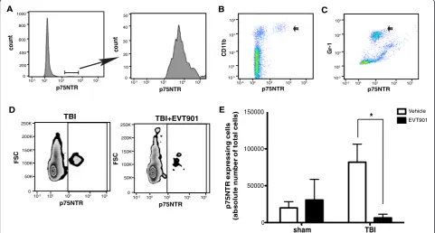
p75NTR may be involved in immune function and that EVT901 may have peripheral effects on inflammation after TBI. Mouse leukocytes express p75NTR (Fig. 1). Both CD11b+ and Gr1+ populations isolated using flow cytometry included p75NTR+ cells, and the number of p75NTR+ cells was increased at 7 days post-TBI (Fig. 1). Treatment for 7 days with EVT901 blocked this in-crease. These data support the possibility that p75NTR could be involved in mediating the early inflammatory responses in the circulation after TBI.
Blocking p75NTR reduces the pro-inflammatory response of monocytes to LPS in vitro
In order to test whether p75NTR might mediate pro-inflammatory responses in the periphery, we asked whether EVT901 would affect the response of pro-inflammatory
monocytes in vitro to LPS stimulation.Since Ly6Chighand
Ly6Cintmonocytes are characterized as the“
pro-inflamma-tory monocytes” [63–68], we evaluated the population of
CD11b + Ly6Cint-high inflammatory monocytes in the
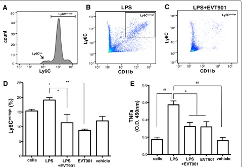
iso-lated leukocytes. Treatment with 10 ng/ml of LPS increased
the population of CD11b + Ly6Cint-high pro-inflammatory
monocytes in the isolated leukocytes (Fig. 2b–d). In order
to determine if p75NTR is involved in the differentiation of
count
0 20 30 40 50
10-3 100 103 104 105
10
count
0 400 600 800 1000
10-3 100 103 104 105
200
10-3 100 103 104 105
10-3
100
103
104
105
p75NTR
b
1
1
D
C
10-3 100 103 104 105
10-3
100
103
104
105
p75NTR
1-r
G
B C
A
p75NTR p75NTR
FSC
0 50K 100K 150K 200K 250K
10-3 100 103 104 105
p75NTR
FSC
0 50K 100K 150K 200K 250K
10-3 100 103 104 105
p75NTR
TBI+EVT901
sham TBI
0 50000 100000 150000
p
75N
T
R
exp
ressi
n
g
cell
s
(a
bs
o
lut
e
nu
m
be
r
o
f
tot
a
l
ce
lls)
*
EVT901Vehicle TBI
D E
Fig. 1EVT901 treatment reduced p75NTR-expressing leukocyte in the circulation at 7 days after TBI.a–cLeukocytes were obtained from normal C57Bl/6 mouse blood, and cell type was identified by flow cytometry. p75NTR is expressed on CD11b+ cells (b) and Gr-1+ cells (c).d,eBlood was obtained from C57Bl/6 mice at 7 days after TBI with or without EVT901 treatment and isolated leukocytes were immuno-stained with p75NTR Ab. TBI increased the absolute number of p75NTR+ cells, while EVT901 treatment significantly reduced the number in the circulation. One-way ANOVA showed a significant effect of condition (F3,9= 4.62,p= 0.03,η2= 0.61, power = 0.71; Tukey’s post hoc test: TBI vehicle vs
CD11b + Ly6Cint-high inflammatory monocyte, cultures were treated with EVT901. Thirty nanometer of EVT901 treatment significantly inhibited the expansion
of CD11b + Ly6Cint-high inflammatory monocytes by
LPS (Fig. 2d).
Elevated TNFα production is a hallmark of
pro-inflammatory stimulation [69–71]. We tested whether
EVT901 treatment inhibits LPS-induced TNFα
produc-tion in the isolated leukocytes. LPS increased TNFα
production, as determined by TNFα ELISA, and
EVT901 treatment significantly reduced TNFα
pro-duction (Fig. 2e). These results suggest that EVT901 has anti-inflammatory effects through blocking the p75NTR signaling pathway in leukocytes and supports the hypothesis that p75NTR is involved in the pro-inflammatory responses of peripheral monocytes. We
next examined the effects of TBI and EVT901 on this population in the circulation in vivo.
EVT901 treatment inhibits the production of CD11b +
Ly6Cint-highinflammatory monocytes in the circulation
after TBI
Ly6Chigh monocytes have been shown to emerge from
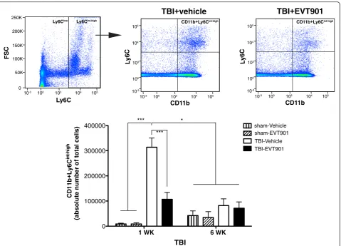
the bone marrow and enter the peripheral blood stream where they are directed to sites of tissue injury and in-flammation [72, 73]. CCI-TBI dramatically increased the
number of circulating CD11b + Ly6Cint-high monocytes
at 7 days after injury. Further, this effect was signifi-cantly reduced by 7-day treatment with EVT901 (Fig. 3). The total absolute number of CD11b + Ly6Cint-high inflammatory monocytes was reduced at 6 weeks after TBI, regardless of treatment, suggesting
cells LPS LPS EVT901 vehicle
0.0 0.2 0.4 0.6 0.8
*
a
F
N
T
)
m
n
0
5
4
.
D.
O(
cells LPS LPS
+EVT901 +EVT901
EVT901 vehicle
0 5 10 15 20 25
L
y6C
int-high
(%)
* **
A
D
count0 20 30 40 50
10-3 100 103 104 105
10
Ly6C
Ly6Cint-high
Ly6Clow
10-3 100 103 104 105
10-3
100
103
104
105
L
y6C
CD11b LPS+EVT901
Ly6Cint-high
E
10-3 100 103 104 105
10-3
100
103
104
105
L
y6C
CD11b LPS
Ly6Cint-high
B
C
##
##
Fig. 2EVT901 treatment inhibited the inflammatory response to LPS in isolated leukocytes in vitro. The isolated leukocytes (1 × 10⁶cells) were
treated with 10 ng/ml LPS, LPS + EVT901 (30 nM), EVT901, or vehicle. After 24-h incubation at 37 °C, cells were isolated by centrifugation and the cell type identified by flow cytometry.aCD11b + Ly6C+ monocytes consisted of two distinct subpopulations: Ly6Clowmonocytes and Ly6Cint-high
pro-inflammatory monocytes.b–dLPS treatment increased the proportion of the population that was the CD11b + Ly6Cint-high, while EVT901
treatment reduced it. Data were obtained from three independent experiments. One-way ANOVA showed a significant effect of condition (F4,14= 7.03,p= 0.007,η2= 0.76, power = 0.92; Tukey’s post hoc test, *p< 0.05, **p< 0.005).eTNFαin the supernatant was measured using
ELISA. EVT901 treatment inhibited TNFαproduction by LPS. Data were obtained from three independent experiments. One-way ANOVA
a normalization of pro-inflammatory cell populations in the blood.
EVT901 treatment reduces the absolute number of total
Ly6Cint-highmonocytes in the injured brain after TBI
In order to examine if the inhibition of peripheral Ly6C
int-high
pro-inflammatory monocytes is reflected in a
reduc-tion of the recruitment of Ly6Cint-highmonocytes in the
in-jured brain after TBI, we used flow cytometry to identify those cells from brain homogenates. CD45 Ab was applied along with Abs to CD11b and Ly6C Ab. We were able to
distinguish leukocytes of peripheral origin (CD45high) and
residential microglia (CD45low) (Fig. 4a). Here, we analyzed
pro-inflammatory monocytes (CD45highCD11b + Ly6C
int-high
), which are increased in the injured brain after TBI
(Fig. 4a–c). There was a significant reduction of
CD45highCD11b + Ly6Cint-high monocytes in the injured
brain after EVT901 treatment compared to vehicle-treated
mice after TBI (Fig. 4a–c). These data suggested that
p75NTR is involved in not only differentiation of Ly6C
int-high
pro-inflammatory monocytes in the circulation but also recruitment of these pro-inflammatory monocytes into the injured brain, perhaps by a variety of potential mechanisms. Further, the TBI-induced inflammatory cell increase was maintained at 6 weeks in the brain as op-posed to the return to baseline seen in the blood (Fig. 4), and the EVT901 treatment during the first week post-injury reduced that sustained effect in brain as well.
EVT901 treatment inhibits the recruitment of CCR2+ pro-inflammatory monocytes into the injured brain at 7 days after TBI and is neuroprotective
CCR2, the chemokine receptor for CCL2, is highly expressed by pro-inflammatory monocytes and promotes
1 WK 6 WK
0 100000 200000 300000 400000
TBI
C
D
11b
+L
y6C
int-hi
g
h
(a
bs
ol
ut
e
n
um
be
r
o
f
tot
a
l c
e
ll
s
)
sham-EVT901 sham-Vehicle
TBI-EVT901 TBI-Vehicle
***
*** *
10-3 100 103 104 105
10-3
100
103
104
105
L
y6C
CD11b
10-3 100 103 104 105
10-3
100
103
104
105
L
y6C
CD11b
TBI+vehicle
TBI+EVT901
FSC
0 50K 100K 150K 200K 250K
10-3 100 103 104 105
Ly6C
Ly6Clow Ly6Cint-high CD11b+Ly6Cint-high CD11b+Ly6Cint-high
Fig. 3EVT901 treatment reduced the number of Ly6Cint-highinflammatory monocytes in the circulation. Blood was obtained from C57Bl/6 WT
mice at 1 and 6 weeks after TBI with or without EVT901. Leukocytes were isolated, stained with CD11b Ab and Ly6C Ab, and run on flow cytometry. TBI significantly increased the absolute number of CD11b + Ly6Cint-highmonocytes in the circulations, while EVT901 treatment reduced
the number. Two-way ANOVA revealed a significant effect of condition (F3,25= 15.85,p< 0.000006,η2= 0.67, power = 1.0), time (F1,32= 5.71,p= 0.025, η2= 0.19, power = 0.63), time × treatment (F
3,32= 9.46,p< 0.0002,η2= 0.54, power = 0.99). Tukey’s post hoc tests showed there was a
their recruitment into sites of tissue injury [67, 73].
CCR2 plays a critical role in recruiting Ly6Chigh
mono-cytes from the bone marrow to the peripheral blood stream, and CCL2 leads monocytes directly to sites of injury and inflammation [72]. Further, a recent study demonstrated that CCR2 signaling is responsible for
80–90 % of the infiltrating monocytes in the injured
brain [19]. To evaluate the effects of p75NTR blockade on the recruitment of this specific population of pro-inflammatory monocytes, we used genetically modified
CCR2-RFP knock-in mice (CCR2+/RFP). The CCR2+/RFP
transgenic is a heterozygous mouse with bi-allelic ex-pression of RFP and CCR2 [20, 50]. Because resident microglia do not express CCR2 [20, 50], we were able to use this reporter to identify monocytes of peripheral origin. We were also able to detect RFP-positive cells in the circulation at 7 days after TBI using flow cytometry (Fig. 5a). We found that RFP-positive cells were
increased in the circulation at 7 days after TBI and that EVT901 treatment reduced that population (Fig. 5a). RFP-positive cells were also detected in the injured brain at 7 days after TBI (Fig. 5c), as previously re-ported [20]. To quantify the infiltrating RFP-positive cells in the injured brain, we applied a proportional area (P.A.) measurement based on previous studies [21, 52]. Brain tissue sections were obtained from the region of interest (ROI) around the lesion (Fig. 5b). The RFP-positive signal was highly increased in the injured brain at 7 days after TBI, while EVT901 treatment significantly
reduced it (Fig. 5b–d), suggesting that EVT901 treatment
inhibits the infiltration of CCR2+ pro-inflammatory monocytes. And consistent with our results in rat [43], we found that treatment with EVT901 for 7 days after TBI in-creased the amount of spared brain tissue (Fig. 5e). These data suggest that blocking p75NTR with EVT901 reduces the peripheral mobilization and expansion of CCR2+
TBI-Vehicle
TBI-EVT901
A
B
C
10-3 100 103 104 105 10-3
100 103 104 105 CD45low
CD45high
CD45
b
1
1
D
C
10-3 100 103 104 105 0
50K 100K 200K 250K
FSC
Ly6C
Ly6Clow Ly6Cint-high 150K
10-3 100 103 104 105 10-3
100 103 104 105
CD45
CD45lowCD1
CD45high
10-3 100 103 104 105 0
50K 100K 200K 250K
FSC
Ly6C
Ly6Clow Ly6Cint-high 150K
b
1
1
D
C
1 WK 6 WK
0 20000 40000 60000 80000
TBI
*
sham-vehicle
sham-EVT901
TBI-vehicle
TBI-EVT901
5
4
D
C
h
gi
h
C
6
y
L
+
b
1
1
D
C
h
gi
h-t
ni
)
sll
e
c
l
at
ot
f
o
r
e
b
m
u
n
et
ul
o
s
b
a(
*
*
*
Fig. 4EVT901 treatment reduced the recruitment of Ly6Cint-highmonocytes into the injured brain at 1 and 6 weeks after TBI. The injured hemisphere
was dissected and homogenized. Single cells were obtained using percoll gradients and then stained with Abs against CD45, CD11b+, and Ly6C.a–c
TBI significantly increased the absolute number of Ly6Cint-highinflammatory monocytes in the injured brain, while EVT901 treatment
reduced the number. Two-way ANOVA revealed significant main effects of treatment (F3,36= 6.19,p= 0.002,η2= 0.4, power = 0.94) and
time (F1,3= 5.10,p< 0.032,η2= 0.15, power = 0.59). Tukey’s post hoc tests showed that TBI significantly increased CD45highCD11b + Ly6Cint-highinflammatory
ROI
*
vehicle
EVT901
stitch images high magnification
CCR2-RFP DAPI MERGE
CCR2-RFP DAPI MERGE
1.5 mm -1.5 mm Propotional Area ) %( l a n gi s + P F R f o A B C D high magnification
E
uninjured TBI-vehicle TBI-EVT901 uninjured 0.00 0.05 0.10 0.15 0.20 0.25 TBI-vehicle TBI-EVT901 contralateral ipsilateral ipsilateral contralateral vehicle EVT901 count 0 500 1000 1500 200010-3 100 103 104 105 RFP count 0 50 100 150 200
10-3 100 103 104 105 RFP 0 1 2 3 4 TBI
vehicle EVT901
sll e c e vit i s o p P F R ) y c n e u q er F( SSC 0 100K 150K 200K 250K
0 50K 100K 150K 200K 250K
FSC 50K SSC 0 100K 150K 200K 250K 50K
10-3 100 103 104 105 RFP vehicle EVT901 R1 R2 R3 * 110 * g ni r a p s e u s si T ) 0 0 1* l ar et al ar t n o c/l ar et ali s pi(
uninjured vehicle EVT901
0 10 20 90 100 † TBI
Fig. 5EVT901 treatment reduced CCR2+ monocytes in the injured brain and circulation and reduced tissue damage at 7 days after TBI.aBlood was obtained from CCR2+/RFPTg mice at 7 days after TBI with or without EVT901. One-way ANOVA showed that there was a significant difference between vehicle and EVT901 after injury (F1,8= 8.12,p= 0.03,η
2
= 0.58, power = 0.66) (n= 3/group).bBrain sections from CCR2+/RFPTg mice were obtained from the lesion area as shown (region of interest (ROI), from 1.5 mm anterior to 1.5 mm posterior of bregma).cCCR2+/RFPcells were observed predominantly in the injured area.dRFP-positive signal was quantified and the proportional area (P.A. = the area of CCR2-RFP+ signal/ total area × 100) is shown. CCR2-RFP-positive signal was highly increased after TBI, and EVT901 treatment significantly reduced it. ANOVA showed a significant effect of side (F1,28= 35.03,p< 0.001,η
2
= 0.83, power = 0.999), condition (F2,7= 10.97,p= 0.007,η 2
= 0.758, power = 0.922), side × condition (F2,28= 7.81,p< 0.02,η
2
= 0.69,p= 0.81), and section (F4,28= 2.83,p< 0.043,η 2
= 0.288, power = 0.695).eEVT901 increased the spared tissue area after TBI. One-way ANOVA showed a significant main effect of condition (F2,7= 12.00,p= 0.005,η
2
= 0.774, power = 0.942). Tukey’s post hoc test comparing TBI-vehicle vs TBI-EVT901 was significant (*p= 0.0374), and comparing UN vs TBI-vehicle was also significant (†p= 0.005).Scale bars
monocytes after injury, perhaps by reducing the chemo-kine signaling from the brain by reducing the amount of injury [43]. However, in light of the effects of EVT901 on the induction of pro-inflammatory monocytes in vitro, EVT901 may also act to reduce CCR2+ monocyte expan-sion more directly by affecting myeloid cell proliferation and differentiation through blocking p75NTR signaling.
Because CCR2+/RFP mice are heterozygotes, there
may be gene-dose-dependent effects that alter recruit-ment of macrophages into the tissue [74]. Therefore, we asked if EVT901 has comparable effects in C57BL/6 WT mice using alternative labeling strategies. To iden-tify peripheral monocytes, we used CD45, which is highly expressed on peripheral leukocytes. As an
alter-native to CCR2+/RFP, we used co-localization of CD45
and CCR2 immunostaining. We found that the
co-localization of positive signal for CD45 and CCR2 is in-creased in the injured brain ROI at 7 days after TBI and that EVT901 treatment significantly reduced this
signal (Fig. 6a, b). Consistent with CCR2+/RFPTg mice,
tissue sparing (measured as the spared tissue area from the lesion ROI sections) was enhanced in C57Bl/6 WT treated with EVT901 compared to vehicle-treated mice (Fig. 6c). Thus, we were able to confirm the effects of EVT901 on CCR2+ cell recruitment using both Tg and wild-type mice.
EVT901 treatment reduces F4/80+ monocytes/ macrophages in the injured brain after TBI
A recent study shows that CCR2 is required for recruit-ing monocytes from the peripheral circulation into the brain in the first few days after TBI [19]. However, since
uninjured vehicl
e
EVT901
0.00 0.05 0.10 0.15 0.20
Propotional Area
of CD45
+CCR2 + (%)
high magnification
CD45 CCR2 DAPI MERGE
CD45 CCR2 DAPI MERGE
vehicle
EVT901
A
B
C
ipsilateral contralateral
vehicle EVT901
0 5 10 15 20 80 90 100 110
g
ni
r
a
p
s
e
u
s
si
T
)
0
0
1*
ar
et
al
ar
t
n
o
c/l
ar
et
ali
s
pi
(
uninjured vehicl
e
EVT901
* *
TBI TBI
Fig. 6EVT901 treatment reduced CD45 + CCR2+ double-positive monocytes in the injured brain in C57Bl/6 WT mice at 7 days after TBI.aBrain tissue sections were immuno-stained with CCR2 Ab and CD45 Ab.bTBI increased the CD45 + CCR2+ double-positive signal in the injured area, while EVT901 treatment abrogated this effect. ANOVA revealed a significant main effect of condition (F2,6= 14.50,p= 0.005,η2= 0.83, power = 0.96)
and section (F4,24= 5.86,p= 0.002,η2= 0.49, power = 0.96). Tukey’s post hoc test showed a significant difference between uninjured and
TBI-vehicle (p= 0.004).cTissue area was significantly reduced by TBI, and this effect was abrogated by EVT901. One-way ANOVA showed a
main effect of condition (F2,6= 14.06,p= 0.003,η2= 0.82, power = 0.96). Tukey’s post hoc tests showed that TBI-vehilce, but not EVT901,
CCR2 is rapidly down-regulated when CCR2 monocytes differentiate into tissue resident macrophages, it is only possible to detect newly arrived CCR2 monocytes in the tissues [20, 50]. To overcome this issue, F4/80, a marker for mature monocytes/macrophages, was used to estimate the total population of infiltrating periph-eral monocytes in the injured brain at 7 days after TBI. We found that the double-positive signal of CD45 and F4/80 was increased in the injured brain at 7 days after
TBI (Fig. 7a) and that EVT901 treatment significantly reduced this signal (Fig. 7a, b).
To confirm the immunohistochemical analysis, we ap-plied flow cytometric analysis of brain homogenates, which allowed us to estimate the absolute total number of infil-trating peripheral monocytes/macrophages in the injured brain. We found that the absolute total number of
CD45highF4/80+ cells was increased in the injured brain at
1 and 6 weeks after TBI, and yet again, EVT901 treatment
Fig. 7EVT901 treatment attenuated the infiltration of peripheral monocytes/macrophages into the injured brain in WT mice.aBrain tissue
sections from C57BL/6 mice at 7 days after TBI were immuno-stained with CD45 and F4/80 Abs.bTBI increased the proportional area of CD45 +
F4/80+ double-positive signal in the injured brain, while EVT901 treatment reduced it. ANOVA indicated that there was a significant treatment effect (F1,6= 14.89,p= 0.005,η2= 0.83, power = 0.97). Tukey’s post hoc test revealed a significant difference between uninjured vs TBI-vehicle (##p
= 0.004) and TBI-vehicle vs TBI-EVT901 (**p= 0.006).c–fCells isolated from C57Bl/6 WT mice brains at 1 and 6 weeks after sham or TBI, with or without EVT901, were stained with Abs against CD45, CD11b+, and F4/80.c,dTBI increased the absolute number of CD45highF4/80+ monocytes,
and EVT901 significantly reduced the number. Two-way ANOVA followed by Tukey’s post hoc tests showed that there was a significant effect of condition (F3,36= 4.06,p= 0.016,η2= 0.31, power = 0.779), but no effects of time. TBI increased CD45highF4/80+ cells in the injured brain compared
to sham, while EVT901 treatment abrogated this effect (*p< 0.05).e,fTBI significantly increased the absolute number of CD45highCD11b+ monocytes
in the injured brain, and EVT901 significantly reduced it. Two-way ANOVA showed a significant effect of condition (F3,35= 9.02,p= 0.0002, η2= 0.5, power = 0.99), but no effect of time. Tukey’s post hoc tests showed a significant effect of injury and EVT901 at both 1 and 6 weeks
significantly reduced these populations (Fig. 7c, d). In addition, we examined total monocyte/macrophage popula-tions in the injured brain using CD11b Ab, a common marker for both immature and mature monocytes and macrophages. We found that there was a significant
in-crease in CD45highCD11b+ monocytes in the injured brain
(Fig. 7e, f), and this increase, present at both 1 and 6 weeks, was abrogated by the 1-week treatment with EVT901. In
contrast, the total number of CD45lowCD11b+ cells, which
represent the resident microglia, was not affected by TBI or EVT901 (data not shown).
Blocking p75NTR signaling by EVT901 treatment improves motor function after TBI and modifies the multivariate syndromic effects of TBI on both immune system and behavior
Previous studies show that prevention of early trafficking of peripheral immune cells into the injured CNS results in better behavioral outcomes and reduction of tissue
damage [13, 19, 20, 22–24].
The CCI-TBI protocol we used in the mouse in the present study resulted in relatively minor motor deficits that largely recovered over the 6 weeks after injury. However, we did see evidence for a drug effect on some measures. To analyze the full set of behavioral and associated inflamma-tory biomarkers, we performed principal components ana-lysis (PCA) to extract the global syndromic outcome patterns. Consistent with these prior studies, we found that
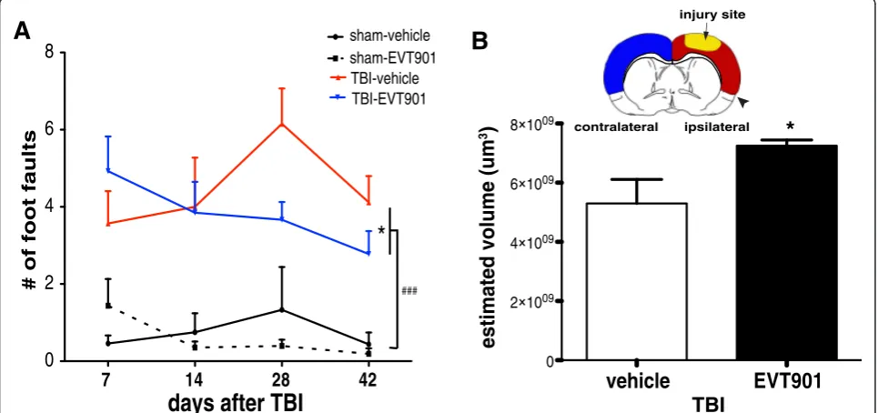
blocking early trafficking of peripheral monocytes into the CNS by EVT901 treatment after TBI reduced neurological deficits. Using a battery of behavioral tests with TBI mice, we found that fine motor functions performed on a beam were significantly improved by EVT901 administered for 1 week after injury (Fig. 8c, d). Further, we found that TBI significantly reduced the total volume of neocortex at 6 weeks, and EVT901 significantly reversed this when com-pared to the vehicle-treated brain (Fig. 8b).
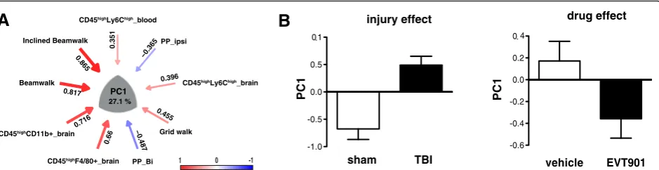
The PCA revealed five orthogonal principal compo-nents that partitioned the shared variance between cellu-lar and behavioral outcome measures at 6 weeks (Fig. 9, and Additional file 1: Figure S1). PC1, which accounted for 27 % of the total variance, appeared to describe the coordinate increase in inflammatory markers and the de-terioration of behavioral function seen after TBI (Fig. 9a), providing a useful composite metric for the syndrome of CCI-TBI in our mice: comparisons of sham and TBI subjects using PC1 as a univariate measure showed a significant increase in value (Fig. 9b). One week of treat-ment with EVT901 dramatically lowered the PC1 z-score values in mice with TBI, supporting the idea that EVT9011 coordinately affected neuroinflammation and behavioral outcomes.
Discussion
Our previous study using the novel p75NTR antagonist EVT901 was initially aimed at blocking the
well-7 14 28 42
0
2
4
6
8
sham-vehiclesham-EVT901TBI-vehicle TBI-EVT901
days after TBI
# of foot faults
*
###
A
B
ipsilateral contralateral
injury site
vehicle
EVT901
0 2 1009 4 1009 6 1009 8 1009
m
u(
e
m
ul
o
v
d
et
a
mit
s
e
3
)
*
TBI
Fig. 8EVT901 treatment improved motor function and reduced brain damage after TBI.aPersistent fine motor coordination was evaluated by beam walking for 6 weeks after TBI. The total number of footfaults was increased by TBI, and EVT901 reduced this deficit. Two-way ANOVA revealed a significant main effect of injury (F1,26= 48.996,p= 0.0000002,η2= 0.65, power = 1), and a significant time × treatment effect
(F3,78= 4.90,p= 0.04,η2= 0.16, power = 0.9). Tukey’s post hoc tests revealed a significant difference between TBI-vehicle and TBI-EVT901 (*p= 0.04) and
sham and TBI (###p< 0.0001).bUnbiased stereological volumetric analysis showed a significant atrophy of cortex on the ipsilateral, but not contralateral
documented effects of p75NTR signaling on apoptotic cell death in CNS neurons and glia, and we showed quite dramatic effects on measures of tissue sparing and a variety of neurological outcomes in multiple models of TBI in the rat [43]. The results also showed very dra-matic reductions in histological measures of neuroin-flammation and microglial activation. We wondered whether EVT901 might be affecting neuroinflammatory signals as well as apoptosis. However, we saw no evi-dence for microglial expression of p75NTR in that study, and aside from a few reports of p75NTR on microglial cells in vitro [75, 76], there is little evidence for micro-glial expression of p75NTR in the adult brain. However, there have been isolated reports of p75NTR expression in leukocytes [45, 46], (reviewed in [44]). We thus raised the question as to whether p75NTR could also be in-volved in the peripheral inflammation seen after TBI, with consequences for the recruitment of monocytes/ macrophages into the injured brain. We found in mouse TBI, a strong pro-inflammatory peripheral acute re-sponse to CNS injury, and the accumulation of periph-eral leukocytes, including pro-inflammatory monocytes and CCR2+ cells, in the brain injury site at 1 and 6 weeks after injury.
Seven days of treatment with EVT901 reduced the ef-fects of each element of the combined blood and brain in-flammatory response to CCI-TBI: it reduced the increased expression of p75NTR on peripheral monocytes, it re-duced the increase in numbers of pro-inflammatory monocytes in the blood at 1 week after injury, and it re-duced the invasion of peripheral myeloid cells (defined by multiple phenotypic markers) into the brain. And consist-ent with our previous work [43], EVT901 was neuropro-tective and reduced post-TBI neurological deficits.
These results are consistent with the hypothesis that EVT901 has both central and peripheral effects on in-flammation by blocking p75NTR signaling in both neural and peripheral immune cells, dampening the ef-fects of central neuroinflammation by reducing both the CNS damage caused by p75NTR apoptotic signaling and the exacerbation of initial microglial activation by the production and invasion of highly pro-inflammatory per-ipheral monocytes. While the reduction in perper-ipheral inflammatory responses might be in part an indirect ef-fect of the reduction of central injury, and therefore of signaling (e.g., by DAMPs, CCL2, cytokines, or other mediators), the strong inhibition by EVT901 of pro-inflammatory responses to LPS in isolated monocytes in vitro provides evidence for a direct effect.
The choice of injury model
We chose to use a controlled-cortical impact (CCI) model of TBI in the mouse for these studies. These in-juries are characterized by frank contusions of the cor-tical mantle and disruption of the local blood-brain barrier, although widespread diffuse injury also has been documented [51, 77]. A significant proportion of human
TBI includes focal contusion injuries that may “bloom”
and expand over the first few days after injury [78, 79]. These injuries, with their frank breach of the blood-brain barrier, are likely to be especially affected by inva-sion of peripheral myeloid cells. Further, the presence of an MRI-defined contusion injury in mild TBI is a strong predictor of negative outcome [80]. Thus, treatments that reduce the evolution of contusion injuries are likely to be translatable to at least an important and potentially treatable subset of human TBI.
sham TBI
-1.0 -0.5 0.0 0.5 1 . 0
injury effect
1
C
P
vehicle EVT901
-0.6 -0.4 -0.2 0.0 0.2 0. 4
1
C
P
drug effect
A
B
PP_Bi PP_ipsi
0.865
0.817
0.716
0.66
−0.487 0.455
0.396 −0.365
0.351
27.1 %
PC1
CD45highLy6Chigh_blood
Inclined Beamwalk
Beamwalk
CD45high rain
CD45high rain
Grid walk
CD45highLy6Chigh_brain
-1 0 1
Fig. 9Multivariate principal component analysis (PCA). To understand the relationship of behavioral outcome and innate immune cell responses at 42 days after TBI, PCA was used. This analysis yielded five dimensional PC-loading patterns accounting for 80.55 % of total variance (details in Additional file 1: Figure S1).aSignificant loading on PC1 indicated that more errors on the beam-walking tasks were associated with the numbers of peripheral inflammatory immune cells in the brain and (less so) with increased pro-inflammatory monocytes in the blood. Bilateral paw placement,
a measure of recovery of cortical function, was inversely correlated between individual variables and PC1.Red arrowindicates a positive