M E T H O D O L O G Y
Open Access
Label-free proteomic methodology for the
analysis of human kidney stone matrix
composition
Frank A. Witzmann
1*, Andrew P. Evan
2, Fredric L. Coe
3, Elaine M. Worcester
3, James E. Lingeman
4and James C. Williams Jr
2Abstract
Background:Kidney stone matrix protein composition is an important yet poorly understood aspect of nephrolithiasis. We hypothesized that this proteome is considerably more complex than previous reports have indicated and that comprehensive proteomic profiling of the kidney stone matrix may demonstrate relevant constitutive differences between stones. We have analyzed the matrices of two unique human calcium oxalate stones (CaOx-Ia and CaOx-Id) using a simple but effective chaotropic reducing solution for extraction/solubilization combined with label-free quantitative mass spectrometry to generate a comprehensive profile of their proteomes, including physicochemical and bioinformatic analysis.`
Results:We identified and quantified 1,059 unique protein database entries in the two human kidney stone samples, revealing a more complex proteome than previously reported. Protein composition reflects a common range of proteins related to immune response, inflammation, injury, and tissue repair, along with a more diverse set of proteins unique to each stone.
Conclusion:The use of a simple chaotropic reducing solution and moderate sonication for extraction and solubilization of kidney stone powders combined with label-free quantitative mass spectrometry has yielded the most comprehensive list to date of the proteins that constitute the human kidney stone proteome.
Keywords:Calcium oxalate, Kidney stone, Label-free quantitative liquid chromatography–tandem mass spectrometry, Matrix protein, Nephrolithiasis, Proteomics
Background
The organic matrix within urinary stones has long been thought to be an important—if poorly understood—part of stone composition. It has been proposed that the process of stone formation involves the primary depos-ition of matrix, with crystal formation occurring second-arily within the matrix layer [1, 2]. Others maintain that crystallization is primary and that most, if not all, of the organics in stones are co-precipitated with the crystals in a manner that is in no way causative [3]. Recent work on the interface of the growth of calcium oxalate (CaOx) stones on Randall’s plaque has suggested that matrix
deposition is the primary event, at least in the formation of CaOx stones over plaque. The first layer covering plaque that has been exposed to urine is an organic layer that contains Tamm Horsfall protein, aka uromodulin [4]. Thereafter, nucleation of apatite begins and depos-ition of CaOx follows [4].
Progress on characterizing the composition of stone matrix has been slow, in part because of its insolubility. The work that has been done also has demonstrated that the matrix composition is remarkably variable and com-plex [5]. However, recent studies of human stone matrix have begun to exploit the power of modern proteomic methods, with some patterns beginning to emerge in the kinds of proteins found [6–17]. Nine of these recent stone proteomic studies contain profiles of CaOx stone-* Correspondence:[email protected]
1Department of Cellular and Integrative Physiology, Indiana University School
of Medicine, 635 Barnhill Drive, Room 362A, Indianapolis, IN 46202-5120, USA Full list of author information is available at the end of the article
or crystal-associated proteins [6, 7, 9, 11–15, 17] but only one study has attempted to quantify them [17].
Thus far, the number of proteins identified in human stone matrices has been relatively modest, ranging from 30 [11] to 242 [10] distinct proteins identified across a var-iety of stone mineral types where the proteome has been investigated. In general, where protein profiles of different types of stones were compared, differences across stone types were minimal whereas protein differences within stone types were quite variable. This is likely due to two factors: 1) only the most abundant, common proteins were identified by these studies and 2) individual kidney stone proteomes are by nature variable and diverse.
We hypothesized that the analysis of human stone matrix should yield significantly more proteins than pre-viously detected and that an improved approach may be useful in studying nephrolithiasis. Using a simple but ef-fective chaotropic reducing solution for extraction and solubilization, one that historically works well for a broad range of protein samples [18–20], combined with gentle, intermittent sonication, we have analyzed ex-tracts of two unique human CaOx stones, CaOx-Ia and CaOx-Id, (of different morphologies of CaOx monohy-drate, according to the morphoconstitutional classifica-tion scheme published by Daudon [21]), using an established label-free quantitative mass spectrometric (LFQMS) method. This method described in detail else-where [22, 23] uses individual three-dimensional align-ment to determine peptide retention time using a clustering method along with peptide ion peak areas cal-culated from the extracted ion chromatogram (XIC) generated by liquid chromatography - tandem mass spectrometry (LC-MS/MS). Only two stone matrices were analyzed in this study, by design. Our intent was to 1) improve and simplify the extraction of protein from the stone powder and 2) apply a novel, more compre-hensive LFQMS approach to identify and quantify as many proteins as possible. The results indicate a stone matrix proteome that is much larger and more complex than previously observed.
Results and discussion
Stone extracts
It is not exactly clear what fraction of the total stone pro-tein can be extracted in a form usable for proteomic ana-lysis. Our extraction method yielded 7.20 μg protein/mg stone powder for CaOx-Ia and 0.81μg/mg for CaOx-Id. The larger value is 3 times the average extraction yield that we have previously reported for calcium oxalate monohydrate (COM) stones using 9 M urea/1 mM DTT [5]. The increased amount of protein in CaOx-Ia was likely due to the presence of the x-ray lucent material identified by micro CT. Note that most researchers, not using micro CT, would likely be unable to detect the
presence of such a non-mineral-rich material, which, in this case, clung tightly to the surface of stone fragments. On the other hand, protein extraction from specimen CaOx-Id yielded a value within the range we found previ-ously for different COM stone specimens [5], though on the low end of that range. The low yield of protein in this specimen might be related to the tightly packed nature of this form of COM stone (Fig. 1d).
The total amount of protein in these types of stones, estimated from complete acid hydrolysis and measure-ment of amino acids, has been reported to be about 17μg/mg [2], but of course, such an acid hydrolysis de-stroys protein identities. It is likely that a considerable amount of stone protein is resistant to extraction by solubilization methods that maintain protein primary structure, as has been previously reported [5]. In com-parison, of the previous studies where proteomic analysis was used and extracted protein yields reported [7, 9, 17], the average was 1.5 μg/mg, probably less than 10 % of the total protein contained within the stones.
Whether the extraction resistant proteins are 1) very low abundance components of the matrix proteome trapped within the crystal matrix [15, 16] and thus not detected by conventional means, 2) chemically more hydrophilic and accordingly more avidly bound to crystal surfaces, or 3) merely unextracted replicates of the pro-teins identified below remains to be determined.
Label-free quantitative mass spectrometry
Using a label-free quantitative mass spectrometry plat-form, we identified and quantified 1,059 unique protein database (UniProt, http://www.uniprot.org) entries in-cluding splice variants or isoforms (809 unique gene names), in the two human kidney stone samples with a false discovery rate (FDR) of ≤0.2 %. These proteins are listed in a table in Additional file 1, along with their Uni-Prot identities, gene symbols, protein names, and abun-dances [22]. As illustrated in Fig. 2, 606 proteins were common to both stone types; 70 proteins were unique to CaOx-Ia, while 383 proteins were detected only in the CaOx-Id stone powder. Specific peptide information for all identified proteins and protein groups including pro-tein coverage, # of unique sequences, # of identified pep-tides, total # of identified sequences, is available in the table found in Additional file 2.
To account for potential bacterial proteins within the stone matrix, the MS data were searched against Coryne-bacterium, Actinomyces, Lactobacillus jensenii, Streptococ-cus anginosus, and Staphylococcus epidermidis protein databases. No proteins were identified. This strongly sug-gests that there is no significant bacterial contribution to the stone matrix proteome in the specimens tested.
matrix proteomes or crystal-associated proteins [6–17]. Compared to those earlier investigations, we found the kidney stone matrix proteome to be larger and much more complex than observed previously. We identified and quantified between 4–35 times as many proteins as found in the other human studies. More specifically, we found 259 proteins that were identified in earlier studies (see the Human Kidney Stone Matrix Proteome Database presented in Additional file 3), and also an additional 577 proteins not identified previously. This database also contains the following information for each protein: iso-electric point (pI), # of negatively charged residues (Asp + Glu), # of positively charged residues (Arg + Lys), Neg/Pos Ratio, aliphatic index, GRAVY score (grand average of hydropathicity), molecular class, biological process, cellu-lar component, and function.
The increased protein identification rate that we achieved in this investigation may be due to two factors. First, several previous studies used a demineralization ap-proach to extract proteins from powdered stones that in-cluded EDTA and/or SDS [7–9, 11, 13–17], both of which were subsequently removed by either centrifugation or dialysis steps. Boonla et al. [6] used a commercially avail-able lithium dodecyl sulfate/glycerol solution at 100 °C to extract proteins. Our use of 8 M urea/10 mM DTT with sonication and two repeated overnight extractions of the stone powder required no additional purification steps
Fig. 1Representative pieces from the two stone specimens used.
aFragment from CaOx-Ia, on mm-grid paper.bMicro CT slice through fragment in A, showing pure calcium oxalate monohydrate (COM).cTwo stones from specimen CaOx-Id; stone on right has shell broken off to reveal interior core, and on left is shell (top) and core from another stone in this specimen. For CaOx-Id, only the shell portions were collected for protein extraction.dMicro CT slice through CaOx-Id stone, showing pure COM
where proteins might be lost. As mentioned earlier, this approach yielded, on average, more protein/mg of stone powder than in previous studies. But that alone does not account for the differences in the number of proteins detected. A second reason may lie in the protein analysis itself. Several previous studies used 1D and/or 2D gel elec-trophoresis and subsequent LC-MS/MS or MALDI-MS/ MS to identify proteins in gel plugs [6, 11, 13, 14, 16, 17]. Others used LC-MS/MS or MALDI-MS/MS of whole ex-tracts [7–10, 14–17], but used LC gradient profiles of 60 min or less to separate the tryptic peptides. Our use of a 190 min LC gradient combined with the rapid scanning features of the Orbitrap Velos Pro mass spectrometer likely underlie our increased protein identification rate. It should be noted that in a proteomic analysis of rat urinary melamine stone matrix (where urea, thiourea, detergent and DTT were used to extract proteins, a 90 min LC gra-dient and a high resolution Orbitrap mass spectrometer were used to separate and analyze tryptic peptides), over 1,000 proteins were identified [24].
Interestingly, there are 204 proteins listed in the data-base in Additional file 3 that were identified in the previ-ous twelve studies, but were not detected in our stone samples. Nearly half of these are accounted for by 96 proteins (of 242) identified by Jou et al. in uric acid stones [10]. The other 146 uric acid stone proteins they detected were also identified in our CaOx stones. This substantial dissimilarity likely is due to the physicochem-ical differences between these various stone types and the extraction methods and analytical approaches used. Additionally, Merchant et al. analyzed the proteome of CaOx stones obtained from human subjects and identi-fied 158 proteins [14]. Forty-five of these were not de-tected in our CaOx stones, suggesting that even within similar stone types, the protein composition may be patient-specific and thus differ significantly.
A brief, non-statistical comparison of CaOx-Ia and CaOx-Id stone matrix protein composition is presented below. At this point, it is important to restate that only two stone matrices were analyzed in this study, by de-sign, and that our intent was to 1) improve and simplify the extraction of protein from the stone powder and 2) apply a novel, more comprehensive LFQMS approach to identify and quantify as many proteins as possible. The following data analysis is by no means intended to imply the existence of significant differences between the unique CaOx stones. It is presented here to compare and contrast the results of our new quantitative ap-proach to previous stone matrix proteome studies and not to make inferences on the pathophysiology of CaOx stone formation.
The 50 most abundant proteins from each stone are listed in Table 1. These correspond to many of the proteins reported in previous studies, in particular: serum albumin,
apolipoproteins, calgranulins, osteopontins (10-12-fold higher in CaOx-Id), prothrombin, alpha and beta hemoglobin, neutrophil defensin I, complement proteins, and alpha-1-antitrypsin. In contrast, proteins such as fatty acid synthase and numerous cytokeratins were rarely ob-served in previous studies. Their absence from previous studies may be accounted for by the fact that these proteins were not disclosed as their occurrence is generally thought to be due to contamination from skin and ambient environ-ment. This may be true of the epidermal and cuticular keratins identified in this study (KRT2, KRT10, KRT31, KRT77, KRT81 KRT84, KRT85), while others are well-established components of epithelial cells throughout the kidney [25–27] (KRT1, KRT3, KRT5, KRT6, KRT7, KRT8, KRT9, KRT13, KRT14, KRT17, KRT18, KRT19, KRT31, KRT80).
When one examines the database in Additional file 3, of the 257 proteins reported in other papers that also were found in our study, albumin, uromodulin (Tamm-Horsfall Protein, THP), calgranulin-A (Protein S100-A8), and calgranulin-B (Protein S100-A9) were common to all studies. Albumin and THP were similarly abundant in both our CaOx stones while calgranulin-A was nearly ten-fold higher in CaOx-Ia. Lactotransferrin, osteopontin, and prothrombin were detected in ten previous studies and Vitamin K-dependent protein Z was detected in nine. Twenty-two immunoglobulin-related proteins and 28 complement-related proteins were identified and quanti-fied, and most were common to both of our CaOx specimens.
Note that many of the proteins listed are undoubtedly from blood or tissue, but this is also true for proteins found in human urine [28]. Some work that has been done on crystallization of calcium oxalate crystals in vitro in human urine has suggested that proteins of blood and tissue are not adsorbed to forming crystals, with the idea that proteins of the blood or tissue are not relevant to stone formation [29]. But simple crystal for-mation is not the same as the forfor-mation of a stone, in which the protein matrix plays an important role [30]. Moreover, injury to the renal papilla may be a normal part of stone formation [31], so the presence of blood and tissue proteins could well be a part of the formation of a true stone.
Physicochemical analysis
as was the average pI of all proteins unique to CaOx-Ia or CaOx-Id stones (6.75 vs. 6.33). In comparison, a slightly more alkaline average pI of 7.2 has been observed in uric acid stone matrix proteins [10]. Conclusive comparison of these and other properties requires additional samples and further study. Nevertheless, negative amino acid/posi-tive amino acid ratios, aliphatic indices (a measure of pro-tein stability) and GRAVY scores were similar across all comparisons in the CaOx stones. The mean GRAVY scores, all considerably negative, indicate overall protein hydrophilicity.
Bioinformatic analysis
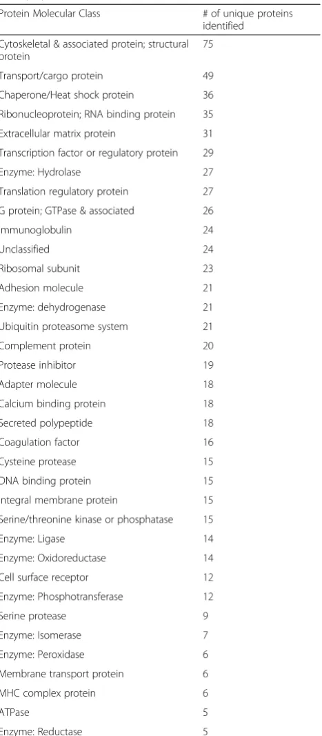
Of the proteins listed in the Human Kidney Stone Matrix Proteome Database (Additional file 3), 680 (82 %) have been detected in kidney cells or tissue (per the Human Proteome Map), 154 (18 %) have not been detected in kidney, so their origin may be considered “extra-renal”, 144 (17 %) are considered to be moder-ately or highly abundant in kidney, 248 (30 %) are con-sidered to be in the “extracellular component” (kidney or otherwise), and 75 (9 %) are considered to be “ cyto-skeletal and/or structural”proteins.
Proteins identified in the CaOx stones also represented a broad variety of molecular classes (Table 3). The distri-bution of classes reflects the prevalence of “cellular proteins” in the matrix, dominated by cytoskeletal & as-sociated proteins, structural proteins, transport/cargo proteins, chaperone/heat shock proteins, and ribonu-cleoprotein/RNA binding proteins, rather than an excess of urine- or plasma-related proteins. Many of these pro-teins may stem from the considerable cellular compo-nents of stone matrices first observed by Boyce in 1956 [32]. Nevertheless, as Table 1 and Additional file 1 indi-cate, the individually most abundant proteins in the Table 1Fifty most-abundant proteins in kidney stones CaOx-Ia
and CaOx-Id
CaOx-Ia CaOx-Id
Hemoglobin subunit beta Prothrombin
Neutrophil defensin 1 (Defensin, Alpha 1)
Hemoglobin subunit beta
Hemoglobin subunit alpha Hemoglobin subunit alpha
Protein S100-A9 (Calgranulin-B) Thrombin light chain
Complement C3 Vitronectin
Hemoglobin subunit delta Complement C3
Protein S100-A8 (Calgranulin-A) Vitamin K-dependent protein Z
Alpha-1-antitrypsin Plasminogen
Keratin, type I cytoskeletal 10 Keratin, type I cytoskeletal 10
Prothrombin Serum albumin
Fibrinogen beta chain Hemoglobin subunit delta
ATP-dependent RNA helicase A C4b-binding protein alpha chain
Apolipoprotein A-IV Alpha-1-antitrypsin
Keratin, type I cytoskeletal 13 Keratin, type II cytoskeletal 1
Serum albumin Fatty acid synthase
Keratin, type II cytoskeletal 1 Apolipoprotein A-I
Keratin, type I cytoskeletal 9 Mannan-binding lectin serine protease 2
Fatty acid synthase Osteopontin, Isoform 5
Nucleolin Myosin-9
Apolipoprotein B-100 Heat shock protein beta-1
Heparin cofactor 2 Fibroleukin (fibrinogen-like 2)
Actin, cytoplasmic 1 Apolipoprotein D
Fibrinogen alpha chain Histidine-rich glycoprotein
Plasminogen Tubulin beta chain
Profilin-1 Fibrinogen beta chain
Eosinophil cationic protein Kininogen-1, Isoform LMW
Coagulation factor XII Keratin, type I cytoskeletal 9
Complement C4-A Heat shock protein HSP 90-beta
Complement C4-B Apolipoprotein B-100
Vitronectin Pro-epidermal growth factor
Myosin-9 Fibrinogen alpha chain
Apolipoprotein A-I Neutrophil defensin 1 (Defensin, Alpha 1)
Leukocyte elastase inhibitor Extracellular superoxide dismutase [Cu-Zn]
Alpha-2-macroglobulin Argininosuccinate synthase
Apolipoprotein D Heparin cofactor 2
Thrombin light chain Complement factor B
Complement factor B Keratin, type I cytoskeletal 19
Histone H4 Apolipoprotein A-IV
Vesicular integral-membrane protein VIP36
Complement C4-A
Fibrinogen gamma chain Coagulation factor X
Table 1Fifty most-abundant proteins in kidney stones CaOx-Ia and CaOx-Id(Continued)
Serum paraoxonase/arylesterase 1 Complement C4-B
Peroxiredoxin-2 Vesicular integral-membrane
protein VIP36
Complement C5 Actin, cytoplasmic 1
Complement component C9 Profilin-1
WD repeat-containing protein 1 Nucleolin
Antithrombin-III Keratin, type II cytoskeletal 7
Mannan-binding lectin serine protease 2
Protein S100-A9 (Calgranulin-B)
Mannosyl-oligosaccharide 1,2-alpha-mannosidase IA
Osteopontin, Isoform B
Vitamin K-dependent protein S Mannosyl-oligosaccharide 1,2-alpha-mannosidase IA
Keratin, type II cytoskeletal 2 Antithrombin-III
CaOx stone matrix are blood/plasma derived–and pre-sumably urine–proteins.
The following calcium binding proteins were detected: annexins A1, A2, A3, A4, A5, A9, A10, and A11; calcy-phosin; calmodulin; calsequestrin-2; cilaggrin-2; cucle obindin-1; osteopontin; Profilaggrin; Protein S100-A2 (S100 calcium-binding protein A2); Protein S100-A6 (Calcyclin); Protein A7 (Psoriasin); Protein S100-A8 (Calgranulin-A); Protein S100-A9 (Calgranulin-B); Protein S100-P (Migration-inducing gene 9 protein); Protein S100-A11 (Calgizzarin); and Protein S100-A12 (Calgranulin-C), and these may have implications in the mineralization process [33, 34]. Additionally, the fol-lowing urinary proteins known to have the potential to modulate crystal formation and retention [35–37] were identified and quantified as prominent constitu-ents of the stone matrix: Tamm-Horsfall protein; osteopontin; Α-1 microglobulin; calprotectin (protein S100-A8 & 9); serum albumin; prothrombin; inter-α trypsin inhibitor (heavy chains H1, H2, and 4); heparin sulphate proteoglycan; bikunin; CD44, fetuin, and various collagens.
As in two previous studies [10, 14] where inflammatory, coagulation, cell adhesion, and acute-phase response path-ways were directly related to high abundance matrix pro-teins, we used pathway analysis to predict with statistical confidence which pathways might be associated with the matrix proteins identified and quantified in our CaOx stones. These results (79 unique pathways) are presented in the Pathway Data found in Additional file 4. Some of the most statistically significant pathways included LXR/ RXR activation, coagulation system, acute phase response signaling, FXR/RXR activation, clathrin-mediated endo-cytosis signaling, intrinsic prothrombin activation path-way, epithelial adherens junction signaling/remodeling, extrinsic prothrombin activation, complement system, and the production of nitric oxide and reactive oxygen species in macrophages. Analogous observations were made via functional annotation clustering of the CaOx proteins presented in the Functional Annotation Clusters found in Additional file 5, where inflammatory response and immune related functions were most notable and cytoskeletal structural molecule activity, extracellular glycoprotein signaling, wound healing, coagulation, and regulation of body fluid levels annotations were signifi-cantly represented.
Conclusions
The proteomic data presented here corroborate and sig-nificantly expand previous observations of the kidney stone matrix protein composition, revealing a more complex matrix proteome than previously reported for human kidney stones of any type. It remains unclear as to whether the identified proteins, their physicochemical properties, and their associated pathways/functions are directly related to mechanisms of stone formation or simply coincident and accumulated through long-term exposure of the growing stone to urine flow. Nonethe-less, the comprehensive approach we have developed and reported here will enable us and others to address such questions by analyzing and comparing comprehen-sive protein profiles in a broad range of stone types in relatively small stone specimens from individual patients who are carefully stratified by phenotype, and for whom important clinical data are known.
Methods
Reagents
Urea, DL-Dithiothreitol (DTT), triethylphosphine (TEP), iodoethanol, and ammonium bicarbonate (NH4HCO3) were purchased from Sigma-Aldrich (St. Louis, MO, USA). LC-MS grade acetonitrile (ACN) with 0.1 % formic acid (v/v) and water (H2O) with 0.1 % formic acid (v/v) were purchased from Burdick & Jackson (Muskegon, MI, USA). Modified sequencing grade porcine trypsin was ob-tained from Princeton Separations (Freehold, NJ, USA). All other reagents used were of the highest quality available.
Stone specimens, preparation of stone matrix protein extracts, and protein assay
Stones were de-identified specimens analyzed as pure COM and obtained from a stone analysis laboratory (Beck Analytical Services, Indianapolis IN). They were chosen so that several grams of material were available within a single specimen. The specimens were scanned using micro CT (SkyScan 1172, Bruker, Kontich, Belgium) to assess overall mineral purity, and mineral composition was confirmed using Fourier-transform infrared spectroscopy [38].
Figure 1 shows representatives of the two specimens used in the present study. The specimen named CaOx-Ia consisted of multiple fragments of COM that mostly had the morphological form Ia, which indicates a tightly Table 2Physicochemical characteristics of stone matrix proteomes
Isoelectric Point Neg/Pos Ratio Aliphatic Index GRAVY Score
CaOx-Ia All Proteins 6.62 ± 1.65 1.16 ± 0.66 79.07 ± 11.17 −0.416 ± 0.229
CaOx-Id All Proteins 6.49 ± 1.65 1.19 ± 0.60 78.92 ± 11.42 −0.438 ± 0.240
CaOx-Ia Only 6.75 ± 1.71 1.08 ± 0.39 80.14 ± 12.26 −0.340 ± 0.171
packed COM form that typically is dark brown in color [21]. The CaOx-Ia specimen also contained some x-ray lucent material, identified by infrared spectroscopy to be non-mineralized protein. Specimen CaOx-Id was also pure COM, but consisted of a dozen smooth stones of the morphological form Id, a distinctly different but still
tightly packed form of COM [21]. Note that CaOx-Id, due to its morphology, contained no extraneous protein, as did CaOx-Ia. It may also be noteworthy that neither of these CaOx specimens contained any apatite that was vis-ible by micro CT (a very sensitive method for detecting this mineral [38]). Thus, these CaOx specimens were not representative of the kinds of stones that would arise from growth on Randall's plaque [4].
CaOx-Ia and CaOx-Id stones were powdered by hand in small portions using an agate mortar and pestle, grinding the powder to the consistency of fine flour. The portions of powder for each specimen were combined and thor-oughly mixed. Aliquots of these stone powders (300 mg) were combined with 1 mL of freshly prepared 8 M urea and 10 mM DTT by vortex mixing (15 s) in Fisherbrand™ skirted microcentrifuge tubes with threaded ends (Catalog No. 02-681-343). The suspension was sonicated (Micro-son™, Misonix, USA) by 10 intermittent 1 s microprobe pulses at an energy output of 15 W, every 30 min for 4 h at room temperature. The tubes were incubated at room temperature overnight on an orbital shaker (200 rpm), vortexed, and subjected to one more sonication step as described above. The suspension was centrifuged at 3,200 x g for 1 h at room temperature (Jouan GR4i centrifuge, ThermoFisher Scientific, USA). The supernatant was col-lected and the pellet subjected to a second protein extrac-tion carried out exactly as the first. Extracts were combined and protein concentration in each pooled extract (~2 mL) was determined by the Bradford assay [39]. The solubilized protein extracts were then stored at
−80 °C until LFQMS analysis.
Proteolysis and LC-MS/MS
A 100μg aliquot of each sample was concentrated using the Vivaspin® 500 Centrifugal Concentrator (Vivaproducts, USA) by centrifugation at 8,000 rpm. The volume of each sample was adjusted to 200 μL (4 M urea) and then re-duced and alkylated by triethylphosphine and iodoethanol as previously described [40]. Briefly, 200μL of the reduc-tion/alkylation cocktail was added to the protein solution. The sample was incubated at 35 °C for 60 min, dried by a Vacuum Concentrator Centrifugal System (RC 10.10, Jouan), and reconstituted with 100 μL of 100 mM NH4HCO3 at pH 8.0. A 150 μL aliquot of a 20 μg/mL trypsin solution was added to the sample and incubated at 35 °C for 3 h, after which another 150μL of trypsin was added, and the solution incubated at 35 °C for an additional 3 h.
Sample digests were analyzed using a ThermoScientific Orbitrap Velos Pro hybrid ion trap-Orbitrap mass spec-trometer coupled with a Surveyor autosampler and MS HPLC system (ThermoScientific). Tryptic peptides were injected as technical replicates onto a C18 reversed phase column (TSKgel ODS-100 V, 3μm, 1.0 mm × 150 mm) at Table 3Representation of CaOx stone proteins by molecular
class (≥5)
Protein Molecular Class # of unique proteins
identified
Cytoskeletal & associated protein; structural protein
75
Transport/cargo protein 49
Chaperone/Heat shock protein 36
Ribonucleoprotein; RNA binding protein 35
Extracellular matrix protein 31
Transcription factor or regulatory protein 29
Enzyme: Hydrolase 27
Translation regulatory protein 27
G protein; GTPase & associated 26
Immunoglobulin 24
Unclassified 24
Ribosomal subunit 23
Adhesion molecule 21
Enzyme: dehydrogenase 21
Ubiquitin proteasome system 21
Complement protein 20
Protease inhibitor 19
Adapter molecule 18
Calcium binding protein 18
Secreted polypeptide 18
Coagulation factor 16
Cysteine protease 15
DNA binding protein 15
Integral membrane protein 15
Serine/threonine kinase or phosphatase 15
Enzyme: Ligase 14
Enzyme: Oxidoreductase 14
Cell surface receptor 12
Enzyme: Phosphotransferase 12
Serine protease 9
Enzyme: Isomerase 7
Enzyme: Peroxidase 6
Membrane transport protein 6
MHC complex protein 6
ATPase 5
a flow rate of 50μL/min. The mobile phases A, B, and C were 0.1 % formic acid in water, 50 % ACN with 0.1 % for-mic acid in water, and 80 % ACN with 0.1 % forfor-mic acid in water, respectively. The gradient elution profile was as follows: 10 % B (90 % A) for 7 min, 10–67.1 % B (90– 32.9 % A) for 163 min, 67.1-100 % B (32.9-0 % A) for 10 min, and 100-50 % B (0-50 % C) for 10 min. It is im-portant to note that the 190 min elution gradient used in this analysis is much longer than conventional 60 min elu-tion profiles used in previous kidney stone proteome stud-ies [7–10, 14–17] where LC-MS/MS was used, and likely accounts for much of the significant improvement in proteome coverage observed here (see Results and Discussion).
The data were collected in the “Data dependent MS/ MS” mode of Fourier transform-ion trap (MS-MS/MS) with the electrospray ionization interface using normal-ized collision energy of 35 % (collision induced dissoci-ation). Dynamic exclusion settings were set to repeat count = 1, repeat duration = 30 s, exclusion duration = 45 s, and exclusion mass width = 10 ppm (low) and 10 ppm (high).
Protein Identification and ouantification
The acquired data were searched against the UniProt protein sequence database of HUMAN (released on 07/09/2014) using X!Tandem algorithms in the Trans-Proteomic Pipeline (TPP, v. 4.6.3) (http://tools.proteo-mecenter.org/software.php). General parameters were set to: parent monoisotopic mass error set as 10 ppm, cleavage semi set as yes, missed cleavage sites set at 2, and static modification set as + 44.026215 Da on Cysteine. The peptide and protein identifications made by X!Tandem were validated by PeptideProphet [41] and ProteinProphet [42] in the TransProteomic Pipeline (http://tools.proteo-mecenter.org). Only validated proteins and peptides with protein probability≥0.9000 and peptide probability≥ 0.8000 were reported. False discovery rate (FDR) was esti-mated by a nonparametric concatenated randomized target-decoy database search [43]. As mentioned earlier, for this experiment and those TPP settings, protein identi-fication FDR was≤0.2 %.
Protein quantity was determined using an in-house label-free quantification software package, IdentiQuantXL [22], developed to individually and accurately align the re-tention time of each peptide and to apply multiple filters for exclusion of unqualified peptides to enhance label-free protein quantification. As previously described in detail [22], peptide retention time determination using cluster-ing, extraction of peptide intensity using MASIC [44], peptide coefficient of variation calculation, and peptides correlation were all conducted within the software plat-form to“filter out”unqualified peptides. Using only quali-fied peptides, protein intensity was calculated using the
formula: Protein Intensity = (intensity of peptide 1)/(pep-tide 1 sharing times) +…+ (intensity of peptide n)/(pep-tide n sharing times). For a pepn)/(pep-tide shared by different proteins, the intensity of this peptide was divided by the number of times the peptide was shared.
Validation of protein identity and quantity
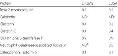
Results of the LFQMS analysis were validated immuno-logically for beta-2-microglobulin, calbindin, clusterin, cystatin-C, glutathione S-transferase P, neutrophil gelatin ase-associated lipocalin, and osteopontin. CaOx-Ia and CaOx-Id protein extracts were processed on a Bio-Plex 200 System with High Throughput Fluidics (HTF) Multiplex Array System (Bio-Rad Laboratories, Hercules, CA). The proteins were quantified simultaneously using Bio-Plex Pro™RBM Human Kidney Toxicity Assays (panels 1 and 2) on the Bio-Plex 200 system (BIO-RAD, USA) according to manufacturer instructions. Results were compared to LFQMS data as the ratio between CaOx-Ia/CaOx-Id and these data are listed in Table 4.
Physicochemical properties of matrix proteins
Grand average of hydropathicity (GRAVY) scores, ali-phatic indices, and number of negatively (Asp and Glu) and positively (Arg and Lys) charged residues for all identified proteins were calculated using the Protein Identification and Analysis Tools (ProtParam) on the ExPASy Server (http://web.expasy.org/protparam/) [45]. The GRAVY score for a peptide or protein is calculated as the sum of hydropathy values of all the amino acids, divided by the number of residues in the sequence. The aliphatic index of a protein is defined as the relative vol-ume occupied by aliphatic side chains (alanine, valine, isoleucine, and leucine) and may be regarded as a posi-tive factor for increased thermostability of globular proteins.
Bioinformatic analysis
To investigate the functional relevance of the proteins identified and quantified in the two stones, we used two common bioinformatic tools. Protein gene-symbol lists were uploaded onto the Ingenuity Pathway Analysis (IPA)
Table 4Validation of LFQMS Results: CaOx-Ia/CaOx-Id Ratio
Protein LFQMS ELISA
Beta-2-microglobulin 0.7 0.2
Calbindin NDa NDa
Clusterin 0.4 0.2
Cystatin-C 0.1 0.4
Glutathione S-transferase P 0.9 0.9
Neutrophil gelatinase-associated lipocalin ND# 8.5
Osteopontin, Isoform 5 0.1 0.1
software server (http://www.ingenuity.com) (Qiagen, US) and analyzed using the Core Analysis module to rank stone-specific proteins into canonical pathways in a statis-tically significant manner. Additionally, UniProt identifiers were submitted to the Human Proteome Map (http:// www.humanproteomemap.org) [46], GeneCards® (http:// www.genecards.org) [47], and the Gene Ontology (GO) database (http://go.princeton.edu/cgi-bin/GOTermMap-per) [48] via Generic Gene Ontology (GO) Term Mapper for information regarding each protein’s molecular class, biological process, cellular component, tissue specificity, and function.
Additional files
Additional file 1:Protein quantitation data for 1,059 human kidney stone proteins. (PDF 418 kb)
Additional file 2:Peptide sequence/Protein Identification, mass spectral data for 5,957 peptides. (PDF 3099 kb)
Additional file 3:Human Kidney Stone Matrix Proteome Database– 1,039 proteins found in human kidney stone matrices from this study and 12 other proteomic papers. Includes details regarding Isoelectric Point, negatively charged residues (Asp + Glu), positively charged residues (Arg + Lys), Neg/Pos Ratio, Aliphatic index, GRAVY score, Molecular Class, Biological Process, Cellular component, and Function. (PDF 316 kb) Additional file 4:Ingenuity Pathway Analysis Results - Canonical Pathways associated with Kidney stone matrix proteins. (PDF 60 kb) Additional file 5:Functional Annotation Clustering of kidney stone matrix proteins obtained from the DAVID database. (PDF 100 kb)
Abbreviations
ACN:acetonitrile; CaOx: calcium oxalate; COM: calcium oxalate monohydrate; CT: computed tomography; DTT: dithiothreitol; EDTA: ethylenediaminetetraacetic acid; GRAVY: grand average of hydropathicity; LC-MS/MS: liquid chromatography tandem mass
spectrometry; LFQMS: label-free quantitative mass spectrometry; MALDI-MS/ MS: matrix-assisted laser desorption ionization tandem mass spectrometry; RNA: ribonucleic acid; SDS: sodium dodecyl sulfate; THP: Tamm-Horsfall Protein; TEP: triethylphosphine.
Competing interests
The authors have no known competing interests either financial or personal between themselves and others that might bias the work.
Authors’contributions
FW and JW were responsible for the conception of the study and performed sample preparation and analysis; FW was responsible for mass spectrometric analysis; FW and JW performed data analyses and prepared figures and tables. AE and FC reviewed the data for significant proteins and critically reviewed the manuscript. EW oversaw the project, compared the data with proteins suspected to be active in stone formation, provided funding for the project, and critically reviewed the manuscript. JL supplied the stone specimens, provided expertise in stone analysis as part of the study, and critically reviewed the manuscript. All authors wrote or contributed to the writing of the manuscript and approved the final version.
Acknowledgements
This project was supported by NIH grant P01 DK-56788. The authors appreciatively acknowledge the technical assistance of Dr. Xianyin Lai and Mrs. Guihong Qi in the mass spectrometric analysis.
Author details
1Department of Cellular and Integrative Physiology, Indiana University School
of Medicine, 635 Barnhill Drive, Room 362A, Indianapolis, IN 46202-5120, USA.2Department of Anatomy and Cell Biology, Indiana University School of Medicine, Indianapolis, IN, USA.3Department of Medicine, Nephrology
Section, University of Chicago, Chicago, IL, USA.4International Kidney Stone
Institute, Methodist Hospital, Indianapolis, IN, USA.
Received: 25 November 2015 Accepted: 19 February 2016
References
1. Boyce WH. Am J Med. 1968;45:673–83. 2. Boyce WH, Garvey FK. J Urol. 1956;76:213–27.
3. Finlayson B, Vermeulen CW, Stewart EJ. J Urol. 1961;86:355–63.
4. Evan AP, Coe FL, Lingeman JE, Shao Y, Sommer AJ, Bledsoe SB, Anderson, J. C., et al. Anat Rec (Hoboken). 2007;290:1315–23.
5. Williams Jr JC, Zarse CA, Jackson ME, Witzmann FA, McAteer JA. J Endourol. 2006;20:560–4.
6. Boonla C, Tosukhowong P, Spittau B, Schlosser A, Pimratana C, Krieglstein K. Clin Chim Acta. 2014;429:81–9.
7. Canales BK, Anderson L, Higgins L, Ensrud-Bowlin K, Roberts KP, Wu B, Kim IW, et al. Urology. 2010;76(1017):e1013–1020.
8. Canales BK, Anderson L, Higgins L, Frethem C, Ressler A, Kim IW, Monga, M. Urol Res. 2009;37:323–9.
9. Canales BK, Anderson L, Higgins L, Slaton J, Roberts KP, Liu N, Monga MJ. Endourol. 2008;22:1161–7.
10. Jou YC, Fang CY, Chen SY, Chen FH, Cheng MC, Shen CH, Liao LW, et al. Urology. 2012;80:260–6.
11. Kaneko K, Kobayashi R, Yasuda M, Izumi Y, Yamanobe T, Shimizu T. Int J Urol. 2012;19:765–72.
12. Kaneko K, Matsuta Y, Moriyama M, Yasuda M, Chishima N, Yamaoka N, Fukuuchi T, et al. Int J Urol. 2014;21:341–6.
13. Kaneko K, Yoshida N, Okazaki K, Yamanobe T, Yamaoka N, Yasuda M, Ogata N, et al. Nucleosides Nucleotides Nucleic Acids. 2011;30:1072–6.
14. Merchant ML, Cummins TD, Wilkey DW, Salyer SA, Powell DW, Klein JB, Lederer ED. Am J Physiol Renal Physiol. 2008;295:F1254–1258.
15. Thurgood LA, Wang T, Chataway TK, Ryall RL. J Proteome Res. 2010;9:4745–57. 16. Thurgood LA, Ryall RL. J Proteome Res. 2010;9:5402–12.
17. Okumura N, Tsujihata M, Momohara C, Yoshioka I, Suto K, Nonomura N, et al. PLoS One. 2013;8, e68624.
18. Valente KN, Choe LH, Lenhoff AM, Lee KH. Electrophoresis. 2012;33:1947–57. 19. Michalski A, Cox J, Mann M. J Proteome Res. 2011;10:1785–93.
20. Wisniewski JR, Zougman A, Nagaraj N, Mann M. Nat Meth. 2009;6:359–62. 21. Daudon M, Bader CA, Jungers P. Scanning Microsc. 1993;7:1081–106. 22. Lai X, Wang L, Tang H, Witzmann FA. J Proteome Res. 2011;10:4799–812. 23. Lai X, Wang L, Witzmann FA. Int J Proteomics. 2013;2013:756039. 24. Liu JD, Liu JJ, Yuan JH, Tao GH, Wu DS, Yang XF, et al. Toxicol Lett.
2012;212:307–14.
25. Skinnider BF, Folpe AL, Hennigar RA, Lim SD, Cohen C, Tamboli P, et al. Am J Surg Pathol. 2005;29:747–54.
26. Xie L, Weichel B, Ohm JE, Zhang K. BMC Syst Biol. 2011;5 Suppl 3:S4. 27. Uhlen M, Fagerberg L, Hallstrom BM, Lindskog C, Oksvold P, Mardinoglu A,
et al. Science. 2015;347:1260419.
28. Thongboonkerd V, McLeish KR, Arthur JM, Klein JB. Kidney Int. 2002;62:1461–9.
29. Morse RM, Resnick MI. J Urol. 1988;139:869–73. 30. Boyce WH, King Jr JS. J Urol. 1959;81:351–65.
31. Stoller ML, Meng MV, Abrahams HM, Kane JP. J Urol. 2004;171:1920–4. 32. Boyce WH, Sulkin NM. J Clin Invest. 1956;35:1067–79.
33. Giachelli CM, Steitz S. Matrix Biol. 2000;19:615–22. 34. Kirsch T. Connect Tissue Res. 2012;53:438–45.
35. Eguchi Y, Inoue M, Iida S, Matsuoka K, Noda S. Kurume Med J. 2002;49:99–107.
36. Khan SR. J Urology. 2013;189:803–11. 37. Khan SR, Kok DJ. Front Biosci. 2004;9:1450–82.
38. Williams Jr JC, McAteer JA, Evan AP, Lingeman JE. Urol Res. 2010;38:477–84. 39. Bradford MM. Anal Biochem. 1976;72:248–54.
40. Hale JE, Butler JP, Gelfanova V, You J-S, Knierman MD. Anal Biochem. 2004;333:174–81.
42. Nesvishskii AI, Keller A, Kolker E, Aebersold R. Anal Chem. 2003;75:4646–58. 43. Elias JE, Gygi SP. Nat Methods. 2007;4:207–14.
44. Monroe ME, Shaw JL, Daly DS, Adkins JN, Smith RD. Comput Biol Chem. 2008;32:215–7.
45. Gasteiger E, Hoogland C, Gattiker A, Duvaud S, Wilkins MR, Appel RD. Bairoch, A. In: Walker JM, editor. The Proteomics Protocols Handbook. Totowa, NJ: Humana Press; 2005. p. 571–607.
46. Kim MS, Pinto SM, Getnet D, Nirujogi RS, Manda SS, Chaerkady R, et al. Nature. 2014;509:575–81.
47. Stelzer G, Dalah I, Stein TI, Satanower Y, Rosen N, Nativ N, et al. Hum Genomics. 2011;5:709–17.
48. Ashburner M, Ball CA, Blake JA, Botstein D, Butler H, Cherry JM, et al. Nat Genet. 2000;25:25–9.
• We accept pre-submission inquiries
• Our selector tool helps you to find the most relevant journal
• We provide round the clock customer support
• Convenient online submission
• Thorough peer review
• Inclusion in PubMed and all major indexing services
• Maximum visibility for your research
Submit your manuscript at www.biomedcentral.com/submit