O R I G I N A L R E S E A R C H
Open Access
Evaluation of D-isomers of 4-borono-2-
18
F-fluoro-phenylalanine and
O
-
11
C-methyl-tyrosine as brain tumor imaging agents:
a comparative PET study with their
L-isomers in rat brain glioma
Masakatsu Kanazawa
†, Shingo Nishiyama
†, Fumio Hashimoto, Takeharu Kakiuchi and Hideo Tsukada
*Abstract
Background:The potential of the D-isomerization of 4-borono-2-18F-fluoro-phenylalanine (18F-FBPA) to improve its target tumor to non-target normal brain tissue ratio (TBR) was evaluated in rat brain glioma and compared with those of L- and D-11C-methyl-tyrosine (11C-CMT).
The L- or D-isomer of18F-FBPA was injected into rats through the tail vein, and their whole body kinetics and distributions were assessed using the tissue dissection method up to 90 min after the injection. The kinetics of L-and D-18F-FBPA or L- and D-11C-CMT in the C-6 glioma-inoculated rat brain were measured for 90 or 60 min, respectively, using high-resolution animal PET, and their TBRs were assessed.
Results:Tissue dissection analyses showed that D-18F-FBPA uptake was significantly lower than that of L-18F-FBPA in the brain and abdominal organs, except for the kidney and bladder, reflecting the faster elimination rate of D-18F-FBPA than L-18F-FBPA from the blood to the urinary tract. PET imaging using18F-FBPA revealed that although the brain uptake of D-18F-FBPA was significantly lower than that of L-18F-FBPA, the TBR of the D-isomer improved to 6.93 from 1.45 for the L-isomer. Similar results were obtained with PET imaging using11C-CMT with a smaller improvement in TBR to 1.75 for D-11C-CMT from 1.33 for L-11C-CMT.
Conclusions:The present results indicate that D-18F-FBPA is a better brain tumor imaging agent with higher TBR than its original L-isomer and previously reported tyrosine-based PET imaging agents. This improved TBR of D-18 F-FBPA without any pre-treatments, such as tentative blood-brain barrier disruption using hyperosmotic agents or sonication, suggests that the D-isomerization of BPA results in the more selective accumulation of10B in tumor cells that is more effective and less toxic than conventional L-BPA.
Keywords:Boron neutron capture therapy, D-isomer,18F-FBPA,11C-CMT, Glioma
* Correspondence:[email protected]
†Masakatsu Kanazawa and Shingo Nishiyama contributed equally to this
work.
Central Research Laboratory, Hamamatsu Photonics K.K., 5000 Hirakuchi, Hamakita, Hamamatsu, Shizuoka 434-8601, Japan
Background
Boron neutron capture therapy (BNCT) is a biologically and physically targeted radiation therapy for tumors with the nuclear capture reaction of boron-10 (10B) specific-ally accumulating in tumor cells. The irradiation of
low-energy thermal neutrons to 10B produces
high-energy α (4He) particles and lithium ions (7Li), leading to cytotoxic effects against tumor cells. Due to the shorter track ranges of these particles (9 and 4 μm for αparticles and7Li ions, respectively) than the diam-eter of tumor cells, adjacent normal cells are expected to remain unaffected when these particles are emitted from tumor cells (for a review, see [1]). Two low molecular
weight10B carrier compounds, sodium sulfhydryl borane
(BSH) [2] and L-4-boronophenylalanine conjugated with fructose (L-BPA-Fr) [3], have been clinically applied. BSH enters tumor cells by passive diffusion through the
cell membrane [4–6]. L-BPA and its F-18-labeled PET
probe L-18F-FBPA [7–12] are transported into tumor
cells through L-type amino acid transporter 1 (LAT1) [11–13] and amino acid transporter B0,+ (ATB0,+) [13], both of which are strongly upregulated in tumor cells [13,14]. Since L-BPA accumulates in tumor cells with a low target tumor to non-target normal brain tissue ratio
(TBR) [6], L-18F-FBPA was developed to predict the
exact concentration of 10B-BPA in tumor cells prior to
BNCT [7–12]. The combined use of PET and inductively
coupled plasma optical emission spectrometry confirmed that L-18F-FBPA-PET has the ability to estimate10B con-centrations delivered by L-BPA in the tumor and normal tissues of rats [10,15] and humans [8].
In order to more effectively and safely perform BNCT, selective10B delivery to tumor tissues with high TBR and sufficient thermal neutrons delivered to the tumor region are critical issues. One of the major limitations of L-BPA is its low TBR [6], which results in difficulties regulating the delivery of an adequate amount of10B to target tumor tissues along with a sufficiently low amount of10B to adja-cent non-target normal tissues in order to avoid an ad-verse reaction. In an attempt to enhance L-BPA uptake into tumor cells, transient blood-brain barrier disruption (BBB-D) using hyperosmotic mannitol has been proposed to facilitate the uptake of L-BPA into F98 glioma in the rat brain, thereby enhancing survival times by BNCT over the
non-BBB-D control [4, 15]. Another BBB-D by focused
ultrasound sonication also increased the uptake of L-18F-FBPA into F98 glioma in the rat brain [16]. Since these BBB-D manipulations require treatments with drugs or sonication prior to the administration of L-BPA, the de-livery of L-BPA into tumor cells may be affected by indi-vidual responses to these pre-treatments.
We previously demonstrated that the D-isomers of O-11C-methyl-tyrosine (11C-CMT),O-18
F-fluoromethyl--tyrosine (18F-FMT), and O-18F-fluoroethyl-tyrosine
(18F-FET) were better tumor imaging agents than their
corresponding L-isomers in HeLa cells inoculated into
the mouse hind leg [17–21]. In contrast to natural
amino acid-based tumor imaging probes such as
D-11C-methionine (D-11C-MET), the D-isomers of
11
C-CMT, 18F-FMT, and 18F-FET were artificial amino
acid-based probes without metabolic conversion from the D-isomer to L-isomer by D-amino acid oxidase
[17, 22]. L-11C-MET converted from D-11C-MET via
α-keto-γ-methiolbutyrate was incorporated not only into proteins via the conversion into amino-acyl-tRNA, but also into non-protein materials such as lipids and RNA by
a transmethylation process via S-adenosyl-L-MET [23],
leading to a worse TBR due to strong uptake into
non-target normal tissues. The D-isomers of 11C-CMT,
18
F-FMT, and18F-FET exhibited higher TBRs than their
corresponding L-isomers in HeLa cells inoculated into the mouse hind leg because of the faster elimination rate of D-isomers than L-isomers from the blood to the urinary tract [17,18].
In the present study, we conducted the
D-isomerization of 18F-FBPA in order to improve the
properties of L-18F-FBPA as a brain tumor imaging
agent with a higher TBR, and the kinetics and distribu-tions of L- and D-18F-FBPA were compared in the whole body organs of normal F344 rats with a tissue dissection method as well as in C6 glioma inoculated into rat brains using PET. Furthermore, in order to establish whether the D-isomerization of amino acid-based PET probes may be generalized to improve TBR in the brain,
the L- and D-isomers of 11C-CMT were also evaluated
in rat brain C6 glioma.
Methods
Animals and chemicals
One hundred (60 for tissue dissection analyses; 32 for PET imaging; 8 for backup) male F344/NSlc rats
(7 weeks of age; body weight of 126–149 g) were
pur-chased from Japan SLC. Isoflurane and medetomidine hydrochloride were purchased from Dainippon Pharma-ceutical and Nippon Zenyaku Kogyo, respectively. Midazolam and butorphanol tartrate were obtained from Sandoz and Meiji Seika Pharma, respectively. L-Tyrosine
was obtained from Nacalai Tesque. AgOTf and
D-tyrosine were from Sigma-Aldrich, Japan. L- and D-4-boronophenylalanine were purchased from Santa Cruz Biotechnology. Standard compounds of L- and D-FBPA were obtained from the NARD Institute. L- and D-isomers of CMT were purchased from Tokyo Chem-ical Industry.
Implantation of tumor cells
Rats (8 weeks of age; body weight of 158–190 g) were
(0.15 mg/kg of medetomidine hydrochloride, 2 mg/kg of midazolam, and 2.5 mg/kg of butorphanol tartrate) and placed in a stereotaxic apparatus. C6 glioma cells (2 × 10⁵ cells/5 μl of Ham’s F-12K) were injected at a rate of 0.5μl/min into the right striatum of the rat (at
the bregma,−3.0 mm lateral to the midline suture, and
−5.0 mm from the cranium) via a microsyringe.
PET ligand synthesis
Carbon-11 (11C) and fluorine-18 (18F) were produced by
14
N(p, α)11C and 20Ne(d, α)18F nuclear reactions, re-spectively, using the cyclotron (HM-18, Sumitomo Heavy Industry). Labeled compounds were synthesized using a modified CUPID system (Sumitomo Heavy In-dustry). Chemical and radiochemical analyses of the la-beled compounds were performed by HPLC consisting of a pump (LC20AT, Shimadzu), UV detector (SPD-20A, Shimadzu), radio detector (US-3000, Universal Giken), and a column (Inertsil ODS-3, GL-Science).
L- and D-18F-FBPA were produced as previously
re-ported with minor modifications [6–12]. 18 F-Acetylhy-pofluorite in Ne gas was bubbled at a flow rate of 50 mL/min at room temperature into 5 mL of
tri-fluoroacetic acid containing 20 mg of L- or
D-4-boronophenylalanine. Trifluoroacetic acid was re-moved under reduced pressure at 80 °C. The residue was also dissolved in 1.8 mL × 2 of water containing 0.1% acetic acid, and the solution was applied to
HPLC (YMC-Pac ODS-A 20*150 mm 5 μm), with
water for injection containing 0.1% acetic acid as the
mobile phase. The fraction of L- or D-18F-FBPA was
collected into an evaporation flask, evaporated to ap-proximately 5 mL. The specific radioactivities of
L-and D-18F-FBPA were 26.4 ± 3.7 and 28.3 ± 1.8 MBq/
μmol, respectively, and the radiochemical purities of
L- and D-18F-FBPA were more than 98%.
L- and D-11C-CMT were prepared by reactions of
11
C-methyl iodide with the corresponding L- and D-tyrosine [17]. The specific radioactivities of L- and D-11C-CMT were 55.7 ± 14.1 and 58.6 ± 11.3 GBq/μmol, respectively, and their radiochemical purities were more than 98%.
Enantiomeric purity was analyzed on HPLC consisting of a pump (DP-8020, Tosoh), UV detector (UV-8020), radiodetector (TCS-913, Aloka), and column (CROWN-PACK CR(+), Daicel) using perchloric acid (pH 2.0) as a mobile phase at a flow rate of 2 mL/min. Enantiomeric purities were more than 99% for the L- and D-isomers of11C-CMT and18F-FBPA.
Tissue distribution assay
A total of 5 MBq of L- or D-18F-FBPA was injected into normal rats (n= 5/each time point) through the tail vein. Animals were killed by decapitation under isoflurane
(1.5–2.0% in O2) anesthesia 1, 5, 10, 30, 60, and 90 min
after the injection; samples of the brain, heart, lung, liver, kidney, spleen, pancreas, stomach, small intestine, colon, bladder, bone, muscle, and blood were rapidly re-moved, and their weights and radioactivities were
mea-sured using aγ-counter (1480 WIZARD, Perkin Elmer).
Standardized uptake values (SUVs) were calculated as radioactivity in tissue divided by the ratio of total injected radioactivity and body weight.
In metabolite analyses, blood samples were centri-fuged to separate plasma and then weighed, and radio-activity was measured; ethanol was added to plasma (sample:ethanol = 1:1) and centrifuged, and superna-tants were developed on a thin-layer chromatography (TLC) plate (TLC Silica gel 60 F254, Merck) using a mobile phase of 1-propanol/30 mM ammonium acet-ate/acetic acid (4/2/0.01). The TLC plate was then transferred to a phosphoimaging plate (BAS-IIIs, Fuji film) for a 10-min exposure, and radioactivity was con-verted to digitalized imaging data using a Fuji BAS sys-tem (FLA-7000, Fuji film). The ratio of radioactivity in the unmetabolized fraction to that in total plasma (me-tabolized plus unme(me-tabolized) was calculated.
PET measurements
Fourteen days after the implantation of glioma cells, rats (n= 4–6/each condition) anesthetized by 1.5–2.0% iso-flurane in O2were positioned prone on a fixation plate
and placed in the gantry hole of the PET scanner (SHR-38000, Hamamatsu Photonics). After a
transmis-sion scan for 15 min using a 68Ge-68Ga rotation rod
source, the L- or D-isomer of 18F-FBPA or11C-CMT at
10 MBq was intravenously injected into each rat via the tail vein, and this was followed by an emission scan for
90 min with 18F-FBPA or for 60 min with 11C-CMT.
The body temperature of each animal was monitored throughout the study using a Thermo-Hygro Recorder (TR-72Ui, T&D Corporation).
PET data obtained were reconstructed using the dy-namic row-action maximum likelihood algorithm method with a 1.0-mm Gaussian post filter [24], with attenuation corrections using transmission scan data. Dynamic images every 5 min for the time activity curve (TAC) as well as
summation images from 60 to 90 min for 18F-FBPA and
from 30 to 60 min for11C-CMT were reconstructed, and
Ex vivo autoradiography imaging of the brain
After PET imaging using L- or D-18F-FBPA, rat was
sacrificed by cervical vertebral dislocation under
anesthesia, and the brain was excised and sliced at a thickness of 2 mm using a brain slicer (Muromachi Kikai). Slices were transferred to a phosphoimaging plate (BAS-IIIs, Fuji film) for 10 min of exposure with an au-thentic standard radioactivity source. Radioactivity was converted to digitalized imaging data using a Fuji BAS system (FLA-7000, Fuji film).
Histological assessment
Rats were sacrificed by cervical vertebral dislocation under anesthesia, and brains were excised. After fixation in 4% neutral-buffered formalin, samples were embedded in paraffin. Thirty-micrometer-thick brain slices were prepared with a cryotome (model CR-502; Nakagawa Seisakusyo), and slices were stained with hematoxylin and eosin (HE) for microscopic examinations.
Statistical analysis
Results are expressed as means ± SD. Comparisons be-tween conditions were performed using an unpaired, two-tailed Student’sttest. A probability level of less than 5% (P< 0.05) was considered to indicate significance.
Results
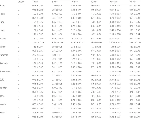
The analyzed data in Fig. 1 confirmed that 18F-FBPA
and 11C-CMT preserved their enantiomeric specificities
for the corresponding precursors after radiolabeling using F-18 or C-11. These retention times of 6.6 and 4.5 min for L- and D-18F-FBPA, respectively (Fig. 1c),
and 11.1 and 9.2 min for L- and D-11C-CMT,
respect-ively (Fig. 1d) were consistent with those obtained by
the corresponding authentic standard compounds of
L-and D-FBPA (Fig. 1a) and L- and D-CMT (Fig. 1b),
respectively.
The kinetics of the L- and D isomers of 18F-FBPA in
normal rats are summarized in Table 1. The blood level
of D-18F-FBPA was significantly lower than that of
L-18F-FBPA from 1 min after the injection throughout
measurements. Metabolic analyses of plasma demon-strated that both isomers of 18F-FBPA were very stable, showing 90.0 ± 0.6 and 89.0 ± 2.4% of unmetabolized L-and D-18F-FBPA, respectively, 90 min after the injection. The SUVs of D-18F-FBPA in the heart, lung, liver, spleen, pancreas, stomach, small intestine, colon, bone, and
muscle were lower than those of L-18F-FBPA throughout
the study after the injection. The uptake of D-18F-FBPA into normal brain tissue was markedly low with 13% of
L-18F-FBPA 90 min after the injection. The SUVs of
D-18F-FBPA in the kidney and bladder were markedly
a
b
c
d
Fig. 1Analyses of enantiomeric purities of L- and D-isomers of18F-FBPA (c) and11C-CMT (d) used in PET measurements. The enantiomeric purity
higher than those of L-18F-FBPA throughout the study after the injection. Differences in whole body distributions
between L- and D-18F-FBPA were generally similar to
those between L- and D-11C-CMT (see Table 2 of [17]).
As shown in Fig. 2, PET imaging of rat C6 glioma
indicated that the L- and D-isomers of 18F-FBPA (a
and b) and 11C-CMT (c and d) facilitated the
detec-tion of the tumor region in the rat brain. Although glioma cells were injected in the striatum as shown in the “Methods” section, the tumor tissues in almost all rat brains were observed in the cortex as shown in
Fig. 2. It is assumed that the injected cells might
draw back from the striatum to the cortex through the track of injection needle due to the high pressure in the brain, and then, the cells that were retained in the cortex proliferated.
The absolute uptakes, expressed as SUV, of the D-isomers of 18F-FBPA (Fig.2b) and 11C-CMT (Fig. 2d) were lower in the tumor and non-target normal tissue regions of the brain than those of the corresponding
L-isomers (Fig. 2a, c); however, the image contrast
reflecting TBR appeared to be markedly better for
D-isomers (Fig. 2b, d) than for the corresponding
L-isomers (Fig.2a,c).
As shown in Fig. 3a, L-18F-FBPA exhibited slow and
gradual uptake over time in tumor and normal re-gions up to 90 min after the injection. In contrast,
al-though the uptake levels of D-18F-FBPA were low,
TAC remained constant in tumor and normal tissues from 10 min and up to 90 min after the injection
(Fig. 3b). The TACs of L- and D-11C-CMT gradually
increased and then remained constant in tumor and
Table 1Distribution and kinetics of L- and D-18F-FBPA in normal rats
Organs 1 min 5 min 10 min 30 min 60 min 90 min
Brain L 0.28 ± 0.20 0.29 ± 0.01 0.41 ± 0.02 0.60 ± 0.02 0.76 ± 0.05 0.77 ± 0.04
D 0.15 ± 0.02 0.07 ± 0.01 0.05 ± 0.01 0.06 ± 0.01 0.07 ± 0.01 0.10 ± 0.01
Heart L 1.23 ± 0.08 1.10 ± 0.03 1.15 ± 0.05 1.17 ± 0.05 1.10 ± 0.04 1.00 ± 0.04
D 0.95 ± 0.08 0.81 ± 0.04 0.36 ± 0.03 0.24 ± 0.02 0.20 ± 0.02 0.21 ± 0.01
Lung L 1.94 ± 0.25 1.56 ± 0.08 1.52 ± 0.15 1.20 ± 0.04 0.94 ± 0.02 0.92 ± 0.05
D 1.92 ± 0.15 1.04 ± 0.07 0.75 ± 0.04 0.48 ± 0.03 0.34 ± 0.03 0.32 ± 0.01
Liver L 1.69 ± 0.06 2.01 ± 0.05 2.16 ± 0.05 1.68 ± 0.07 1.40 ± 0.04 1.27 ± 0.06
D 1.16 ± 0.07 1.42 ± 0.04 1.64 ± 0.09 1.67 ± 0.04 1.19 ± 0.08 0.80 ± 0.04
Kidney L 10.56 ± 0.65 11.57 ± 0.69 10.88 ± 0.97 9.57 ± 0.47 9.11 ± 0.77 8.15 ± 0.62
D 18.37 ± 1.13 37.61 ± 1.66 47.82 ± 1.17 38.30 ± 0.81 25.56 ± 3.32 19.97 ± 1.76
Spleen L 1.83 ± 0.07 2.08 ± 0.08 2.16 ± 0.21 1.77 ± 0.13 1.46 ± 0.04 1.33 ± 0.05
D 0.88 ± 0.06 0.66 ± 0.04 0.44 ± 0.02 0.44 ± 0.01 0.43 ± 0.04 0.44 ± 0.02
Pancreas L 2.67 ± 0.35 2.80 ± 0.88 3.05 ± 0.29 2.40 ± 0.22 2.00 ± 0.24 1.75 ± 0.21
D 1.08 ± 0.15 0.94 ± 0.14 1.24 ± 0.13 1.15 ± 0.08 0.80 ± 0.12 0.73 ± 0.04
Stomach L 1.26 ± 0.16 1.62 ± 1.05 1.14 ± 0.08 1.12 ± 0.08 0.94 ± 0.04 0.86 ± 0.05
D 1.06 ± 0.07 0.81 ± 0.05 0.53 ± 0.06 0.35 ± 0.02 0.28 ± 0.04 0.28 ± 0.02
S. intestine L 1.64 ± 0.09 1.51 ± 0.07 1.58 ± 0.04 1.33 ± 0.08 1.20 ± 0.06 1.08 ± 0.07
D 0.90 ± 0.02 0.51 ± 0.02 0.50 ± 0.04 0.69 ± 0.06 0.78 ± 0.03 0.73 ± 0.07
Colon L 0.73 ± 0.19 0.51 ± 0.04 0.61 ± 0.08 0.62 ± 0.08 0.57 ± 0.01 0.54 ± 0.02
D 0.48 ± 0.04 0.33 ± 0.03 0.26 ± 0.03 0.16 ± 0.01 0.15 ± 0.02 0.15 ± 0.03
Bladder L 0.85 ± 0.19 1.29 ± 0.12 1.17 ± 0.22 1.60 ± 0.46 1.75 ± 0.33 1.84 ± 0.33
D 0.98 ± 0.26 1.46 ± 0.24 1.92 ± 0.63 5.16 ± 2.13 4.79 ± 2.37 4.46 ± 1.82
Bone L 1.00 ± 0.09 1.05 ± 0.05 1.09 ± 0.04 1.04 ± 0.09 0.97 ± 0.01 0.94 ± 0.04
D 1.01 ± 0.09 1.01 ± 0.05 0.71 ± 0.03 0.70 ± 0.03 0.61 ± 0.02 0.57 ± 0.01
Muscle L 0.33 ± 0.02 0.36 ± 0.02 0.48 ± 0.01 0.65 ± 0.05 0.75 ± 0.02 0.78 ± 0.04
D 0.28 ± 0.06 0.27 ± 0.03 0.22 ± 0.01 0.12 ± 0.01 0.09 ± 0.01 0.11 ± 0.01
Blood L 2.96 ± 0.04 2.09 ± 0.04 1.86 ± 0.02 1.03 ± 0.04 1.10 ± 0.02 0.99 ± 0.05
D 0.55 ± 0.06 1.15 ± 0.07 0.84 ± 0.05 0.56 ± 0.02 0.42 ± 0.03 0.38 ± 0.01
Rats (n= 5/each time point) were injected intravenously with 5 MBq of L- or D-18
normal tissues 40 min and more after the injection (Fig. 3c, d).
The uptakes of the D-isomers of 18F-FBPA and
11
C-CMT into brain glioma were 68 and 89% of the cor-responding L-isomers, while the uptakes of D-isomers into non-target normal brain tissues were only 15 and
68% of the corresponding L-isomers (Fig. 4a, c). The
important results of the present study are shown in
Fig. 4b, d, namely, TBRs were improved by
D-isomerization from 1.45 to 6.93 for 18F-FBPA
(TBR-D/TBR-L = 4.76) (b) and from 1.33 to 1.75 for
11
C-CMT (TBR-D/TBR-L = 1.38) (d).
The rat brains were dissected into slices for an auto-radiographic examination just after the PET imaging. As shown in Fig. 5, both L- (a) and D-18F-FBPA (b) accu-mulated less in the normal region of the brain, and thus, the tumor regions corresponding to HE-positive area (c) were clearly imaged. As expected from the PET imaging
data, although the uptake of D-18F-FBPA into glioma
was lower than that of L-18F-FBPA, D-isomer uptake
into non-target normal tissue was markedly lower than that of the L-isomer, resulting in TBR values of 6.12 and
1.74 for D-18F-FBPA and L-18F-FBPA, respectively
(Fig. 5d). These results were consistent with those ob-tained in PET imaging analyses shown in Fig.4.
Discussion
The present study demonstrated that the D-isomers of
18
F-FBPA and 11C-CMT had higher TBRs in rat brain
C6 glioma than the corresponding L-isomers. In tumor imaging using amino acid-based PET probes, their
trans-port through upregulated LAT1 [13, 19, 25, 26] and
ATB0,+ [13] is important for their high uptake into
tumor cells, and the uptake of D-18F-FBPA into tumor
cells may depend on the activity of these transporters, as
a
b
c
d
Fig. 2PET imaging using L- and D-18F-FBPA (a,b) and L- and
D-11C-CMT (c,d) in C6 glioma-bearing rat brains. Rats anesthetized by isoflurane were positioned prone on a fixation plate. After a transmission scan for 15 min, 10 MBq of each PET probe was intravenously injected, and an emission scan was performed for 90 min with18F-FBPA or for 60 min with11C-CMT. Summation
images from 60 to 90 min for18F-FBPA and from 30 to 60 min for
11
C-CMT were reconstructed, and SUV images were created
a
b
c
d
Fig. 3Time activity curves (TACs) of L- and D-18F-FBPA (a,b) and L- and D-11C-CMT (c,d) in C6 glioma-bearing rat brains. PET scans were
reported previously for L-18F-FBPA [11, 12] and L- and D-11C-CMT [19]. Our previous study using mice inocu-lated with HeLa cells into their hind legs demonstrated
that the uptakes of D-11C-CMT into the normal brain
region and abdominal organs, except for the kidney and
bladder, were significantly lower than those of
L-11C-CMT [17], which was consistent with the present results obtained using L- and D-18F-FBPA. In contrast to
the similar uptake levels of L- and D-11C-CMT into
HeLa cells inoculated into the mouse hind leg [17], the
uptakes of D-11C-CMT and D-18F-FBPA into glioma
cells inoculated into the rat brain were lower than those of the corresponding L-isomers. There were several dif-ferences between the two assay conditions: the species used (mouse vs. rat), tumor cells (HeLa vs. C6 glioma), and inoculated regions (hind leg vs. brain). Among these differences, we confirmed that HeLa and C6 glioma cells
both strongly expressed LAT1 [19]. In addition to the
species difference, the involvement of BBB needs to be taken into account for the different kinetic properties between the L- and D-isomers. The effects of the D-isomerization of amino acid-based PET probes on BBB penetration appear to be controversial; one study
showed no selective transport between L- and
D-11C-MET in normal and glioma regions [27], whereas
another indicated the lower uptake of D-11C-MET than
L-11C-MET in normal and astrocytoma regions [28].
Furthermore, the K1 (rate constant of amino acid
a
b
c
d
Fig. 4Uptakes (a,c) and TBRs (b,d) of L- and D-18F-FBPA (a,b) and L- and D-11C-CMT (c,d) into C6 glioma-bearing rat brains. PET scans were performed as shown in the legend of Fig.2, and the VOIs of glioma and contralateral normal regions were set on the summation SUV images from 60 to 90 min for18F-FBPA (a) and from 30 to 60 min for11C-CMT (c). The target tumor (glioma) to non-target (normal) ratios of L- and D-18F-FBPA (b) and L- and D-11C-CMT (d) were calculated. *P< 0.05 vs. vehicle.#P< 0.05 vs. L-isomer
a
b
d
c
Fig. 5Ex vivo autoradiography and histochemical imaging of C6 glioma-bearing rat brains. After PET imaging using L- (a) or D-18F-FBPA
transport) of D,L-18F-FBPA in normal and glioma re-gions was lower than that of L-18F-FBPA [8]. It is im-portant to note that these three studies found no significant differences in TBRs between the L- and D (D,L) isomers [8, 27, 28]. The present results
demon-strated that the uptake levels of D-18F-FBPA and
D-11C-CMT into normal brain regions were 15 and 68%,
respectively, of their corresponding L-isomers and also that the uptakes of these D-isomers into brain C6 glioma were 69 and 89%, respectively, of the corresponding L-isomers. These different uptake profiles between the L- and D-isomers in normal and tissue regions resulted
in improved TBRs from 1.45 (L-18F-FBPA) to 6.93
(D-18F-FBPA) (TBR-D/TBR-L = 4.76) and from 1.33
(L-11C-CMT) to 1.75 (D-11C-CMT) (TBR-D/TBR-L =
1.38). The faster washout of D-isomers than L-isomers from blood resulted in limited tumor uptake due to shorter available time for its uptake into tumor, while the lower background activity was the advantage for
higher TBRs. The TBR of D-18F-FBPA (6.93) was
mark-edly higher than that of L-18F-FET ranging from 2.0
(U87 glioblastoma) [29] to 3.1 (F98 glioma) [9] in rat brain glioma.
A critical point for more beneficial BNCT is to achieve a high delivery amount of10B to tumor cells; however, it is considered to be more important to maximize the TBR of
10
B uptake for proper therapeutic efficacy in tumors with less damage to normal tissues. L-BPA is currently used as a10B carrier molecule to tumors, and although some pre-liminary evidence for the therapeutic effectiveness of BNCT using L-BPA has been reported, clinical outcomes are still considered to be unsatisfactory [30, 31]. The major limitation of BNCT efficacy is the relatively low TBR of 10B uptake [6]. Tentative BBB-D using hyperos-motic agents or sonication has been proposed to facilitate the brain tumor uptake of L-BPA/18F-FBPA [4,15]. When an intracarotid injection of L-BPA was combined with a pretreatment with hyperosmotic mannitol, the TBR of L-BPA was 1.3–1.6-fold higher in F98 glioma than in the
non-treated control [4, 15]. However, in addition to
BBB-D, the administration of mannitol increases the glomerular filtration rate and renal blood flow [32], which may affect the kinetics and delivery of L-BPA to tissues due to a change in the excretion rate from the blood to the urinary tract. Another pretreatment with focused ultrasound sonication also enhanced the TBR of18
F-FBPA into the brain tumor by up to 1.75-fold that of the non-treated control [16]. One of the difficulties associated with BBB-D is possible neurotoxicity that may result in the exposure of normal brain tissues to toxic materials. Although L-BPA is not known to be neurotoxic, BBB-D further disseminates10
B-BPA as well as18F-FBPA not only to tumor cells, but also to normal brain tissues that have an intact BBB before manipulations. Furthermore, since
these BBB-D manipulations require pre-treatments with drugs or sonication prior to the administration of BPA, the delivery of BPA into tumor cells may be affected by in-dividual responses to these pre-treatments, which are dif-ficult to predict prior to BNCT. In contrast, the present results suggested that D-isomerization improved the TBRs
of 18F-FBPA in C6 glioma without any pre-treatments.
Based on the higher TRB of D-18F-FBPA than
L-18F-FBPA, the D-isomerization of BPA may have poten-tial as a method to improve the TBR of10B uptake. Fur-ther animal studies are needed not only to investigate the effects of the D-isomerization of BPA on TBR improve-ment, but also to evaluate treatment efficacy with BNCT.
Conclusions
The present study demonstrated the potential of
D-18F-FBPA as a better brain tumor imaging agent with
higher TBR than its original L-isomer and previously
re-ported tyrosine-based PET imaging agents.
D-isomerization was more effective for 18F-FBPA than
11
C-CMT to improve TBR for brain tumor imaging
agents. This improved TBR of D-18F-FBPA without any
pre-treatments, such as tentative BBB-D using
hyperos-motic agents or sonication, suggests that the
D-isomerization of BPA results in the more selective ac-cumulation of 10B in tumor cells that is more effective and less toxic than conventional L-BPA.
Acknowledgements
We gratefully acknowledge the technical assistance of Dai Fukumoto, Hiroyuki Ohba for PET measurements, and Yuji Nozaki for the preparation of C6 glioma rats and histological analyses.
Funding
All authors are employees of Hamamatsu Photonics K.K., and this research was supported by the company’s budget.
Availability of data and materials Please contact the author for data requests.
Authors’contributions
MK participated in the radiosyntheses of L- and D-18F-FBPA and drafted the manuscript. SN performed the radiosyntheses of L- and D-11C-CMT. FH performed
the PET data analyses. TK performed the animal care and PET measurements. HT participated in the study coordination and the design of the study and drafted the manuscript. All authors read and approved the final manuscript.
Ethics approval
All applicable international, national, and/or international guidelines for the care and use of animals were followed. Experiments were approved by the Ethical Committee of the Central Research Laboratory, Hamamatsu Photonics K.K. (Approval number HPK-2016-08).
Competing interests
The authors declare that they have no competing interests.
Publisher’s Note
Received: 15 March 2018 Accepted: 1 June 2018
References
1. Pisarev MA, Dagrosa MA, Juvenal GJ. Boron neutron capture therapy in cancer: past, present and future. Arq Bras Endocrinol Metabol. 2007;51:852–6. 2. Soloway AH, Hatanaka H, Davis MA. Penetration of brain and brain tumor. VII.
Tumor-binding sulfhydryl boron compounds. J Med Chem. 1967;10:714–7. 3. Mishima Y, Honda T, Ichihashi M, Obara H, Hiratsuka J, Fukuda H, Karashima
H, Kobayashi T, Kanda K, Yoshino K. Treatment of malignant melanoma by single thermal neutron capture therapy with melanoma-seeking10
B-compound. Lancet. 1989;2:388–9.
4. Barth RF, Yang W, Bartus RT, Moeschberger ML, Joel DD, Nawrocky MM, Goodman JH, Soloway AH. Boron neutron capture therapy of brain tumors: enhanced survival following intracarotid injection of either sodium borocaptate or boronophenylalanine with or without blood-brain barrier disruption. Cancer Res. 1997;57:1129–36.
5. Yoshida F, Matsumura A, Yamamoto T, Kumada H, Nakai K. Enhancement of sodium borocaptate (BSH) uptake by tumor cells induced by glutathione depletion and its radiobiological effect. Cancer Lett. 2004;215:61–7. 6. Yokoyama K, Miyatake S, Kajimoto Y, Kawabata S, Doi A, Yoshida T, Asano T,
Kirihata M, Ono K, Kuroiwa T. Pharmacokinetic study of BSH and BPA in simultaneous use for BNCT. J Neuro-Oncol. 2006;78:227–32.
7. Ishiwata K, Ido T, Mejia AA, Ichihashi M, Mishima Y. Synthesis and radiation dosimetry of 4-borono-2-[18F]fluoro-D,L-phenylalanine: a target compound
for PET and boron neutron capture therapy. Int J Rad Appl Instrum A. 1991; 42:325–8.
8. Imahori Y, Ueda S, Ohmori Y, Kusuki T, Ono K, Fujii R, Ido T. Fluorine-18-labeled fluoroboronophenylalanine PET in patients with glioma. J Nucl Med. 1998;39:325–33.
9. Wang H-E, Wu S-Y, Chang C-W, Liu R-S, Hwang L-C, Lee T-W, Chen J-C, Hwang J-J. Evaluation of F-18-labeled amino acid derivatives and [18F]FDG as PET probes in a brain tumor-bearing animal model. Nucl Med Biol. 2005; 32:367–75.
10. Hanaoka K, Watabe T, Naka S, Kanai Y, Ikeda H, Horitsugi G, Kato H, Isohashi K, Shimosegawa E, Hatazawa J. FBPA PET in boron neutron capture therapy for cancer: prediction of10B concentration in the tumor and normal tissue
in a rat xenograft model. EJNMMI Res. 2014;4:70.
11. Watabe T, Ikeda H, Nagamori S, Wiriyasermkul P, Tanaka Y, Naka S, Kanai Y, Hagiwara K, Aoki M, Shimosegawa E, Hatazawa J.18F-FBPA as a tumor-specific probe of L-type amino acid transporter 1 (LAT1): a comparison study with18F-FDG and11C-methionine PET. Eur J Nucl Med Mol Imaging.
2017;44:321–31.
12. Yoshimoto M, Kurihara H, Honda N, Kawai K, Ohe K, Fujii H, Itami J, Yasuaki Arai Y. Predominant contribution of L-type amino acid transporter to 4-borono-2-18F-fluoro-phenylalanine uptake in human glioblastoma cells. Nucl
Med Biol. 2013;40:625–9.
13. Wongthai P, Hagiwara K, Miyoshi Y, Wiriyasermkul P, Wei L, Ohgaki R, Kato I, Hamase K, Nagamori S, Kanai Y. Boronophenylalanine, a boron delivery agent for boron neutron capture therapy, is transported by ATB0,+, LAT1
and LAT2. Cancer Sci. 2015;106:279–86.
14. Kanai Y, Endou H. Heterodimeric amino acid transporters: molecular biology and pathological and pharmacological relevance. Curr Drug Metab. 2001;2: 339–54.
15. Hsieh C-H, Chen Y-F, Chen F-D, Hwang J-J, Chen J-C, Liu R-S, Kai J-J, Chang C-W, Wang H-E. Evaluation of pharmacokinetics of 4-borono-2-18 F-fluoro-L-phenylalanine for boron neutron capture therapy in a glioma-bearing rat model with hyperosmolar blood-brain barrier disruption. J Nucl Med. 2005; 46:1858–65.
16. Yang F-Y, Chang W-Y, Li J-J, Wang H-E, Chen J-C, Chang C-W. Pharmacokinetic analysis and uptake of18F-FBPA-Fr after
ultrasound-induced blood-brain barrier disruption for potential enhancement of boron delivery for neutron capture therapy. J Nucl Med. 2014;55:616–21. 17. Tsukada H, Sato K, Fukumoto D, Nishiyama S, Harada N, Kakiuchi T.
Evaluation of D-isomers ofO-11C-methyl tyrosine andO-18F-fluoromethyl
tyrosine as tumor-imaging agents in tumor-bearing mice: comparison with L- and D-11C-methionine. J Nucl Med. 2006;47:679–88.
18. Tsukada H, Sato K, Fukumoto D, Kakiuchi T. Evaluation of D-isomers of O-18F-fluoromethyl,O-18F-fluoroethyl andO-18F-fluoropropyl tyrosine as
tumour imaging agents in mice. Eur J Nucl Med Mol Imaging. 2006;33: 1017–24.
19. Urakami T, Sakai K, Asai T, Fukumoto D, Tsukada H, Oku N. Evaluation ofO -[18F]fluoromethyl-D-tyrosine as a radiotracer for tumor imaging with
positron emission tomography. Nucl Med Biol. 2009;36:295–303. 20. Murayama C, Harada N, Kakiuchi T, Fukumoto D, Kamijo A, Kawaguchi AT,
Tsukada H. Evaluation of D-18F-FMT,18F-FDG, L-11C-MET, and18F-FLT for
monitoring the response of tumors to radiotherapy in mice. J Nucl Med. 2009;50:290–5.
21. Zitzmann-Kolbe S, Strube A, Frisk A-L, Kakonen S-M, Tsukada H, Hauff P, Berndorff D, Graham K. D-18F-Fluoromethyl tyrosine imaging of bone
metastases in a mouse model. J Nucl Med. 2010;51:1632–6.
22. Stegink LD, Moss J, Printen KJ, Cho ES. D-methionine utilization in adult monkeys fed diets containing DL-methionine. J Nutr. 1980;110:1240–6. 23. Ishiwata K, Vaalburg W, Elsinga PH, Paans AMJ, Woldring MG. Comparison of
L-[1-11C]methionine and L-methyl-[11C]methionine for measuring in vivo
protein synthesis rates with PET. J Nucl Med. 1988;29:1419–27. 24. Tanaka E, Kudo H. Optimal relaxation parameters of DRAMA (dynamic
RAMLA) aiming at one-pass image reconstruction for 3D-PET. Phys Med Biol. 2010;55:2917–39.
25. Vaalburg W, Coenen HH, Crouzel C, Elsinga PH, Långström B, Lemaire C, Meyer GJ. Amino acids for the measurement of protein synthesis in vivo by PET. Nucl Med Biol. 1992;19:227–37.
26. Ishiwata K, Kawamura K, Wang WF, Furumoto S, Kubota K, Pascali C, Bogni A, Iwata R. Evaluation of O-[11C]methyl-L-tyrosine and O-[18
F]fluoromethyl-L-tyrosine as tumor imaging tracers by PET. Nucl Med Biol. 2004;31:191–8. 27. Schober O, Duden C, Meyer GJ, Muller JA, Hundeshagen H. Non selective
transport of [11C-methyl]-L- and D-methionine into a malignant glioma. Eur
J Nucl Med. 1987;13:103–5.
28. Bergström M, Lundqvist H, Ericson K, Lilia A, Johnström P, Långström B, von Holst H, Eriksson L, Bromqvist G. Comparison of the accumulation kinetics of L-(methyl-11C)-methionine and D-(methyl-11C)-methionine in brain tumor
studies with positron emission tomography. Acta Radiol. 1987;28:225–9. 29. Stegmayr C, Oliveira D, Niemietz N, Willuweit A, Lohmann P, Galldiks N, Shah NJ, Ermert J, Langen K-J. Influence of bevacizumab on blood-brain barrier permeability and O-(2-18F-fluoroethyl)-L-tyrosine uptake in rat
gliomas. J Nucl Med. 2017;58:700–5.
30. Nakagawa Y, Hatanaka H. Boron neutron capture therapy: clinical brain tumor studies. J Neuro-Oncol. 1997;33:105–15.
31. Joensuu H, Kankaanranta L, Seppala T, Auterinen I, Kallio M, Kulvik M. Boron neutron capture therapy of brain tumors: clinical trials at the Finnish facility using boronophenylalanine. J Neuro-Oncol. 2003;61:123–34.