Introduction
Dental caries is a multifactorial, non-communicable disease which has afflicted mankind, since the prehistoric era; however, the incidence of dental caries has increased in modern times.[1] Dental caries is overtaking communicable diseases as the leading health problems in all but a few parts of the world. This rapidly changing global disease pattern is closely linked to changing lifestyles, which include diets rich in carbohydrates, widespread use of tobacco and increased consumption of alcohol. Like all diseases, dental caries affect primarily the disadvantaged and socially marginalized populations, causing severe pain and suffering, impairing function, and impacting on quality of life. The prevalence of dental caries is 60–70% among
ABSTRACT
Dental caries is breakdown of teeth due to activities of bacteria. The presentation of caries is highly variable. Sometimes caries may be directly visible. However, other methods of detection such as X-rays are used for less visible areas of teeth and to judge the extent of destruction. Lasers for detecting caries allow detection without ionizing radiation and are now used for detection of interproximal decay (between the teeth). Disclosing solutions are also used during tooth restoration to minimize the chance of recurrence. The modern era has seen the rise of microdentistry which is associated with the use of magnification for many procedures. The aim of the study was to compare the effectiveness of visual examination and examination under low magnification in detection of dental caries in dental outreach program. A total of 54 subjects attending the dental outreach program were included in the study. All the 54 subjects were examined both by normal visual examination and vision under low magnification. One examiner used dental loupes and the other examiner examined by normal visual examination. Dental caries was recorded based on the World Health Organization (WHO) criteria in a WHO oral health assessment form. Data were entered in Microsoft Excel spreadsheet and analyzed using SPSS software (version 20). The inter-examiner kappa value for maxilla and mandible is calculated as 0.7841 and 0.705, respectively. Hence, there is a substantial agreement between the examiners using dental loupes and normal visual examination in detecting dental caries both in maxillary and mandibular teeth.
Keywords: Dental camp, dental loupes, dental caries, visual examination
Comparing the effectiveness of vision under low
magnification and normal visual examination in detecting
dental caries in a dental outreach program in India
Shivashankar Kengadaran, R. Pradeep Kumar, I. Meignana Arumugham, D. Sri Sakthi
Department of Public Health Dentistry, Saveetha Dental College, Saveetha University, Chennai, Tamil Nadu, India
Correspondence: Dr. R. Pradeep kumar, Saveetha Dental College and Hospitals, 162, Poonamallee high road, Velappanchavadi, Chennai - 600 077, Tamil Nadu, India. E-mail: [email protected]
Original Article
school going children and the vast majority of adults in industrialized countries. The treatment of dental caries is extremely costly even in industrialized countries and is unaffordable in most low- and middle-income countries.[2]
Dental caries is the most common oral disease that affects significant number of Indian population. The prevalence of caries in India is reported as 31.5–89%.[3-9] Early diagnosis of the carious lesion can prevent the incidence of dental caries. Occlusal caries detection constitutes a clinically difficult task due to the complex morphology of the groove fossa system and the frequent presence of staining. In addition, the extensive use of fluoride and remineralizing agents seems to delay cavitation, altering the characteristics of the carious process and hampering the detection of incipient occlusal caries.[10] Previous data indicate that visual examination is possible only in more advanced stages, highlighting the need for reliable detection in the pre-cavitation stages of the carious process.[11] The early detection, assessment and correct diagnosis of caries lesions are key targets in the overall effort to move away from operative toward non-operative preventive dentistry.
Clinically, there are many methods developed to detect dental caries at its earliest stages. Processes aiming to detect carious lesions
How to cite this article: Kengadaran S, Kumar RP, Arumugham IM, Sakthi DS. Comparing the effectiveness of vision under low magnification and normal visual examination in detecting dental caries in a dental outreach program in India. J Adv Pharm Edu Res 2017;7(2):110-114.
Source of Support: Nil, Conflict of Interest: None declared.
Access this article online
in the initial stage with optimum efficiency employ a variety of technologies such as dental loupes which use low magnifying lens such as 4× for magnification, trans-illumination, and light and laser fluorescence (QLF® and DIAGNOdent®) which works by absorption of laser and spontaneous emission of fluorescence and auto-fluorescence which is natural emission of light by biological structures when they absorbed light and is used to distinguish light originating from artificially added fluorescent markers (Soprolife® and VistaCam®), electric current/impedance (CarieScan®), tomographic imaging, and image processing.[12-15] Among these methods dental loupes can be used in dental outreach programs as they do not need a complex procedure.
In spite of these developments, visual examination still is the most commonly used method for detecting caries in dental outreach programs. However, to reach the goals set by the World Health Organization (WHO) to reduce dental caries experience, effective diagnostic aids must be employed in dental outreach programs. Using dental loupes in clinical conditions have been proved to be an effective method of caries detection. However, the effectiveness of low magnification dental loupes has not been tested in compromised conditions like dental outreach cams. Hence, the aim of this study is to compare the effectiveness of visual and vision under low magnification in detecting dental caries in dental outreach programs.
Materials and Methods
Study type: Diagnostic assay
Study area: Chennai.
Visual examination is the most conventional method of dental examination, where the dentist detects the presence of caries and diseases in the mouth through sight (visual inspection). This is the most commonly used method of caries detection in dental outreach programs. Magnifying loupes are a low magnification modality and due to their lower cost may be of interest for the majority of clinicians. Loupes are capable of achieving a significantly higher sensitivity and comparable specificity with unenhanced vision.[15,16]
Study population
Subjects from Chennai who reported to the dental outreach program conducted by Department of Public Health Dentistry.
Inclusion criteria
• All the subjects who reported to the dental outreach program
conducted by Department of Public Health Dentistry were included in the study.
• Subjects in the age group between 35 and 44 years are included
in the study.
Exclusion criteria
• Subjects those who are not willing are not included in the study.
Ethical clearance
• Before the start of the study ethical clearance was obtained from
the Institutional Ethics Committee, Saveetha University.
• Written informed consent was obtained from the study
participants.
• The anonymity of the participants was maintained.
Scheduling
Data collection was scheduled in the month of January 2016.
Sample size
The sample size was calculated using G-Power as 60 (N = 60) with a power of 95% as per study conducted by Kuhnisch et al. in the year 2008.
Total sample size =60
Group 1: Visual examination n = 30
Group 2: Dental loupes n = 30
All the people attending the dental outreach program conducted by the Department of Public Health Dentistry, Saveetha Dental College, was included in the study (n = 108).
Survey instrument
A well-structured, pre-tested pro forma was adopted from the WHOs oral health survey’s pro forma.
The first part of the questionnaire consisted of questions recording the demographic details such as name, age, and gender; questions to assess the oral hygiene status of the subjects like frequency of brushing and materials used for brushing. The second part consisted of the WHO pro forma for recording dental status of the individual.
Information on the decayed, missing, and filled teeth index (DMFT) is recorded. The D component includes all teeth with codes 1 or 2. The M component comprises teeth coded 4 or 5, i.e. missing due to caries or for any other reason. The F component includes teeth only with code 3. Teeth coded 6 (fissure sealant) or 7 (fixed dental prosthesis/bridge abutment, special crown, or veneer/implant) are not included in calculations of the DMFT index.
Study methodology
The study included two examiners; one examiner who was trained in the use of dental loupes for examination of dental caries and another examiner for examination with normal visual examination was involved in the study. Disposable mouth mirror, 4× magnification dental loupes with LED light, torch light was used in the study. After a brief introduction on the purpose and intent of the study the subjects were screened with both vision under low magnification (dental loupes) and normal visual examination for dental caries
the examiners were trained to reduce the inter-examiner and intra-examiner variability. The kappa value was found to be k = 0.875 which is almost perfect.
Statistical analysis
Data were entered in Microsoft Excel spreadsheet and analyzed using SPSS software (version 20).
Descriptive statistics were used.
For significance level, P <0.05 was considered statistically significant.
Kappa statistics were calculated manually using the formula:
− =
−
0 e
e
P P 1 P
P0= Proportion of observed agreement;
Pe = Proportion of agreement that could be expected by chance for sound teeth, and for caries
The Kappa statistic is interpreted as follows:
<0.20: Poor agreement
0.21–0.40: Fair agreement
0.41–0.60: Moderate agreement
0.61–0.80: Substantial agreement
0.81–1.00: Almost perfect agreement
Results
Table 1 summarizes the calculation of the kappa score in examining for dental caries in maxilla. The kappa value for maxilla is calculated as 0.7841. Hence, there is a substantial agreement between the examiners using dental loupes and normal visual examination in detecting dental caries.
Table 2 summarizes the calculation of the kappa score in examining for dental caries in mandible. The kappa value for mandible is calculated as 0.705. Hence, there is a substantial agreement between the examiners using dental loupes and normal visual examination in detecting dental caries.
Table 3 summarizes the 2 × 2 table for calculation of sensitivity, specificity, positive likelihood ratio, negative likelihood ratio, positive predictive value, and negative predictive value of normal visual examination. The sensitivity and specificity is 83.7% and 95.8%, respectively. The positive and negative likelihood ratio was found to be 19.94 and 0.17, respectively. The positive and negative predictive values are 77.4% and 97.16%, respectively.
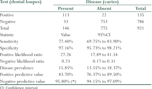
Table 4 summarizes the 2 × 2 table for calculation of sensitivity, specificity, positive likelihood ratio, negative likelihood ratio, positive predictive value, and negative predictive value of dental loupes. The sensitivity and specificity is 77.4% and 97.16%, respectively. The positive and negative likelihood ratio was found to be 27.26 and 0.23, respectively. The positive and negative predictive values are 83.70% and 95.80%, respectively.
Table 5 summarizes the comparison of mean DMF(T) scores using two different methods of examination. The mean DMF(T) score of the subjects examined using dental loupes is 3.778 ± 2.60670 and the mean DMF(T) score of subjects examined under normal vision is 3.778 ± 2.80567. The difference is not statistically significant.
Discussion
The diagnosis of caries is unique in that it is lacking the hypothetico-deductive reasoning.[17] Rather it is based on experiences of an individual. This highlights the subjective nature of occlusal caries detection.[18,19] In clinical conditions, there are various diagnostic aids to assist detection of dental caries, whereas in dental outreach program where the conditions are compromised there are no definitive
Table 1: Calculation of the kappa score in examining for dental caries in maxilla
Dental loupes Normal vision
Sound Caries Total
Sound 388 13 401
Caries 9 45 54
Total 39s7 58 455
Kappa score for maxilla=0.7841
Table: 2 Calculation of the kappa score in examining for dental caries in mandible
Dental loupes Normal vision
Sound Caries Total
Sound 365 18 383
Caries 13 68 81
Total 378 86 464
Kappa score for mandible=0.705
Table 3: 2×2 table for calculation of sensitivity, specificity, positive likelihood ratio, negative likelihood ratio, positive predictive value, and negative predictive value of normal visual examination
Test (visual examination) Disease (caries) Present Absent Total
Positive 113 33 146
Negative 22 753 775
Total 135 786 921
Statistic Value 95%CI
diagnosis aids for dental caries. There are many studies suggesting that vision under low magnification aids in better detection of dental caries.
As there is a paucity of literature in use of dental loupes in detection of dental caries in dental outreach camps, this study was conducted to compare the effectiveness of dental loupes with normal visual examination in detection of dental caries in dental outreach programs.
A total of 54 subjects attending the dental outreach program conducted by the Department of Public Health Dentistry, Saveetha Dental College were included in the study. All the 54 subjects were examined both by normal visual examination and vision under low magnification.
Two pre-trained examiners were involved in this study. One examiner used low magnification dental loupes (4× magnification) with LED light, mouth mirror, and explorer for detection of dental caries. The other examiner used torch light, mouth mirror, and explorer. Dental caries was recorded based on the WHO criteria in a WHO oral health assessment form.
In the current study, the kappa value for maxilla and mandible is calculated as 0.78 and 0.70, respectively. Hence, there is a substantial agreement between the examiners using dental loupes and normal visual examination in detecting dental carries, whereas in the study conducted by Mitropoulos et al.[16] the degree of intra-examiner agreement for the two examiners without and with magnification was almost perfect for Grades 0, 2, and 3 and substantial for Grade 1 irrespective of the use of magnification the difference in the variability between the studies is due to the reason that our study is conducted in a dental outreach program whereas the other study is an in vitro study conducted in ideal
conditions. In the study conducted by Kuhnisch et al.,[14] there was an inter-examiner and intra-examiner variability of 0.88 and 0.90 using dental loupes for detection of dental caries among children in a dental outreach program. In this study, two different criteria for detecting dental caries were used, which included ICDAS criteria using dental loupes and DMFT index without dental loupes whereas in study conducted by Timucin et al.[20] ICDAS criteria are evaluated using unaided visual examination and low magnification dental loupes showed excellent intra-examiner reproducibility (0.75–0.96). In the study conducted by Sisodia et al.,[13] the highest values for both inter- and intra-examiner reproducibility were obtained for Surgical Operating Microscope (0.63), compared to unenhanced visual-tactile and loupes. This could be attributed to coherent illumination provided by microscope that would allow for better visualization of defects.
In our study, the sensitivity of normal visual examination is 83.7% whereas the sensitivity of dental loupes is 77.4% which makes normal visual examination a better screening test in outreach programs for detection of dental caries. On the contrary, the specificity of vision under low magnification is 97.16% which is more than the specificity of normal visual examination 95.8% which makes it a better confirmatory test of detection of dental caries in dental outreach programs. The positive and negative likelihood ratio of normal visual examination is 19.94 and 0.17, respectively whereas; the positive and negative likelihood ratio of vision under low magnification is 27.26 and 0.23, respectively. The positive likelihood ratio of both the examination method is more than 1 which indicates that the probability of presence of caries among those tested positive is more than 50% and the presence of disease among those tested negative are <30%. The positive and negative predictive values of normal visual examination are 77.4% and 97.16%, respectively whereas; the positive and negative predictive values using dental loupes are 83.70% and 95.80%, respectively.
In our study, the mean DMF(T) score of the subjects examined using dental loupes is 3.77 ± 2.60 and the mean DMF(T) score of subjects examined under normal vision is 3.77 ± 2.80. The difference was not statistically significant. This implies that there is very mild difference in detection of dental caries using dental loupes and normal vision in a dental outreach program. Dental caries levels (DMFT) of 35–44 years olds in India as given by the WHO were 5.0–8.9[2] whereas in our study the (DMFT) dental caries levels of 35–44 years were 3.778. This reduced caries levels are due to less sample size in our study.
It can be concluded that there is no statistically significant difference between the effectiveness of vision under low magnification and normal visual examination in detecting dental caries in a dental outreach program. Hence, the null hypothesis is accepted.
It is recommended that better diagnostic aids must be adopted in detection of dental caries in dental outreach programs as it paves a way in prevention of early caries lesions and treatment of advanced lesions. Further studies have to be done employing other clinical aids in dental outreach programs to improve diagnosis of early caries lesion.
Table 4: 2×2 table for calculation of sensitivity, specificity, positive likelihood ratio, negative likelihood ratio, positive predictive value, and negative predictive value of dental loupes
Test (dental loupes) Disease (caries)
Present Absent Total
Positive 113 22 135
Negative 33 753 786
Total 146 775 921
Statistic Value 95%CI
Sensitivity 77.40% 69.75% to 83.90% Specificity 97.16% 95.73% to 98.21% Positive likelihood ratio 27.26 17.89 to 41.54 Negative likelihood ratio 0.23 0.17 to 0.31 Disease prevalence 15.85% 13.55% to 18.37% Positive predictive value 83.70% 76.37% to 89.50% Negative predictive value 95.80% (*) 94.15% to 97.09% CI: Confidence interval
Table 5: Comparison of mean DMF (T) scores using two different methods of examination
Diagnostic method N Mean±SD P value Dental loupes 54 3.7778±2.60670 0.496 Visual examination 54 3.7778±2.80567
Conclusion
The difference in the effectiveness of vision under low magnification and normal visual examination in detecting dental caries in a dental outreach program is not significant.The use of dental loupes and normal visual examination in dental outreach programs is effective in detection of dental caries. However, normal visual examination is more sensitive and can be used as a screening test whereas vision under low magnification has more specificity and can be used as a confirmatory test.
References
1. Amaechi B, Amerongen J, Loveren CV, Kidd E. Caries management, diagnosis
and treatment strategies. In: Summits’ Fundamentals of Operative Dentistry. A Contemporary Approach. Vol. 4. United States of America: Quintessence
Books; 2013. p. 200-40.
2. Petersen PE. Challenges to improvement of oral health in the 21st century - the
approach of the WHO global oral health programme. Int Dent J 2004;54 6 Suppl 1:329-43.
3. Shourie KL. Dental caries in Indian children. Indian J Med Res 1941;29:709-21. 4. Damle SC, Patel AR. Caries prevalence and treatment need amongst children
of Dharavi, Bombay, India. Community Dent Oral Epidemiol 1994;22:62-3.
5. Antia FE. The dental caries experience of school going children in the city of
Bombay. J Indian Dent Assoc 1962;39:325.
6. Tewari A, Chawla HS. Study of prevalence of dental caries in an urban area of India (Chandigarh). J Indian Dent Assoc 1977;49:231-9.
7. Dash JK, Sahoo PK, Bhuyan SK, Sahoo SK. Prevalence of dental caries and
treatment needs among children of Cuttack (Orissa). J Indian Soc Pedod Prev Dent 2002;20:139-43.
8. Dhar V, Jain A, Van Dyke TE, Kohli A. Prevalence of dental caries and treatment needs in the school-going children of rural areas in Udaipur district. J Indian Soc Pedod Prev Dent 2007;25:119-21.
9. Saravanan S, Kalyani V, Vijayarani MP, Jayakodi P, Felix J, Arunmozhi P,
et al. Caries prevalence and treatment needs of rural school children
in Chidambaram Taluk, Tamil Nadu, South India. Indian J Dent Res
2008;19:186-90.
10. Sheehy EC, Brailsford SR, Kidd EA, Beighton D, Zoitopoulos L. Comparison
between visual examination and a laser fluorescence system for in vivo diagnosis of occlusal caries. Caries Res 2001;35:421-6.
11. Ricketts DN, Ekstrand KR, Kidd EA, Larsen T. Relating visual and radiographic ranked scoring systems for occlusal caries detection to histological and microbiological evidence. Oper Dent 2002;27:231-7.
12. Forgie AH, Pine CM, Pitts NB. The use of magnification in a preventive
approach to caries detection. Quintessence Int 2002;33:13-6.
13. Sisodia N, Manjunath MK. Impact of low level magnification on incipient occlusal caries diagnosis and treatment decision making. J Clin Diagn Res
2014;8:ZC32-5.
14. Kühnisch J, Berger S, Goddon I, Senkel H, Pitts N, Heinrich-Weltzien R.
Occlusal caries detection in permanent molars according to WHO basic methods, ICDAS II and laser fluorescence measurements. Community Dent Oral Epidemiol 2008;36:475-84.
15. Hintze H, Wenzel A, Danielsen B, Nyvad B. Reliability of visual examination,
fibre-optic trans illumination, and bite-wing radiography, and reproducibility of direct visual examination following tooth separation for the identification of cavitated carious lesions in contacting a proximal surfaces. Caries Res 1998;32:204-9.
16. Mitropoulos P, Rahiotis C, Kakaboura A, Vougiouklakis G. The impact of magnification on occlusal caries diagnosis with implementation of the ICDAS II criteria. Caries Res 2012;46:82-6.
17. Bader JD, Shugars DA. What do we know about how dentists make caries-related
treatment decisions? Community Dent Oral Epidemiol 1997;25:97-103.
18. Nyvad B, Fejerskov O. Assessing the stage of caries lesion activity on the basis
of clinical and microbiological examination. Community Dent Oral Epidemiol 1997;25:69-75.
19. Maupome E. Cumulative assessment of factors leading to restorative demonstrations in an educational setting. A graphical demonstration of an
in-vivo care. Oper Dent 2000;25:336-43.
20. Ari T, Kofman SH, Ari N. In vitro evaluation of magnification and LED illumination for detection of occlusal caries in primary and permanent molars using ICDAS criteria. Dent J 2013;1:19-30.