This is an open access journal, and articles are distributed under the terms of the Creative Commons Attribution-Non Commercial-Share Alike 4.0 License, which allows others to remix, tweak, and build upon the work non-commercially, as long as appropriate credit is given and the new creations are licensed under the identical terms.
Serum Hepatocytes growth factor in acute and chronic Kidney
diseasepatients and its relation to disease activity
Ghada Khalifa Zayet
Nephrology Department, Theodor Bilharz Research Institute, Egypt.
Correspondence: GhadaKhalifa Zayet, Nephrology Department, Theodor Bilharz Research Institute, Egypt, E-mail: [email protected].
ABSTRACT
Background: Chronic kidney disease (CKD) is a major health problem, it is characterized by progressive destruction of functional nephrons, which finally cause end stage kidney disease. Also acute kidney disease (AKD) is a serious condition characterized by acute loss of kidney function with variable degree up to renal failure. One of the important factors in the regulation of cell growth is the hepatocyte growth factor (HGF) which is well known as a mesenchyme-derived pleiotropic, in addition, HGF is playing a role in the morphogenesis of different kinds of cells and cell motility. HGF is a potent renotropic protein that promotes tubular repair and renal regeneration after injurious stimuli. Aim: The aim of the work was to study the correlation between serum HGF level and renal functions of patients with AKD and CKD, including hemodialysis patients. Materials: The study included 62 patients that divided into five groups (of different stages of chronic CKD ((I – V) and AKD in compare to patients with normal renal functions (not including patients with; renal transplantation, malignancies, liver diseases, Pregnancy and lung diseases). All patients were subjected to through case history, clinical examination, chest X ray, abdominal U/S, examination, (BUN, serum creatinine, liver function tests), estimation of GFR by MDRD equation, and serum level of HGF. Results: There was significant increase of serum HGF level in AKI and CKD patients of stages I, II, III and IV, and prompt in patients of stage V specifically patients on regular hemodialysis. Also there was significant correlation between renal sonographic appearance and the serum HGF level as well as the degree of nephropathy and age.
Keywords:CKD, AKI, Hepatocyte growth factor (HGF), renal, fibrosis
Introduction
Worldwide, Chronic kidney disease (CKD) represent a major health crisis because it has elevated incidence during early and final (renal failure) phases, and also due to the elevation in hospital coasts beside improper response to the therapy [1].
The pathogenesis of CKD is illustrated by continuous increase in the loss of the parenchyma of kidney and the increase in the destruction of active nephrons, which leads to failure of the kidney function and ended with chronic kidney disease [2]. The
mechanism of action of CKD is not known completely, but the disease slowly progress, and leads to the loss of active nephrons,
so the body try to compensate the activity of nephrons through stimulation of both molecular and cellular procedures, leading to increase in the growth of the inactive or the rest of nephrons
[3].
Chronic renal failure is considered as one of the major risk factors that threaten the life of peoples, worldwide. The rising rate of peoples suffering yearly from end stage kidney disease carry a most important public and economic load on most countries and there are three principal choices for renal replacement therapy (RRT), comprising peritoneal dialysis (PD), hemodialysis (HD) and finally renal transplantation [4].
The method of hemodialysis aims to clear the body from waste products like, urea and creatinine and withdrawn free water from the blood in cases of renal failure [5].
Hepatocyte Growth Factor (HGF) consists of 69 kDaα-chain and a 34 kDaβ-chain, a heterodimer molecule derived from the mesenchyma, possess both pleiotropic, and multifunctional cytokines [6].
HGF is produced from different tissues such as liver, kidney, lung and spleen. It exerts multiple biological activities on different cell types as in injured tissues. It helps tissue repair and regeneration rapidly as it has great role in stimulation of
Access this article online
Website:www.japer.in E-ISSN: 2249-3379
How to cite this article: GhadaKhalifa Zayet. Serum Hepatocytes growth
factor in acute and chronic Kidney disease patients and its relation to disease activity. J Adv Pharm Edu Res 2018;8(3):74-80.
hepatocytes and renal cells’ proliferations especially in acute kidney disease [7].
The role of HGF is dual, where it carry anti-apoptotic, morphogenic and mitogenic behavior on kidney cells and subsequent acute kidney damage, the HGF receptor c met is released primarily in kidney tubular epithelium together with ablation of c – met in renal tubules enhance renal inflammation and chemokine appearance post acute kidney infection (AKI) [8].
It appears to stimulate renal repair in acute renal failure and it may play a role in chronic kidney disease by counteracting tissue fibrosis with elevation of serum HGF levels in patients with chronic kidney disease, and it has been proposed that the altitude of HGF is interrelated with renal functions [9].
Patients and Methods
The current study was conducted in the period extending from July 2017 to April 2018 in Internal Medicine Department, Nephrology and Dialysis Unit of Theodor Bilharz Research Institute (TBRI) , Egypt.
Subjects:
62 patients with acute, chronic kidney disease and end stage kidney disease on regular hemodialysis with (age between 18-80 years; Males and females), patients with AKI due to either glomerular or tubular interestial or vascular causes, patients with chronic kidney diseases due to either glomerular or tubular or vascular and poly cystic kidney disease, patients with end stage kideny disease on regular hemodialysis receiving 3 sessions of hemodialysis per week, each session was 4 hours and duration of dialysis more than 2 months.
Patients with renal transplantation, malignancies, liver diseases, Pregnant women, severe inflammatory or severe concomitant diseases, and lung diseases were not included.
Operational design:
The study is a case control study in which 62 patients are divided into four groups: Group 1 (control group): Including 10 patients (4 males and 6 females), with normal renal functions: normal serum creatinine (0.3-1.1 mg/dl), normal BUN (6-20 md/dl) and eGFR (>90 mL/min/1.73 m2). Group
2: Including 6 patients (5 males and 1 females) of satges I and II of chronic kideny disease: with GFR of ≥60 mL/min/1.73 m2.
Group 3:10 patients (3 males and 7 females) of stages III and IV of chronic kideny disease: with GFR of 15- 59 mL/min/1.73 m2. Group 4: 26 patients (14 males and 12 females) with
chronic kidney disease of stage V and ESKD patients with regular hemodialysis including 6 patients of chronic kidney disease stage V and not on regular hemodialysis and 20 patients of end stage kidney disease and on regular hemodialysis receiving 3 sessions of hemodialysis per week, each session 4 hours, duration of dialysis more than 2 months, and the type of hemodialysis solution is bicarbonate and GFR of <15 mL/min/1.73 m2 or dialysis. Group5: 10 patients (5 males and
5 females) of Acute renal failure with percentage increase in
serum creatinine of ≥ 50% and GFR of 15- 59 mL/min/1.73 m2.
Methods
All patients were subjected to full history, full clinical examination, laboratory investigations (serum creatinine, serum urea, serum sodium and serum potassium); estimation of GFR was done by MDRD equation:
MDRD equation=186 × Scr-1.154 × Age-0.203 × (1.212 if black) ×
(0.742 if female). Liver functions (liver enzymes - serum albumin) and serum hepatocyte growth factor were also calculated. Also, patients were subjected to pevli-abdominal ultrasonography, and Chest X-ray. Renal biopsy was done for patients with glomerulonephritis.
Human HGF Assay
Serum samples were collected from participants and stored at -70 °C. We use BioSource international, Inc. hHGF kits which is a solid phase sandwich Enzyme Linked Imluo-Sorbent Assay (ELISA).
Principle of the method
A monoclonal antibody specific for hHGF coated onto the wells of the micro-titer strips were provided. Samples including standards of known hHGF content, control specimens, and unknowns, were pipetted into these wells, followed by the addition of a second monoclonal biotinylated antibody.
During the first incubation, the hHGF antigen binds simultaneously to the immobilized (capture) antibody on one site, and to the solution phase biotinylated antibody on a second site. After removal of excess second antibody, Streptavidin- peroxidase (enzyme) is added. This binds to the biotinylated antibody to complete the four- member sandwich. After a second incubation and washing to remove the entire unbound enzyme, a substrate solution is added, which acts upon by the bound enzyme to produce color. The intensity of this colored product is directly proportional to the concentration of hHGF present in the original specimen.
Reagents
Human HGF assay procedure
Thereagents and standards were prepared. 50 µl of sample (serum and standard) was added to wells; then a- 50 µl of incubation buffer, b- 50 µl of biotin conjugate, c- 50 µl of biotin conjugate were added, and incubated for 2 hours at room temperature, aspirated and washed 4 times. Then, 100 µl of stabilized Chromogen was added and incubated for 30 minutes at room temperature. 100 µl of stop solution was added, then plate is read at 450 nm by ELIZA reader.
Administration design
Approvals were obtained from governmental departments, ethical committee of TeodorBilharz Research Institute institutional review board (IRB) and from patients included in the study.
Results
In our study, we found that there is significant increase of serum HGF in the patients with CKD (all stages) as well as the patients with chronic renal failure on regular hemodialysis and patients with AKD.
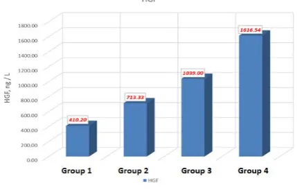
In control group was with mean value of 410.2 ± 87.5ng/L. There were normal levels of HGF in correlation to normal kidney function and liver functions.
In group 2 (CKD stages I and II) there were mild to moderate increase of HGF levels with mean serum HGF value of 713.3 ± 123.9 ng/L, while there were more increase of the concentration of HGF in the serum in group 3 (CKD stages III and IV) with mean value of 1039 ± 165.8 ng/L. In group 4 (CKD stage V including hemodialysis patients) there were prompt increase of levels of serum HGF with mean value of 1616.5 ± 324.5 ng/L, especially in hemodialysis patients. Our study results showed significant correlation between different stages with different eGFR of CKD with serum HGF levels. The increase of HGF levels on Hemodialysis patients was more than the patients with chronic kidney stages I, II, III and IV. In group 5 (AKD) there was mild to moderate increase of HGF levels with mean serum HGF value of 600.2 ± 98.57ng/L.
Table 1: Gender distribution among studied groups
Stage
χ2 P
G
roup1 Contr
ol
G
roup 2 (I – II) Group 3 (III–I
V)
G
roup 4 (V) Group5 (AKI)
N=10 N= 6 N=10 N=26 N=10
G
en
der Female
No. 6 1 7 12 5
4.82 .185 % 60.0% 16.7% 70.0% 46.2% 50%
Male No. % 40.0% 4 83.3% 5 30.0% 3 53.8% 14 50% 5
This table showed that there were no statistical significant differences between two groups regarding sex.
Table 2: Age and Weight of the studied groups
G
roup1 Contr
ol
G
roup 2 (I – II) Group 3 (III-IV) Group 4 (V) Group 5 AKI F Sig.
(N=10) (N=6) (N=10) (N=26) (N=10)
Age ±8.5 52.2 ±11.9 31.3 ±12.9 59 ±12.9 59.1 ±12.9 49.1 9.200 <0.001**
Weight ±13.5 89.1 77.2 ±16 ±15.1 80.2 ±13.6 89.3 75.2 ±15 1.982 .129
This table showed that there were high statistical significant differences between different groups as regarding age, while there was no statistical significance as regarding weight.
Table 3: Mean±SD value of HGF in ng/dl in each group
G
roup1 Contr
ol
G
roup2 (I – II) Group3
(III
–
IV)
G
roup4 (V)
G
roup5 AKD F Sig.
(N=10) (N=6) (N=10) (N=26) (N=26)
H
GF
,
ng/
L 410.2
± 87.5 ± 123.9 713.3 ± 165.8 1039 ± 324.5 1616.5 ± 98.5 65.48 <0.001** 600.2
This table shows significant difference among different groups as regarding serum level of HGF.
Table 4: Post-hoc test using least significant difference (LSD), to indicate which groups were significantly different
from which others
LSD (Control) Group1 Group 2 (I – II) Group 3 (III – IV)
(N=10) (N=6) (N=10)
(Group 4) V < 0.001** < 0.001** < 0.001** ( Group 3) III – IV < 0.001** 0.015*
(Group 2) I – II 0.024*
** Highly Significant
This table shows high significant difference of group 4 from others groups, while there was high significant difference of group 3 with control group, significant difference with group 2, and least significance difference between group 2 and group 1 (control).
Figure 1:Comparison between the different groups regarding the mean level of serum HGF
Table 5: Comparison among the five groups regarding mean± SD value of certain studied parameters in each
C
on
tr
ol
I –
II
III
–
IV
V AKI F Sig.
(N=10) (N=6) (N=10) (N=26) (N=10) S.ALT
U/L ± 4.4 15.1 ± 1.9 16.8 ± 4 14 ± 4.2 18.1 ± 2.4 16.1 102 3. .035
S.AST
U/L ± 2.8 14.7 ± 2.7 16.5 ± 5.1 14.6 ± 7.2 19.1 ± 2.8 15.7 261 2. .093
S. K+
mEq/L ± 0.5 4.3 ± 0.3 4.2 ± 0.8 4.4 ± 0.6 4.7 ± 0.3 4.2 125 2. .109
S. Na+
mEq/L ± 5.2 141.7 140.2 ± 2.1 140.5 ± 4.7 139.6 ± 4.5 140.2 ± 2.4 .525 .667
S. Alb
g/dL ± 0.4 4.8 ± 0.8 3.6 ± 0.5 3.6 ± 0.3 3.5 ± 0.5 3.6 240 <0.001** 21.
eGFRml/
min/1.73m² ± 14.8 113.6 ± 27.5 87 ± 9.1 29.1 ± 3.1 6.3 233.490 <0.001**
BUN
mg/dL ± 3.1 14.2 ± 5.1 17.5 ± 15.7 36.5 ± 10.1 73.7 ± 15.7 40.5 113.971 <0.001**
S. Cr
mg/dL 1 ± 0 ± 0.2 1.1 ± 0.6 2.1 ± 3.3 9.8 ± 0.5 2.5 106 <0.001** 54.
This table shows high significant difference among different groups as regarding serum albumin, eGFR, BUN and serum creatinine but no significant difference regarding serum ALT, serum AST, serum K+ and serum Na+.
Table 6: Mean value of S.HGF (ng/L) in the different sonographic groups
No
rm
al
Ki
dne
ys
gr
ad
e I
ne
ph
ropa
th
y
gr
ad
e
II
ne
ph
ropa
th
y
gr
ad
e III
ne
ph
ropa
th
y
Sm
all
shu
rn
ke
n
Ki
dne
y
Number of values N=9 N=9 N=8 N=9 N=2
Minimum 590.0 820.0 890.0 1400 1780
25% Percentile 730.0 885.0 1163 1615 1780
Median 920.0 1060 1340 1770 1800
75% Percentile 1250 1620 1878 1840 1820
Maximum 1600 2000 2100 2050 1820
Mean 1004 1233 1464 1739 1800
Std. Deviation 327.7 420.2 416.5 186.1 28.28 Std. Error of Mean 109.2 140.1 147.3 62.04 20.00
P value
This table showed that there were high statistical significant differences between different sonographic groups as regarding mean value of serum HGF of each group.
Figure2:Comparison between the different sonographic groups regarding mean value of serum HGF (ng/L) of each group.
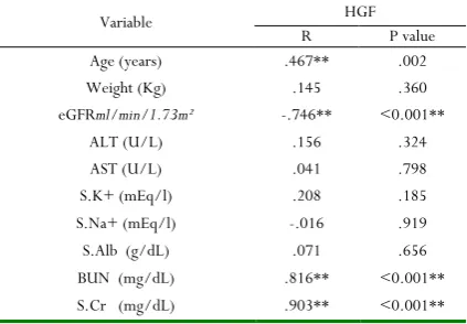
Table 7: Pearson’s correlation between S.HGF (ng/L) and other studied parameters in CKD groups
Variable HGF
R P value
Age (years) .467** .002
Weight (Kg) .145 .360
eGFRml/min/1.73m² -.746** <0.001**
ALT (U/L) .156 .324
AST (U/L) .041 .798
S.K+ (mEq/l) .208 .185
S.Na+ (mEq/l) -.016 .919
S.Alb (g/dL) .071 .656
BUN (mg/dL) .816** <0.001**
S.Cr (mg/dL) .903** <0.001**
r= correlation coefficient
**Correlation is significant at the 0.01 level (2-tailed). *Correlation is significant at the 0.05 level (2-tailed).
Figure 4:Correlation between age and serum HGF (ng/L) in different groups.
Figure 5:ROC curve of HGF CKD groups
Area under the ROC curve (AUC)
Area under the ROC curve (AUC) 0.919
Standard Error a 0.0412
95% Confidence interval b 0.793 to 0.981
z statistic 10.167
Significance level P (Area=0.5) <0.001**
HGF cut-off Sensitivity Specificity +LR -LR +PV -PV >920 ng/dl 90.32 63.64 2.48 0.15 87.5 70.0
Discussion
In the present work, we studied the levels of HGF on the patients with chronic kidney diseases, patients on regular hemodialysis and AKD patients. The aim was to study the correlation between serum HGF level and renal functions of patients with CKD and AKD
In our study, we found that there is significant increase of serum HGF in the patients with CKD (all stages) as well as the patients with chronic renal failure on regular hemodialysis and patients with AKD.
In control group with mean value of 410.2 ± 87.5 ng/L there were normal levels of HGF in correlation to normal kidney function and liver functions, which was in agreement with the study of Lohr et al. in which the mean value of serum HGF in control group with normal kidney functions was 460 ± 40 ng/L
[9].
In group 2 (CKD stages I and II) there were mild to moderate increase of HGF levels with mean serum HGF value of 713.3 ± 123.9 ng/L, while there was more increase of the concentration of HGF in the serum in group 3 (CKD stages III and IV) with mean value of 1039 ± 165.8 ng/L. These results were consistent with a study which revealed that in non-dialysis subjects with renal insufficiency serum HGF was increased significantly (p<0.001) compared to normal individuals, and the increased serum HGF was associated with level of creatinine in the serum [10].
In group 4 (CKD stage V including hemodialysis patients) there were prompt increase of levels of serum HGF with mean value of 1616.5 ± 324.5 ng/L, especially hemodialysis patients. Our study results showed significant correlation between different stages with different eGFR of CKD with serum HGF levels. This coincides with Libetta et al. [11] who demonstrated that
there is very significant increase of HGF levels in hemodialysis patients with low eGFR, which was in agreement with Zhang Y
et al. (2015) who showed that dialysis method act as stimulator to release HGF, like tumor necrosis factor and interleukin-1 (IL-1), which may play a role for HGF release (the so-called injurins). Moreover, the processes of dialysis may lead to the discharge of coagulation signals that change HGF from inactive to active structure. Then, at least hypothetically, elevated HGF action persuaded by dialysis may be pathophysiologically significant in individuals demanding renal replacement treatment [12].
The increase of HGF levels on hemodialysis patients was more than the patients with chronic kidney stages I, II, III and IV. These findings coincide with study done by Libetta et al., in 2013, who stated that hemodialysis causes a rapid and extended discharge of HGF into the blood stream, elevating HGF serum level up to 30 episodes [11]. Many authors postulated variable
mechanisms for explaining the high levels of serum HGFin HD patients, such as activation of leukocytes and production of cytokines in the blood as a result of circulation of extracorporeal during renal hemodialysis. The activation in the peripheral leukocytes in the circulation are considered as a potential source of HGF, since HGF is created by leukocytes in the hepatic tissues of liver-diseased patients, then cytokines may act as injurins. Therefore, dialysis may convince the discharge of HGF either due to stimulation of tissue cells by cytokines or due to activation of leucocytes in the peripheral circulation. Also, renal dialysis may lead to discharge of coagulation factors that change HGF into an active form. Also Alvarez et al. confirmed that the discharge of HGF does not depend on heparin-induced detaching of receptor bound HGF from peripheral tissues, but can be released during dialysis without
0 20 40 60 80 100
HGF
0 20 40 60 80 100
100-Specificity
S
e
n
s
it
iv
it
addition of heparin. Therefore, high serum HGF level appears soon after the initiation of extracorporeal blood circulation [13].
In group 5 (AKD) there were mild to moderate increase of HGF levels with mean serum HGF value of 600.2 ± 98.57ng/L.
Flaquer et al., in 2012 in an in vitro experiment noticed during dialysis that the peripheral blood mononuclear cell (PBMC) contained a high concentration of HGF, this revealed that: (a) PBMC are capable of releasing HGF and (b) dialysis solution can stimulate HGF discharge by PBMC, possibly due to the bio-incompatibility of HD therapy. Additionally, the connection among inflammatory processes and level of HGF in HD demonstrate that serum HGF is strictly correlated with IL-6 and C-reactive protein (CRP); it is also possible that HD-related HGF release might be induced by elevated circulating levels of inflammatory cytokines, which could characterize a powerful stimulus for HGF discharge [14].
A study done by Ciro et al. [15], suggested that hemodialysis
causes increase of serum HGF concentration in majority of patients with ESKD on regular hemodialysis (74 out of 76), however that effect was not uniform in all patients. Also, the increase of serum HGF could be due to application of heparin and it wasn’t excluded that process of hemodialysis itself causes such change, due to stimulation of peripheral blood mononuclear [15]
On the other hand, there was also significant increase of serum HGF levels with increasing the age. This is in agreement with Mizuno et al., who stated that the age plays an important role, where higher ages are generally associated by higher incidence of comorbidities comprising CVD and higher circulating levels of many inflammation markers in the serum such as HGF. The relation between the age and level of HGF is reinforced by some researches reporting that the stimulation of an inflammatory cytokines induces up-regulation of HGF [16], and
HGF levels increased with increasing age; this may elucidate the positive correlation between inflammation and the increasing in age [17].
In our study there was significant correlation between renal sonographic appearance and the levels of serum HGF with increasing the serum HGF level with increasing the degree of nephropathy (fibrosis) as the mean level of HGF in normal kidneys ultrasonography was 1004 ng/L, in the grade I nephropathy was 1233 ng/L, in the grade II was 1464 ng/L, in the grade III was 1739 ng/Land reaching to highest level in marked fibrosis when kidneys became shrunken up to 1800 ng/L.
Our study showed that there was statistically no significant correlation found among the 4 groups, regarding gender. These results agree with Libetta et al., who mentioned that there was no difference between the groups of CKD patients regarding sex during study of serum HGF [11].
Some trials were carried out in various animal models of renal failure, hepatic diseases, cardiovascular diseases (CVD) and diabetes mellitus by direct treatment with gene therapy and HGF, verifying its efficacy in prompting renal and hepatic
regeneration post acute and chronic inflammation and employing cardio-protective effects post acute myocardial infarction [18].
This study gives evidence that serum HGF increase in ACD, CKD and hemodialysis patients, and HGF is a strongrenotropic protein that shows a serious part in stimulating tubular healing and renal restoration after exposure to harmful agents. This deserves consideration for several reasons. First, hemodialysis is frequently necessary to treat individuals with toxic-ischemic acute kidney injury, possibility if HGF plays an important role in recovery from this disease. Second, HGF is a multitropic element that induces the production of many cell kinds such as distribution and arrangement of cells in complex structures for example, tubular, fibroblasts, angiogenesis and contributes in inflammatory processes. In spite of several effects of HGF corresponding repair and regeneration of tissues, undesirable actions of sustained HGF stimulation are probable, e.g., in the mobilization and proliferation of cancerous cells or initiation of exaggerated fibrosis in inflamed tissues. Accordingly, the release of HGF should be taken into consideration in evaluating the biocompatibility of renal dialysis. Hemodialysis denotes a tedious and reproducible disorder of activation of HGF and may be a suitable model to further study its physiology. Finally, exogenous HGF can be introduced as therapeutic agent for patients with acute kidney injury and patients with CKD.
Conclusion
From the previously mentioned results we can conclude that there were increase of serum HGF levels in patients with Acute kidney injury and chronic kidney disease with prompt increase of the levels of serum HGF in CKD stage V especially ESKD on regular hemodialysis, so serum HGF may be a good indicator for degree of renal fibrosis in CKD patients.
Acknowledgement:
Thanks to stuff and patients of TeodorBilharz Research Institute for helping me to accomplish this work
References
1. Liangos O and Jaber B: Chronic Kidney Disease, Dialysis, and Transplantation. Sctionlll; 24: 354-370 (2012).
2. Amandine V, Khalil E and Denise L: J Clin Invest, (2010), Lipocalin 2 is essential for chronic kidney disease progression in mice and humans.; 120 (11): 4065–4076.
4. Kikuchi H, Kanda E, Mandai S, et alClinical and Experimental Nephrology, (2016): Combination of low body mass index and serum albumin level is associated with chronic kidney disease progression: the chronic kidney disease-research of outcomes in treatment and epidemiology (CKD-ROUTE) study. 37: 1-8
5. K/DOQI clinical practice guidelines for chronic kidney disease, Am J Kidney Dis (2012): Evaluation, classification, and stratification. Kidney DiseaseOutcome Quality Initiative; 39: S1–S246.
6. Boros P and Miller CLancet; (2011): Hepatocyte growth factor: A multifunctional cytokine. 345:293– 295
7. Dong Z, Roderick J, Lin L, Lili Z and YouhuaL Kidney International (2013): Activation of hepatocyte growth factor receptor, c-met, in renal tubules is required for renoprotection after acute kidney injury; 84: 509–520 8. Christophe Q, Sylvain M and AurelieF Am J Physiol
Lung Cell MolPhysiol; (2008): Regulation of hepatocyte growth factor secretion by fibroblasts in patients with acute lung injury. 294:334-343.
9. Lohr J, Lee T, Farooqui M and Mookerjee B. Med (2010): Increased levels of serum hepatocyte growth factor in patients with end-stage renal disease; 31(3-4): 131-141.
10. Sugimura K, Kim T, Goto T, Kasai S, Takemoto Y, Matsuda J, Yoshimoto M, Yamagami S and KishimotoT Serum hepatocyte growth factor levels in patients with chronic renal failure Nephron (2009); 70(3): 324-328. 11. Libetta C, Esposito P, Sepe V, Rampino T, Zucchi M,
Canevari M and Dal Canton A: Acute kidney injury effect of hemodialysis membrane on HGF and recovery of renal function. Clin Biochemistry (2013); 46:103– 108
12. Zhang Y, Jain RK and Zhu M: Recent progress and advances in HGF/MET-targeted therapeutic agents for cancer treatment biomedicines (2015); 3(1):149–181 13. Alvarez V, Pulido R, Campaner O, Paraiso V,
De-Landazuri MO, Sanchez FKidneyInt (2010): Differentially regulated cell surface expression of leukocyte adhesion receptors on neutrophils.; 40:899 – 905
14. Flaquer M, Franquesa M, Vidal A, et al: Hepatocyte growth factor gene therapy enhances infiltration of macrophages and may induce kidney repair in db/db mice as a model of diabetes. Diabetologia (2012); 55:2059–2068
15. Ciro E, Bina P, Andreana D, Flavia C, Gianluca F, Annalisa F, Tizziana M Kideny international (2014): Hepatocyte growth factor (HGF) modulates matrix turnover in human glomeruli.; 67: 2143-2150
16. Mizuno S, Nakamura T, Cowin A, Kallincos N and Hatzirodos N: Improvement of sepsis by hepatocyte growth factor, an anti-inflammatory regulator: emerging insights and therapeutic potential. Gastroenterol Res Pract (2012);909350: 14-18
17. Stenvinkel P, Heimburger O, Paultre F, et al KidneyInt; (2010): Strong association between malnutrition, inflammation, and atherosclerosis in chronic renal failure; 55:1899-1911.