Victor V. Evstigneev
1, a, E, f, Volha V. Kistsen
1, B–D, Igor V. Bulaev
2, B,
Ruslan a. Sakovich
3, BThe Effect of Structural White Matter Abnormalities
on the Clinical Course of Epilepsy*
Wpływ zaburzeń strukturalnych istoty białej
na kliniczny przebieg padaczki
1 Department of Neurology and Neurosurgery, Belarusian Medical academy of Postgraduate Education,
Minsk, Belarus
2 Department of Radiology, Republican Scientific and Practical Center of Traumatology and Orthopedics,
Minsk, Belarus
3 Department of Radiology, City Clinic Hospital No 2, Minsk, Belarus
A – research concept and design; B – collection and/or assembly of data; C – data analysis and interpretation;
D – writing the article; E – critical revision of the article; F – final approval of article; G – other
Abstract
Objectives.The aim of this study was to assess the impact of the extent of brain white matter lesions on the devel-opment of cognitive and psychoemotional disorders, and to investigate correlations between the degree of integra-tion of brain pathway structures and the clinical features of epilepsy.
Material and Methods.forty-six epileptic patients (36 with pharmacoresistant epilepsy and 10 who had been in remission for over a year) and 10 normal volunteers (the control group) were examined. To evaluate diffusion tensor MRI findings, the index of fractional anisotropy (fa) and index of apparent diffusion coefficient (aDC) were used. for an intergroup comparison of DTI data, the Mann-Whitney test was used, criterion; correlation analysis was per-formed using the Spearman rank correlation coefficient. The threshold of statistical significance was set at р < 0.05.
Results.a significant difference was noted in the aDC data on the side of the epileptic focus in the patients in per-sistent remission as compared to the pharmacoresistant patients (р < 0.05). No differences were found between the patient groups’ fractional anisotropy data. In cases of mesial temporal sclerosis in patients with pharmacoresistant epilepsy, a “weakening” of the tractography pattern in the opposite hemisphere was found (r = 0.66, p = 0.0005). Decreases in the tracts appearing in brain temporal lobes was typical of patients with pharmacoresistant forms of epilepsy (r = 0.46, p = 0.0005). a pathological decrease in fa and an increase in aDC correlated with the results on the Beck scale and the Spielberger-Khanin anxiety scale (r = –0.2, p < 0.001) as well as with P300 peak latency data (r = 0.23, p < 0.001). analyses of the peculiarities of EEG patterns and fa data demonstrated a correlation between the existence of epileptic activity and a decrease in fa (r = –0.7, t = –2.44, p = 0.01).
Conclusions. Microstructural brain matter changes make it possible to assess the course of epilepsy to predict the outcomes of medicamental correction of paroxysmal states (Adv Clin Exp Med 2013, 22, 4, 529–537).
Key words:epilepsy, diffusion tensor magnetic resonance tomography, tractography.
Streszczenie
Cel pracy. Ocena wpływu stopnia uszkodzenia istoty białej mózgu na rozwój zaburzeń poznawczych i psycho-emocjonalnych oraz zbadanie korelacji między stopniem integracji struktur szlaku mózgu i cech klinicznych padaczki.
Materiał i metody. Do badań włączono 46 chorych na padaczkę (36 z padaczką oporną na leczenie i 10, który byli w remisji od ponad roku) i 10 zdrowych ochotników (grupa kontrolna). aby ocenić wyniki obrazowania
tenso-adv Clin Exp Med 2013, 22, 4, 529–537 ISSN 1899–5276
ORIGINal PaPERS
© Copyright by Wroclaw Medical University
Significant progress in studying and diagnos-ing a number of neurological diseases has been achieved. This is due, in the first place, to the wide-spread introduction of up-to-the-minute neuroim-aging techniques into clinical practice. X-ray com-puter and magnetic resonance imaging, magnetic resonance spectroscopy, single photon emission computed tomography and positron emission to-mography enable medical professionals to discov-er many of the mechanisms of degendiscov-erative and dystrophic processes and to study the structure of many pathological states [9, 11, 12, 18].
The importance of computed tomography (CT) in diagnosing local lesions causing the de-velopment of epilepsy has significantly decreased, mainly because it provides images of sufficient quality only in the case of high density tissues. One should take into account that CT sensitivity in di-agnosing structural lesions in patients with epi-lepsy does not exceed 30%. CT is practically use-less in diagnosing mesial temporal sclerosis, which is the most common cause of pharmacoresistant epilepsy [9].
The introduction of magnetic resonance im-aging (MRI) into clinical practice significantly fa-cilitated the identification of various structural changes in the brain tissue underlying the forma-tion of an epileptic focus that subsequently results in the development of epilepsy. The detection rate for mesial temporal sclerosis, tumors and trauma, which more often occur in adults, is high. In in-fancy and childhood, the most common causes of epilepsy are malformations of the brain. MRI per-mits the identification of such malformations as lissencephaly, nodular periventricular heterotopia, schizencephaly, hemimegalencephaly and others, with focal cortical dysplasia being an important an-atomic substrate of extratemporal epilepsies [7].
Compared with standard MR images, diffu-sion weighted images are much more sensitive to structural and metabolic changes that occur in the brain. In the course of changes in normal cerebral
metabolism new pathways appear, and simulta-neously new metabolic products. These in turn change normal cell composition and cause an oc-currence of some cell components and, as a con-sequence, result in the formation of pathological brain micro- and macrostructures. all these pro-cesses can be described in terms of quantity, ac-cording to the change of fractional anisotropy and diffusion coefficients in different brain struc-tures [5, 14].
Temporal lobe epilepsy (TlE) is the most fre-quent form of focal epilepsy in which there are structural and functional disturbances both in the ictal onset zone and at a distance from it. The use of diffusion tensor imaging (DTI) on patients with temporal epilepsy permits the detection of the brain white matter lesion that is disturbing different struc-ture connections. This is more evident in the hemi-sphere ipsilateral to an epileptic focus [1, 2, 16, 21]. The most important DTI parameter defining white matter integration is the index of fractional anisotropy (fa), which is assessed as a magnitude of the water diffusion path in tridimensional space. Densely spaced fascicles of white matter provide structural coherence due to which water diffusion has a definite direction and the fa is rather high. In cases of white substance structure failure, the water diffusion is less ordered, resulting in a de-crease in the fa [21]. To evaluate diffusion ten-sor MRI findings, one uses the index of the appar-ent diffusion coefficiappar-ent (aDC), increases in which are related to a neurogenesis defect or a cell loss with a subsequent increase in extracellular space. DTI can be used to map specific brain white mat-ter tracts (tractography).
There are sporadic contradictory data on the role of structural changes in brain white matter tracts in the clinical course of epilepsy and the for-mation of cognitive and psychoemotional disor-ders [6, 13, 19, 22].
In the present study, data were analyzed from brain DTIs from patients with pharmacoresistant
ra, wykorzystano wskaźnik frakcyjnej anizotropii (fa) i wskaźnik pozornego współczynnika dyfuzji (aDC). aby porównać dane DTI między grupami, zastosowano test Manna-Whitneya. analizę korelacji przeprowadzono za pomocą współczynnika korelacji rang Spearmana. Próg istotności statystycznej ustalono na р < 0,05.
Wyniki. Zauważono znaczącą różnicę w danych aDC po stronie ogniska padaczkowego u pacjentów w remisji w porównaniu z pacjentami opornymi na leki (р < 0,05). Nie stwierdzono różnic między danymi o anizotropii grup pacjentów. W przypadku stwardnienia centralno-skroniowego u chorych na padaczkę oporną na leczenie wykryto „osłabienie” wzoru traktografii w przeciwnej półkuli (r = 0,66; p = 0,0005). Zmniejszona emisja w szla-kach znajdujących się w płatach skroniowych mózgu była typowa dla chorych na padaczkę oporną na leczenie (r = 0,46; p = 0,0005).Patologiczne zmniejszenie fa i zwiększenie aDC korelowało z wynikami w skali Becka i skali lęku Spielbergera-Khanina (r = 0,2; p < 0,001), a także dla maksymalnego późnienia P300 (r = 0,23; p < 0,001). analiza cech szczególnych zapisu EEG i danych fa wykazała korelację między istnieniem aktywności padaczkowej i zmniejszeniem fa (r = –0,7; t = –2,44; p = 0,01).
Wnioski. Zmiany mikrostruktury istoty mózgu pozwalają ocenić przebieg padaczki i przewidzieć wyniki farmako-logicznej korekty stanów napadowych (Adv Clin Exp Med 2013, 22, 4, 529–537).
epilepsy and patients in persistent remission, as well as from healthy controls. The impact of the extent of brain white matter lesions on cognitive and psychoemotional disorder development was assessed. Correlations between the degree of in-tegration of brain pathway structures and clinical features of the course of epilepsy course were also investigated.
Material and Methods
forty-six epileptic patients – 36 with phar-macoresistant epilepsy and 10 who had been in remission for over a year, average age 28.1±1.2 (mean ± SEM) – and 10 normal volunteers as a control group (average age 27.4±3.1) were exam-ined. The diagnosis of epilepsy was made on the basis of the clinical presentation of the disease, the findings of multiple EEG-investigations mapping the main indices and processing the data with a di-pole source program as well as DTI data. Detailed anamneses were taken from all patients, neuropsy-chological tests were carried out (the Beck scale and the Spielberger-Khanin anxiety scale) along with Quality of life in Epilepsy (QOlIE) and Sei-zure Severity Questionnaires (SSQ) aimed at as-sessing the severity of attacks. Cognitive event-ed potentials (P300) were registerevent-ed.The authors used a standard, auditory two-tone oddball par-adigm with targets of 2000 Hz and standards of 1000 Hz. The functional state of brain stem struc-tures was assessed on the basis of blink reflex pa-rameters. The characteristics of the formation of the reflex electromyographic components of the blink reflex of the orbicularis oculi muscle were studied during electrical stimulation of the supra-orbital nerve in all patients.
Various combinations of anticonvulsants were prescribed to the patients with pharmacoresis-tant epilepsy; however, despite careful selection of medications, the frequency of attacks did not change significantly. Subsequently, along with me-dicamental therapy, the 36 with pharmacoresistant epilepsy patients underwent a course of repetitive transcranial magnetic stimulation that led to a de-crease in the number of attacks, with 26.7% of them having been in remission for six months [15].
The selection criteria for the control group in-cluded a lack of neurological and mental diseases, a lack of family members with epilepsy, no events of loss of consciousness and no contraindications to MRI.
The MRI investigations were carried out with a Philips tomograph with a magnetic field intensity of 1.5 T using a head radio frequency coil consist-ing of 18 elements. The investigation proceedconsist-ings
included standardized brain MRI programs as well as 3D neuroimaging of the mediobasal parts of the temporal lobes through thin sections, with the possibility of post-processing and imaging in different planes. among the task-level programs, T2-weighted fast spin-echo sequence was used perpendicular to the longitudinal axis of the hip-pocampus, with a section thickness of 2 mm. The diffusion tensor was calculated for each voxel, and as a result the fa and aDC were mapped. Trac-tography was made for all the participants exam-ined, assessing the number of tracts for the anteri-or and posterianteri-or quadrants in axial sections. Visual identification of the tractography results was made on the basis of an MRI atlas of brain white mat-ter [17]. The fa and aDC indices were calculated for the same sections for all the obtained images. The MRI results were compared with EEG data to identify the side of the epiactivity focus.
The statistic analysis for the groups compared was carried out using STaTISTCa 6.0 software. The results are given as median (Ме) (25÷75 per-centiles). for the intergroup comparison of DTI data, the Mann-Whitney test was used; the corre-lation analysis was performed using the Spearman rank test. Differences were considered significant when p < 0.05.
Results
The analysis of the MRI findings revealed sub-arachnoid space dilatation in 97.8% of the patients; 63.0% of them had subarachnoid space dilatation above the temporal brain lobes; and in 96.4% asym-metry was noted, with the prevailing pathological changes on the focus side. asymmetry of the later-al ventricles was observed in 33.3% of the patients, among whom 80% showed dilatation of the tem-poral horn of the lateral ventricle on the side of the epileptogenic focus, and dilatation of central parts of the lateral ventricles was noted in 61.9%. The third ventricle was abnormally dilated in 83.3%, and the fourth in 90.5% of cases. Visualized fields of brain lesions (sporadic small gliotic foci, small cysts in the globus pallidus and thalamus) were re-vealed in only six patients (13%).
Diffusion tensor MRI with tractography re-vealed microstructural alterations in all the pa-tients examined.
Decreases in the White Matter
Integration Level
group, the fa index was 0.56 (0.54÷0.57) for an-terior quadrants and 0.565 (0.56÷0.57) for poste-rior quadrants. The examined patients with phar-macoresistant epilepsy had a bilateral decrease in fa: The fa index for anterior brain parts was 0.52 (0.50÷0.55), and for posterior brain parts it was 0.53 (0.52÷0.54). Interhemispheric asymmetry in the fa index was revealed only for the posterior quadrants, mainly the temporal lobes (Z = –2.4, р = 0.015), with the predominant fa reduction being noted in the hemisphere with the epilep-tic focus (р < 0.05). In patients in remission for over a year, the fa data was lower than that of the control group, except for the data for the poste-rior quadrant contralateral to the epileptic focus (р < 0.05).
To study the mechanisms of the microstruc-tural changes, the aDC data was calculated. In the control group this index was 0.83 (0.80÷0.86) for the anterior quadrants and 0.85 (0.80÷0.88) for the posterior quadrants. The aDC in the group of pa-tients with pharmacoresistant epilepsy was high-er than that of the control groupfor the anterior hemisphere quadrant with the epileptic focus and for the posterior parts of both brain hemispheres (р < 0.05). The aDC index in the group of patients in remission for over a year was higher than in the controls in the posterior hemisphere quadrant that had the epifocus (Z = 2.48, p = 0.013).
The results obtained showed a significant dif-ference in the aDC data on the side of the epileptic focus in the patients in persistent remission com-pared to the pharmacoresistant patients: for anteri-or quadrants 0.87 (0.84÷0.90) and 0.90 (0.86÷0.93), for posterior quadrants 0.88 (0.86÷0.89) and 0.90 (0.88÷0.91), respectively (р < 0.05). No differences were found between the patient groups’ fa data.
The correlation analysis revealed a significant association between the fa of the anterior parts of the hemisphere with the epileptic focus and the central part of the lateral ventricle (r = –0.52, p = 0.009), as well as between the fa of posteri-or quadrant of that hemisphere and the size of the third ventricle (r = –0.28, p = 0.0001). The aDC data of the anterior and posterior parts of both hemispheres had a positive correlation to the sizes of the lateral ventricles and the third one, which is more important for the anterior parts of the hemi-sphere on the side of the epileptic focus (r = 0.58, p < 0.0001). a connection was found between dis-ease duration and a decrdis-ease in posterior quadrant fa data (r = –0.46, p < 0.0001) and an increase in the aDC index (r = 0.35, p < 0.0001) homolat-erally to the epileptogenic focus. The enlargement of the fourth ventricle had no correlation with the fa and aDC parameters. There was a certain in-terrelation between fractional anisotropy in the
hemisphere with the epileptic focus and attack se-verity index (r = 0.38, p < 0.05).
Tract Disorder Zones and
Pathologically Altered Regions
of Brain White Matter
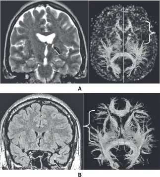
Visual assessment of tract asymmetry revealed that a decrease in their presentation on the focus side was typical of patients with cortical dysplasia or extra-hippocampal lesion foci visualized (fig. 1a). In cases of mesial temporal sclerosis in the patients with phar-macoresistant epilepsy, a „weakening” of the tracto-graphic pattern was revealed in the opposite hemi-sphere (r = 0.66, t = 4.07, p = 0.0005) (fig. 1В), which that can be related to the kindling effect and the phe-nomenon of contralateral hippocampal deafferenta-tion (a reducdeafferenta-tion of the tract presentadeafferenta-tion on the side opposite to the lesion focus).
The occurrence of tract asymmetry appeared to be related to the fa and aDC data – on the side of imaged abnormalities where the number of tract lines was lower, a decrease in the fa and an in-crease in the apparent diffusion coefficient were noted (r = 0.99, p < 0.05).
In the control group, the tract length in anteri-or quadrants was 27.9 (25.4÷29.7) mm, and in pos-terior quadrants it was 46.0 (43.1÷47.7) mm. The data for tract lengths in the group of patients with epilepsy did not significantly differ from data for the controls (р > 0.05).
according to the results obtained, four pat-terns of zones were identified with reduced frac-tional anisotropy data (as compared to the con-trols) imaged through tractography:
1) a decrease in tract presentation in the brain frontal lobes,
2) a decrease in tract presentation in the exte-rior parts of the brain hemispheres, either mono- or bilaterally,
3) a reduction in anterior and/or posterior commissure,
4) a combination of the changes listed above. To some extent, these patterns are predictors of the course of the disease and also reveal the “contribution” of various brain regions to the oc-currence of an epileptic attack.
recorded bilaterally (r = 0.7, p < 0.0001); and an increase in average theta-rhythm amplitude was noted (r = 0.58, p = 0.0003). This pattern type ap-peared to be the only one exhibiting a correlation with the indices of the blink reflex (short latency of peak R1 less than 11.5 msec, latency of peak R2 less than 35.0 msec, and prolongation of peak R2 more than 39.9 msec; r > 0.5, p < 0.05) that can testify to the involvement of frontal lobe structures in regulating brain stem activity. In the patients with this pattern type, repetitive transcranial magnetic stimulation ap-peared less evident in comparison with other changes in the tractographic pattern (r = 0.46, p = 0.005).
Decreases in tract presentation in the exterior parts of the brain hemispheres correlated with par-ticular types of debut-stage epilepsy: Bilateral tract “weakening” was typical of the disease onset as a generalized tonico-clonical attack; a monolater-al decrease in the presentation of neurmonolater-al pathways was typical of a focal onset (r = 0.42, p = 0.03).
The latent period of cognitive potential Р300 in patients was 346 ± 7.7 msec. The lack of anterior and posterior commissures of the cerebral hemi-spheres correlated only to the latent period of cog-nitive potential Р300 (r = 0.39, p = 0.029) with no connection to other neurophysiological and clini-cal features (p > 0.05) (fig. 2).
Integration of Brain White
Matter in Relation to
Psychoemotional Abnormalities
and Cognitive Functions
Thirty percent of the patients had a total Beck scale score greater than 12, and 50% of the patients were considered a high level of anxiety, which concurs with data reported in the literature [20]. a correlation was identified with a pathological decrease in fa and an increase in aD, with test-ing accordtest-ing to the Beck scale and Spielberger-Khanin anxiety scale (r = –0.2, p < 0.001) as well as peak P300 latency data (r = 0.23, p < 0.001). The P300 latent period correlated with the aDC (r = 0.36, р < 0.05) of the anterior quadrant of the epileptogenic hemisphere.
Correlation Analysis
of Diffusion Tensor MRI Indices
and Electroencephalography
Results
an analysis of the peculiarities of EEG patterns and fa data demonstrated a correlation between the existence of epileptic activity and a decrease in
Fig. 1. Peculiarities of the tractographic pattern depending on brain lesion type:
А)a decrease in tract presen-tation (bracket) on the side of the lesion focus (arrow); В) a decrease in tract presen-tation (bracket) on the side opposite to the lesion focus in a patient with a gliotic zone (arrow) in the medio-basal parts of the left tempo-ral lobe
Ryc. 1. Specyfika obrazu traktograficznego w zależ-ności od rodzaju uszkodzeń mózgu:
А) zmniejszenie prezenta-cji połączenia (nawias) po stronie zmiany chorobowej (strzałka);
В) zmniejszenie prezenta-cji połączenia (nawias) od strony przeciwnej do zmia-ny chorobowej u pacjenta z obszarem glejozy (strzałka) w przyśrodkowej podstawnej części lewego płata skronio-wego
А
Fig. 2. Various reductions in tractographic visualization of anterior and posterior com-missures:
А) a decrease in the presenta-tion of anterior commissure tracts in a patient with mesial temporal sclerosis;
В) a decrease in the presenta-tion of posterior commissure tracts in a female patient with a perinatal pathology; С) a decrease in the presenta-tion of anterior and posterior commissure tracts in a patient with cortical dysplasia (Dandy-Walker syndrome); D) a combination of various patterns of tract reduction: a lack of visualization of both commissures, an abrupt decrease in the presentation of tracts in the frontal and exter-nal parts of temporo-parieto-occipital sections
Ryc. 2. Różne ograniczenia wizualizacji traktograficznych spoidła przedniego i tylnego: А) zmniejszenie prezentacji połączenia spoidła przedniego u chorego ze stwardnieniem przyśrodkowej części płata skroniowego;
В) zmniejszenie prezentacji połączeń tylnego spoidła u pacjentki z patologią około-porodową,
С) zmniejszenie prezentacji przedniego i tylnego spoidła u pacjenta z dysplazją korową (zespołem Dandy-Walkera), D) połączenie różnych sche-matów ograniczenia wizuali-zacji połączeń: brak wizualiza-cji obu spoideł, nagłe zmniej-szenie prezentacji połączeń części czołowej i zewnętrznej obszaru skroniowo-ciemienio-wo-potylicznego
А
В
С
fa (r = –0.7, t = –2.44, p = 0.01). Normal ampli-tude, frequency and alpha rhythm index data were typical for patients with unchanged fa parame-ters (p < 0.05). alpha rhythm inversion and an in-crease in the theta rhythm index were accompa-nied by a decrease in fa data (r = –0.61, t = –28, p = 0.001). It should be noted that the occurrence of theta rhythm outbreaks had no correlation with any of the diffusion tensor MRI indices. The dis-covery of a positive correlation of focusing the-ta rhythm and normal fa dathe-ta (r = 0.27; t = 7.1; p = 0.00001) makes it possible to posit that the oc-currence of this EEG phenomenon is typical of relatively stable brain structures with the poten-tial possibility of engaging inhibitory mechanisms effectively.
Discussion
at present, MRI is the leading method of neuroimaging in diagnosing epilepsy that per-mits the detection of the foci of abnormal chang-es in brain tissuchang-es. However, ordinary MRI dochang-es not give information about all types of structur-al damage [9, 10]. In connection with this, the ne-cessity for functional techniques of neuroimaging has arisen. These techniques facilitate the study of pathological changes in different structural brain parts in vivo, and also permit the identification of some mechanisms of epileptogenesis. These meth-ods include diffusion tensor MRI with tractogra-phy, which is the technology of the future for mor-phological imaging. The interpretation of changes in inter-, peri- and postictal diffusion is rather dif-ficult, but has a potential to provide deeper insight into the mechanisms of an epileptic attack.
Brain tract configuration is a fundamental fac-tor for understanding cerebral function. MRI en-hancement due to diffusion tensor imaging with post-processing and tract visualization in vivo al-lows abnormalities not only of grey matter but of white matter as well to be objectified. DTI data changes have been shown in a number of studies. In mesial temporal sclerosis a decrease in fa data has been noted not only in the epileptogenic hippocam-pus and temporal lobe, but also in the posterior ex-tratemporal zones ipsilaterally, as well as a decrease in diffusibility in the contralateral hippocampus, cerebellar tonsil and temporal pole [8, 23].
In the current study, comparing the group of patients with epilepsy to the controls revealed the presence of discrete regions of abnormally altered brain white matter, with pathological changes lo-calized not only in the epileptogenic hemisphere but in the opposite hemisphere as well. The most specific DTI feature for detecting structural lesions
is the fa data. The most significant features for predicting the course of the disease are the aDC data and the “weakening” of tract patterns in ex-terior parts of the temporal lobes, especially the phenomenon of contralateral hippocampal deaf-ferentation. The aDC on the epileptic focus side in patients with pharmacoresistant epilepsy is sig-nificantly higher than in patients in persistent re-mission. The lack of aDC changes in the anterior hemisphere parts (frontal brain lobes) in the group of patients in remission is noteworthy; the aDC in-dex can be a significant factor relating to the prog-nosis of the clinical course of the disease.
The data obtained in the current study support the opinion of Bonilha et al. that hippocampal de-afferentation plays an important role in brain ex-trahippocampal lesions. The data in this study shows a connection between the development of resistance and the presence of contralateral hippo-campal deafferentation [4].
The interrelation between fractional anisotro-py in the hemisphere with the epileptic focus and the attack severity index can be a prognostic cri-terion in estimating the probable effectiveness of pharmacotherapy. This issue needs further studies and analyses of larger samples.
The regions of white matter with pathologi-cal fa and aDC data corresponded to the zones with altered tracts not only in the limbic circle but also to zones of intra- and interhemispheric con-nections integrating frontal, temporal, parietal and occipital brain lobes. The data obtained indicate a correlation between the fa in the hemisphere with the epileptic focus and the severity of attacks, which can be a prognostic criterion in estimating the efficacy of medical measures. In the patients with a decrease in tract presentation in the fron-tal brain parts, repetitive transcranial magnetic stimulation appeared to be the least effective com-pared to patients with other tractographic pattern changes.
The integrity of the corpus callosum contrib-utes to the stability of patients’ cognitive and emo-tional status. among the features of mental func-tions that are highly sensitive to brain white matter changes is the P300 latent period, which is proba-bly an effect of disturbances in the interhemispher-ic integral interaction provided by the integrity of the corpus callosum.
anterior and posterior commissures of the cerebral hemispheres correlated only with the Р300 latent period, with no connection to other neurophysi-ological and clinical features. Thus, the integrity of the corpus callosum correlates with cognitive functions.
a decrease in tract presentation in the exterior parts of the brain hemispheres correlated with de-but-stage epilepsy: as noted above, bilateral tract “weakening” was typical of the disease onset as a generalized tonico-clonical attack, and a mono-lateral decrease in the presentation of neural path-ways was typical of a focal onset. The data obtained makes it possible to hypothesize that the debut of temporal epilepsy as a generalized attack may be related to bilateral lesions of brain white matter.
Pathological changes in EEG patterns in epi-lepsy are to a large extent conditioned by disor-ders in brain tract integration. Diffusion tensor MRI with tractography broadens the concept of the role of microstructural alterations of grey and
white matter integrity in epilepsy and facilitates the specification of the etiologic structural-met-abolic subtype of the disease, in accordance with the recommendations of the International league against Epilepsy [2].
The current investigation shows that epilep-sy is characterized by various changes in brain white matter pathways, resulting in disconnections among different brain regions that lead in turn to further disruption of connections between the cor-tical and subcorcor-tical parts and alters the transfer of information. Specifically, a lack of corpus callosum commissures in tractography is a predictor of sig-nificant cognitive abnormalities developing later.
The role of DTI with tractography is steadily becoming wider in clinical practice. However, in-terpreting the data obtained is complicated and de-tailed standardization is needed, which will allow precise criteria to be established for subtle struc-tural lesions and the identification of the location of the epileptogenic focus.
References
[1] Ahmadi ME, Hagler DJ JR, McDonald CR, Tecoma ES, Iragui VJ, Dale AM, Halgren E: Side Matters: Diffusion Tensor Imaging Tractography in left and Right Temporal lobe Epilepsy. aJNR am J Neuroradiol 2009, 30, 1740– –1747.
[2] Basser PJ, Pajevic S, Pierpaoli C., Duda J, Aldroubi A:In vivo fiber tractography using DT-MRI data. Magn Reson Med 2000, 44, 625–632.
[3] Berg AT, Berkovic SF, Brodie MJ, Buchhalter J: Revised terminology and concepts for organization of seizures and epilepsies: Report of the IlaE Commission on Classification and Terminology, 2005–2009. Epilepsia 2010, 51, 676–685.
[4] Bonilha L, Edwards JC, Kinsman SL, Morgan PS, Fridriksson J, Rorden C, Rumboldt Z, Roberts DR, Eckert MA, Halford JJ: Extrahippocampal gray matter loss and hippocampal deafferentation in patients with temporal lobe epilepsy. Epilepsia 2010, 51, 519–528.
[5] Catani M, Jones DK, Donato R, Flytche DH: Occipito-temporal connections in the human brain. Brain 2003, 126, 2093–2107.
[6] Catani M, Ffytche DH: The rises and falls of disconnection syndromes. Brain 2005, 128, 2224–2239.
[7] Colombo N, Citterio A, Galli C, Tassi L, Lo Russo G, Scialfa G, Spreafico R: Neuroimaging of focal cortical dysplasia: neuropathological correlations. Epileptic Disord 2003, 5, 67–72.
[8] Concha L, Beaulieu C, Collins DL, Gross DW: White-matter diffusion abnormalities in temporal-lobe epilepsy with and without mesial temporal sclerosis. J Neurol Neurosurg Psychiatry 2009, 80, 312–319.
[9] Daniel KH: Investigation Epilepsy: CT and MRI in Epilepsy. Nepal J Neurosci 2004, 1, 64–72.
[10] Doelken MT, Stefan H, Pauli E, Stadlbauer A, Struffert T, Engelhorn T, Richter G, Ganslandt O, Doerfler A, Hammen T:1H–MRS profile in MRI positive versus MRI negative patients with temporal lobe epilepsy. Seizure
2008, 17, 490–497.
[11] Duncan JS: Neuroimaging methods to evaluate the etiology and consequences of epilepsy. Review. Epilepsy Res 2002, 50, 131–140.
[12] Garnett MR, Blamire AM, Corkill RG, Cadoux-Hudson TAD, Rajagopalan B, Styles P: Early proton magnetic resonance spectroscopy in normal appearing brain correlates with outcome in patients following traumatic brain injury. Brain 2000, 123, 2046–2054.
[13] Hermann BP, Bayless K, Hansen R, Parrish J, Seidenberg M: Cerebellar atrophy in temporal lobe epilepsy. Epilepsy Behav 2005, 7, 279–287.
[14] Kier EL, Staib LH, Davis LM, Bronen RA: anatomic dissection tractography: a new method for precise MR local-ization of white matter tracts. am J Neuroradiol 2004, 25, 670–676.
[15] Kistsen VV, Evstigneev VV: Repetitive transcranial magnetic stimulation can be adjunctive method to low anti-convulsant doses. Epilepsia 2012, 53, 154.
[16] Lin JJ, Riley JD, Juranek J, Cramer SC: Vulnerability of the frontal–temporal connections in temporal lobe epi-lepsy. Epilepsy Res 2008, 82, 162–170.
[17] Moris, WS, Nagae-Poetscher LM, Van Zijl PCM: MRI atlas of human white matter. amsterdam: Elsevier; 2005.
[19] Oyegbile TO, Dow C, Jones J, Bell B, Rutecki P, Sheth R, Seidenberg M, Hermann BP: The nature and course of neuropsychological morbidity in chronic temporal lobe epilepsy. Neurology 2004, 62, 1736–1742.
[20] Reuber M, Andersen B, Elger CE, Helmstaedter C: Depression and anxiety before and after temporal lobe epi-lepsy surgery. Seizure Eur J Epiepi-lepsy 2004, 13, 129–135.
[21] Riley JD, Franklin DL, Choi V, Kim RC, Binder DK, Cramer SC, Lin JJ: altered white matter integrity in tem-poral lobe epilepsy: association with cognitive and clinical profiles. Epilepsia 2010, 51, 536–545.
[22] Schoene-Bake JC, Faber J, Trautner P, Kaaden S, Tittgemeyer M, Elger CE, Weber B: Widespread affections of large fiber tracts in postoperative temporal lobe epilepsy. Neuroimage 2009, 46, 569–576.
[23] Thivard L, Lehericy S, Krainik A, Adam C, Dormont D, Chiras J, Baulac M, Duponta S: Diffusion tensor imag-ing in medial temporal lobe epilepsy with hippocampal sclerosis. NeuroImage 2005, 28, 682–690.
Address for correspondence:
Victor V. Evstigneev
Department of Neurology and Neurosurgery
Belarusian Medical academy of Postgraduate Education P. Brovka str. 3/3
220000 Minsk Belarus
Tel.: +375 29 8748704 E-mail: [email protected]
Conflict of interest: None declared Received: 19.02.2013