Jacek Szymański
1, Sylwia Konarska
2, Michał Polguj
3, Piotr Oszukowski
2Pelvi-caliceal Collecting System in Swine
– Authors Own Anatomical Classification
Układ kielichowo-miedniczkowy u świni – własny podział anatomiczny
1 Department of Normal and Clinical Anatomy, Medical University of Łódź, Poland2 Department of Clinical Morphology, Medical University of Łódź, Poland 3 Department of Angiology, Medical University of Łódź, Poland
Abstract
Background. Experiments on humans and their organs produce legal, moral and ethical dilemmas. Nowadays, the use of domestic swine as an experimental animal is increasing steadily. Thus it is important to know the structure and characteristics of swine organs.
Objectives. The purpose of the study was to investigate the shape, dimensions and volume of the renal collecting system in swine, based on authors own outline of division of this anatomical structure, as well as comparing some features of the pelvi-caliceal collecting system for swine and man.
Material and Methods. The study was carried out on 36 kidneys of adult swine (Sus scrofa domestica) of both sexes. The authors generated the corrosion specimens by applying Plastogen G. The authors took the density of the stained and hardened mass into consideration and weighed the corrosion casts. Then the authors could estimate the volume of the pelvi-caliceal collecting system by applying the formula which states that volume is a quotient of weight and density.
Results. The mean dimensions of the pelvi-caliceal collecting system in swine are: length – 69.2 mm, width – 23.5 mm, thickness – 15.8 mm; the volume of this system is on average 7.0 cm3. The renal collecting system originates
from the minor calices which are in the shape of flattened cones. Their mean dimensions are: diameter – 9.3 mm, height – 3.3 mm. The major calices, taking the form of a cylindrical body, emerge from the minor calices. The mean dimensions of the primary major calices inserted in the renal pelvis are: length – 18.0 mm, diameter – 6.6 mm; the primary calices which come into secondary ones: length – 6.7 mm, diameter – 7.3 mm; the secondary major calices: length – 16.3 mm, diameter – 8.0 mm. The renal pelvis is the last part of the pelvi-caliceal collecting system and takes the form of a distorted triangle. Its mean dimensions are: width – 14.1 mm, height – 13.7 mm.
Conclusions. The pelvi-caliceal collecting system in swine is a massive structure of high volume, with a well-devel-oped structure of the renal calices and swollen renal pelvis. The studied structure is more massive and its volume is about three times larger than that in man. Despite some disadvantages, perhaps in the future a swine kidney will be transplanted into a man, but nowadays due to lack of dissection material it may be used as a teaching model or in experimental research (Adv Clin Exp Med 2012, 21, 1, 27–33).
Key words: pelvi-caliceal collecting system, renal calices, renal pelvis, swine.
Streszczenie
Wprowadzenie. Eksperymenty na ludziach i ich narządach wywołują prawne, moralne oraz etyczne dylematy. W obecnych czasach wykorzystanie świni domowej jako zwierzęcia doświadczalnego systematycznie rośnie. Warto więc znać budowę oraz właściwości narządów świni.
Cel pracy. Zbadanie kształtu, wielkości i objętości układu kielichowo-miedniczkowego nerek u świni na podstawie własnego schematu podziału tej struktury anatomicznej, a także porównanie niektórych cech układu kielichowo- -miedniczkowego świni i człowieka.
Materiał i metody. Badania przeprowadzono metodą korozyjną na 36 nerkach, pochodzących od dorosłych świń domowych (Sus scrofa domestica) obu płci, z użyciem Plastogenu G. Znając gęstość zabarwionego i utwardzonego tworzywa, po zważeniu odlewu, obliczano objętość preparatu, korzystając ze wzoru, który mówi, że objętość jest ilorazem masy i gęstości.
Wyniki. Średnie wymiary układu kielichowo-miedniczkowego u świni wynoszą: długość – 69,2 mm, szerokość – 23,5 mm, grubość: – 15,8 mm; objętość układu wynosi średnio – 7,0 cm3. Za początek układu
kielichowo-mied-niczkowego uznaje się kielichy nerkowe mniejsze, u świni przyjmujące formę spłaszczonych stożków. Ich średnie
Adv Clin Exp Med 2012, 21, 1, 27–33 ISSN 1899-5276
OrIGINAl PAPErS
wymiary wynoszą: średnica – 9,3 mm, wysokość – 3,3 mm. Kielichy nerkowe mniejsze uchodzą do kielichów ner-kowych większych, jawiących się jako walcowate twory. Średnie wymiary kielichów nerner-kowych większych pierw-szorzędowych uchodzących do miedniczki nerkowej wynoszą: długość – 18 mm, średnica – 6,6 mm; kielichów pierwszorzędowych uchodzących do kielichów drugorzędowych: długość – 6,7 mm, średnica – 7,3 mm; kielichów nerkowych większych drugorzędowych: długość – 16,3 mm, średnica – 8 mm. Ostatnim odcinkiem opisywanego układu jest miedniczka nerkowa, występująca u świni w formie zniekształconego trójkąta. Jej średnie wymiary wynoszą: szerokość – 14,1 mm, wysokość – 13,7 mm.
Wnioski. Układ kielichowo-miedniczkowy u świni jest strukturą masywną o dużej objętości, bogato rozbudowanej strukturze kielichowej i rozdętej miedniczce nerkowej. Badana struktura jest znacznie masywniejsza, a jej objętość jest trzykrotnie większa od objętości podobnego układu u człowieka. Mimo wad być może w przyszłości nerki świni będą przeszczepiane człowiekowi, ale obecnie z powodu braku materiału prosektoryjnego mogą służyć jako mate-riał dydaktyczny lub do badań eksperymentalnych (Adv Clin Exp Med 2012, 21, 1, 00–00).
Słowa kluczowe: układ kielichowo-miedniczkowy, kielichy nerkowe, miedniczka nerkowa, świnia.
Nowadays, the use of domestic swine as an experimental animal is increasing steadily. This is because experiments on humans and their organs produce legal, moral and ethical dilemmas [1]. The size and structure of internal organs in swine (espe-cially the heart and kidneys) are similar to organs in man. Moreover, this species quickly reaches sexual maturity, has a short breeding season and is easy to breed. Thus the organs can be cheaply and easily obtained [2–4].
The implementation of new surgical tech-niques and their importance can be tested on ap-propriate animal models beforehand. Animal or-gans are suitable for testing less invasive surgical interventions, such as laparoscopy [3]. Addition-ally, organs of swine could be used as a model for teaching and practicing – students can gain experi-ence by learning simpler surgical techniques on an animal model [2].
In view of the insufficiency of transplants re-ceived from both alive and dead donors, more and more often medicine is considering the idea of ani-mals providing compatible organs for humans; so-called xenotransplantations [5, 6].
Animal organs are therefore very helpful and useful. Thus it is important to know their structure and characteristics which are similar to human or-gans, but also the differences between them.
The pelvi-caliceal collecting system is a con-tractual clinical concept, including urinary tracts from minor calices, through major calices, to the renal pelvis. In humans, there are many types of pelvi-caliceal collecting systems which have been classified mainly according to the shape of the
renal pelvis. Miękoś et al. [7] and Kosiński [8]
distinguish six types of renal pelvises: ampullary, intermediate ampullary, bifurcated ampullary, linear, duplicated calicopelvic system with a bifur-cated ureter and complete duplication of calicopel-vic system and ureters. Niżankowski [9], in turn, proposes the division into four types of pelvises: primitive, intermediate ampullary, intermediate dendritic and progressive. According to the
classi-fication of Sokołowska-Pituchowa [10], Piasecki et al. [11, 12] and Augustyn [13] there are three types of pelvises: ampullary, intermediate and bifurcat-ed. The ampullary type is characteristic for swol-len pelvises. The place where the renal pelvis joins the ureter is narrowed. There are two polar major renal calices and a variable number of middle ma-jor renal calices. Some authors call it a primitive or secondary form [9, 13]. The intermediate type is characterized by well-developed major renal cal-ices, whose renal pelvis is also swollen. The crite-rion for distinguishing the bifurcated type is visible bifurcation of the major renal calices.
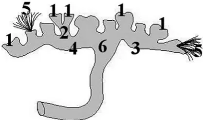
The authors decided to simplify the classifica-tion of the renal collecting system in swine to the one with the first-order or primary and the second-order or secondary major calices. Figure 1 presents the individual structures of the pelvi-caliceal col-lecting system with the particular nomenclature.
The purpose of the study was to investigate the shape, dimensions and volume of the renal collect-ing system in swine, based on our own outline of division of this anatomical structure, as well as comparing some features of the pelvi-caliceal col-lecting system of swine and man.
Material and Methods
The study was carried out with a corrosion method on 36 kidneys, 18 right and 18 left, which
came from adult domestic swine (Sus scrofa
performed under pressure, and it thus made it possible to fill up the lumen of the specimen of the pelvi-caliceal collecting system. In order to re-ceive the consistency enabling the penetration of all structures, Plastogen G was thinned down with a diluter. So as to make individual parts more vis-ible, the renal collecting system was also stained with a yellow pigment for acrylic paints. After fill-ing up the specimen with Plastogen G, the stump of the ureter was tied off. The specimen was then submerged for 24 hours in a 0.9% solution of
so-dium chloride at a temperature of 40°C in order
to harden the Plastogen. Next, the specimens were subjected to digestion in 10% potassium hydroxide
in a laboratory thermostat, at 40°C, for 24 hours.
The corrosion casts obtained were subjected to a rinse with normal tap water. After drying off, suitable measurements were made with calipers. Photographic documentation was made with digi-tal electronics.
The volume of the studied pelvi-caliceal col-lecting system was calculated with an indirect method. The corrosion casts of all the studied pel-vi-caliceal collecting systems were weighed, after cutting away the cast of the stump of the ureter. Their volume was calculated on the known den-sity (ρ) of hardened and stained Plastogen G, ac-cording to the formula:
Table 1. Dimensions and volume of the pelvi-caliceal collecting system in swine. Arithmetic mean (X) and stan-dard deviation (SD)
Tabela 1. Wymiary i objętość układu kielichowo-mied-niczkowego u świni. Średnia arytmetyczna (X) oraz odchylenie standardowe (SD)
Measured parametr
(Zmierzony wskaźnik) X SD
length (Długość) [mm] 69.2 12.3
Width (Szerokość) [mm] 23.5 5.9
Thickness (Grubość) [mm] 15.8 3.5
Volume (Objętość) [cm3] 7 3.7
Table 2. The number of minor calices in one kidney (A) and percentage of kidneys with the particular number of minor calices (B)
Tabela 2. liczebność kielichów nerkowych mniejszych w jednej nerce (A) oraz odsetek nerek z określoną liczbą kielichów nerkowych mniejszych (B)
Measured parameter (Zmierzony wskaźnik)
Obtained results (Uzyskane wyniki)
A 4 5 6 7 8 9 10 11 12 13 14 15 16
B 2.8 0 2.8 8.3 11.1 13.9 16.7 13.9 16.7 8.3 0 2.8 2.8
V = m ρ
where V means volume, m – weight of cast and ρ its density.
Results
It was stated that the pelvi-caliceal collecting system in swine has the shape of an elongated, ir-regular solid. Its length ranges from 47 to 92 mm, the width – from 15 to 36 mm and the thickness – from 7 to 21 mm. The arithmetic mean of these measurements is presented in Table 1.
Minor calices should be regarded as the begin-ning of the pelvi-caliceal collecting system (Fig. 2). Their number ranges from 4 to 16 (Table 2), and they take the form of flattened cones, with the diameter ranging from 3 mm to 22 mm and the
Fig. 1. Scheme of the pelvi-caliceal collecting system in swine. 1 – minor calices, 2 – primary major calices inserted in secondary major calyx, 3 – primary major calyx inserted in renal pelvis, 4 – secondary major calyx, 5 – renal collecting tubules, 6 – renal pelvis
height from 1 mm to 10 mm. The arithmetic mean of these measurements is presented in Table 3.
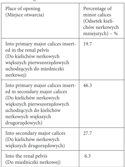
Minor calices come into major calices or di-rectly to the renal pelvis. Between 1 and 5 minor calices join the primary calyx (inserted in the re-nal pelvis), whereas 2 to 4 join those inserted in the secondary calices. From 1 to 4 primary calices, and also from 0 to 6 minor calices, join second-ary calices. In some cases, the minor calices (in the number from 1 to 5) come directly into the renal pelvis. The frequency of appearance of the minor calices orifices opening into individual structures of the pelvi-caliceal collecting system is presented in Table 4. The highest percentage of the minor
Table 3. Dimensions of minor calices. Arithmetic mean (X) and standard deviation (SD)
Tabela 3. Wymiary kielichów nerkowych mniejszych. Średnia arytmetyczna (X) oraz odchylenie standardowe (SD)
Measured parameter
(Zmierzony wskaźnik) X SD
largest diameter
(Największa średnica) [mm] 9.3 1.7
Height (Wysokość) [mm] 3.3 0.7
Table 4. Distribution (%) of minor calices inserted in particular structures of the pelvi-caliceal collecting system in swine
Tabela 4. Odsetek kielichów nerkowych mniejszych uchodzących do określonych struktur układu kielichowo- -miedniczkowego u świni
Place of opening
(Miejsce otwarcia) Percentage of minor calices (Odsetek kieli-chów nerkowych mniejszych) – % Into primary major calices
insert-ed in the renal pelvis (Do kielichów nerkowych większych pierwszorzędowych uchodzących do miedniczki nerkowej)
19.7
Into primary major calices insert-ed in secondary major calices (Do kielichów nerkowych większych pierwszorzędowych uchodzących do kielichów nerkowych większych drugorzędowych)
46.3
Into secondary major calices (Do kielichów nerkowych większych drugorzędowych)
27.7
Into the renal pelvis
(Do miedniczki nerkowej) 6.3
Table 5. Dimensions of major calices. Arithmetic mean (X) and standard deviation (SD)
Tabela 5. Wymiary kielichów nerkowych większych. Średnia arytmetyczna (X) oraz odchylenie standardowe (SD) Measured parameter (Zmierzony wskaźnik) largest dia-meter (Największa średnica) length (Długość)
X SD X SD
Primary major cali-ces inserted in the renal pelvis (Kielichy nerkowe większe pierwszorzędowe uchodzące do mied-niczki nerkowej) [mm]
6.6 2.5 18 7.3
Primary major calices inserted in secondary major calices (Kielichy nerkowe większe pierwszorzędowe uchodzące do kielichów nerkow-ych większnerkow-ych drugorzędowych) [mm]
7.3 2.6 6.7 2.6
Secondary major calices (Kielichy nerkowe większe drugorzędowe) [mm]
8 2.9 16.3 6.2
Table 6. The frequency of appearance of individual types of the major calices orifices opening into the renal pelvis Tabela 6. Częstotliwość występowania ujść kielichów nerkowych większych bezpośrednio do miedniczki nerkowej
Types of major calices open-ing into the renal pelvis (rodzaje ujść kielichów nerkowych większych bezpośrednio do miedniczki nerkowej)
Percentage of particular types of opening
(Odsetek występo-wania poszczególnych typów ujść) – % Two secondary major calices
(Dwa kielichy nerkowe większe drugorzędowe)
52.8
Primary and second-ary major calices (Kielich nerkowy większy pierwszo- i drugorzędowy)
36.1
Two primary major calices (Dwa kielichy nerkowe większe pierwszorzędowe)
calices (46.3%) comes into the primary major ca-lices inserted in the secondary major caca-lices. The percentages of the minor calices which are joined to the secondary major calices (27.7%) and those joined to the primary major calices inserted in the
renal pelvis (19.7%) are similar. The smallest per-centage of minor calices (6.3%) comes directly into the renal pelvis.
Primary or secondary major calices, which take on a cylindrical form, constitute the indirect stage of the renal collecting system between minor calices and the renal pelvis (Fig. 3). The primary major calices inserted in the renal pelvis are be-tween 7 and 34 mm long, their diameter ranges from 4 to 11 mm; the calices which come into sec-ondary ones are from 3 to 14 mm long, and their diameter ranges from 3 to 15 mm. The length of the secondary major calices ranges from 5 to 30 mm, and their diameter – from 4 to 14 mm. The arithmetic mean of these measurements is pre-sented in Table 5.
Two major calices (primary, secondary or both), mainly the secondary ones, come into the renal pelvis (Fig. 3). The frequency of appearance of individual types of the major calices orifices opening into the renal pelvis is presented in Table 6. Mainly (52.8%) the two secondary calices, or more seldom (36.1%) one primary and one sec-ondary calyx, go into the renal pelvis. Occasion-ally (11.1%) two primary renal calices end in the renal pelvis. The renal pelvis (Fig. 3) is the last part of the pelvi-caliceal collecting system which takes the form of a distorted triangle, whose width ranges from 6 to 28 mm and height from 6 to 25 mm. The arithmetic mean of these measurements is presented in Table 7.
The volume of the pelvi-caliceal collecting
sys-tem ranges from 1.6 cm3 to 16.2 cm3, on average
7.0 cm3. Significant differences between the
vol-Table 7. Dimensions of renal pelvis. Arithmetic average (X) and standard deviation (SD)
Tabela 7. Wymiary miedniczki nerkowej. Średnia arytme-tyczna (X) oraz odchylenie standardowe (SD)
Measured parameter
(Zmierzony wskaźnik) X SD
largest width
(Największa szerokość) [mm] 14.1 5.5
Height (Wysokość) [mm] 13.7 5.4
Fig. 2. Minor calyx (arrow). Corrosion cast. Plastogen G. Magnification × 3
Ryc. 2. Kielich nerkowy mniejszy (strzałka). Preparat korozyjny. Plastogen G. Powiększenie × 3
Fig. 3. Pelvi-caliceal collecting system in swine. 1 – primary major calyx inserted in renal pelvis, 2 – primary major calyx inserted in secondary major calyx, 3 – secondary major calyx, 4 – renal pelvis. Corrosion cast. Plastogen G. Natural size
ume of the studied structures, according to sex and the side of the animal which the studied kidney came from, were not observed.
While analyzing the pelvi-caliceal collecting system of swine it was stated that 47.2% of the studied structures can be compared to the interme-diate type in humans, however there are 30.6% of pelvises similar to the ampullary type. The smallest percentage (22.2%) appears amongst pelvises simi-lar to the bifurcated type.
Discussion
Because of the variable anatomical structure of pelvises in humans, a few types have been distin-guished and different classifications made. How-ever, it seems that, in spite of the different criteria of identity accepted by different authors [7–15], it is possible to compare the structure of particular types. For example, on the basis of the characteristics of the two types, bifurcated ampullary and linear, de-scribed by Miękoś [7] and Kosiński [8], it is possible to compare them to the bifurcated type classified by Sokołowska-Pituchowa [10]. The intermediate ampullary type can be compared to the intermedi-ate type. According to Niżankowski [9], there are four types of pelvises. It is possible to identify the primitive type with the ampullary type and the in-termediate ampullary type is similar to the interme-diate type. However, the intermeinterme-diate dendritic and progressive varieties are very close to the bifurcated type, as described by Sokołowska-Pituchowa [10]. According to all of the quoted authors [7–15], the ampullary type (or similar) definitely constitutes the smallest percentage of pelvises. For most of the au-thors [10–14], the prevailing type is the bifurcated one (or alternatively its counterpart/counterparts). Some other authors [7–9, 15] indicate that the in-termediate type (or similar) is in the majority.
It seems that present classification can be ana-lyzed in a similar way. Despite the fact that the authors simplified the division and decided not to distinguish types, it is possible to compare some characteristics of swine pelvises with those of hu-man pelvises. The renal pelvises of a domestic swine are more massive than those in man. As was men-tioned, among the studied structures of the swine, pelvises similar to the intermediate type in humans, as well as to the ampullary type, were prevailing. The smallest percentage of swine renal pelvises demonstrates features of the bifurcated type.
Kupeli et al. [16] compared the dimensions and the structure of the stone-bearing kidneys in humans with the normal contralateral ones. The mean volume of the stone-bearing pelvi-caliceal
collecting system was 2.6 cm3; as for the normal
contralateral kidneys, it was 1.8 cm3. Studies
con-ducted by Acar et al. [17] showed similar results: the volume of the stone-bearing pelvi-caliceal
col-lecting system was on average 2.4 cm3, while in
normal contralateral kidneys also 1.8 cm3. The
vol-ume of the pelvi-caliceal collecting system in swine is much larger than in humans. According to our studies, the mean volume of the pelvi-caliceal
col-lecting system in swine is 7 cm3.
Piasecki et al. [11, 12], having analyzed differ-ent material, state that there are from 7 to 12 (as well as from 7 to 14) minor calices in man. In cur-rent material, the number of minor calices ranges from 4 to 16 in one kidney, most frequently from 7 to 13 (Table 2).
Having analyzed the results, the authors can conclude that the pelvi-caliceal collecting system in swine is a massive structure of high volume. The renal pelvis is swollen and well-developed. Most frequently it is formed as a result of the anasto-mosis of two secondary major calices, and often one secondary and one primary major calices. The secondary major calices have a large diameter and many structures are inserted into them (both pri-mary major calices and minor calices). The result is that the pelvi-caliceal collecting system in swine has a well-developed structure of the calices.
What is characteristic about the minor re-nal calices is the fact they are inserted into all the structures that form the pelvi-caliceal collecting system in swine – into the primary and secondary major calices, and even into the renal pelvis.
The pelvi-caliceal collecting system in swine is more massive and its volume is about three times larger than that in man. It should be con-cluded that, in swine, the most frequent type is the one which is the most similar to the intermediate type. The ampullary type can be observed less fre-quently. The smallest percentage appears among the renal pelvises similar to the bifurcated variety. In man, however, the smallest percentage refers to the ampullary type.
References
[1] Gillespie JI: A developing view of the origins of urgency: the importance of animal models. BJU International 2005, 96, Suppl. 1, 22–28.
[2] de Sá Earp PP: Percutaneous renal surgery – new model for learning and training. Int Braz J Urol 2003, 29, 151–154.
[3] Sampaio FJB, Pereira-Sampaio MA, Favorito LA: The pig kidney as an endourologic model: Anatomic contribu-tion. J Endourol 1998, 12, 45–50.
[4] Wanzel K R, Matsumoto E D, Hamstra S J, Anastakis DJ: Teaching technical skills: training on a simple, inex-pensive, and portable model. Plas reconstr Surg 2002, 109, 258–263.
[5] Gonwa TA: Transplantation. Am J Kidney Dis 2000, 35, Suppl. 1, S 153–S 159.
[6] Skuciński J, Nowak W, Wieczorek J, Solecki R: Wykorzystanie nerek transgenicznych świń do przeszczepów allogenicznych i ksenogenicznych. Med Wet 2007, 63, 941–945.
[7] Miękoś E, Zapędowski Z, Kosiński H, Schreiber K: Miedniczki nerkowe człowieka. Biul WAM 1974, 17, 263–269.
[8] Kosiński H: Variations in shape and position of human renal pelvis. Folia Morphol (Warsz) 1987, 46, 207–214.
[9] Niżankowski C: Suggestion of new classification of the shape of human renal pelvis with consideration of the number of renal papillae. Folia Morphol (Warsz) 1978, 37, 367–380.
[10] Sokołowska-Pituchowa J: Formy miedniczek nerkowych u człowieka, ich odmiany, najczęstszy typ. Folia Morphol (Warsz) 1956, 7, 53–62.
[11] Piasecki Z, Piotrowski J, Jugowski F: rozmieszczenie naczyń tętniczych w nerce ludzkiej. Folia Morphol (Warsz) 1962, 13, 183–193.
[12] Piasecki Z, Jugowski F, Piotrowski J: Formy miedniczek nerkowych u człowieka w odlewach i radiogramach. Folia Morphol (Warsz) 1969, 28, 177–187.
[13] Augustyn M: Variation of the calicopelvic system of the human kidney in ontogenic development. Folia Morphol (Warsz) 1978, 37, 157–165.
[14] Czerwiński F, Mierzwa J, Mierzwa A, Michalczyk K, Glińska B: Miedniczka nerkowa a nerka płodu ludzkiego. Urol Pol 1995, 48, 230–236.
[15] Niżankowski C: Miedniczki nerkowe. Wiad lek 1958, 11–12, 527–528.
[16] Kupeli B, Tunc L, Acar C, Gurocak S, Alkibay T, Guneri C, Bozkirli I: The impact of pelvicaliceal anatomical variation between the stone-bearing and normal contralateral kidney on stone formation in adult patients with lower caliceal stones. Int Brazil J Urol 2006, 32, 287–294.
[17] Acar C, Kupeli B, Gurocak S, Alkibay T, Guneri C, Ozkan S, Bozkirli I: Is pelvicaliceal anatomy a risk factor for stone formation in patients with solitary upper caliceal stone? Urology 2006, 67, 1159–1163.
Address for correspondence:
Jacek Szymański
Department of Normal and Clinical Anatomy Medical University of Łódź
Narutowicza 60 90-136 Łódź Poland
Tel.: +48 42 630 49 49
E-mail: [email protected] Conflict of interest: None declared received: 28.03.2011