UDC: 636.3.082.853.5.09:616-089.819
Review Article
ABSTRACT
INTRODUCTION
The assisted reproduction technologies (ART), used in animal breeding, includes various methods for improvement of the genetic value of the produc-tion animals as well as for promoproduc-tion of superior genotypes. ART represent a combination of scien-tifi c knowledge in reproductive physiology, endo-crinology, cellular and molecular biology as well as genomic techniques. (1). In the last 30 years this technology experienced a dynamic development, especially in the dairy cows Embryo transfer
indus-LAPAROSCOPY-PROMISING TOOL FOR IMPROVEMENT OF
REPRODUCTIVE EFFICIENCY OF SMALL RUMINANTS
Dovenski Toni
1, Trojacanec Plamen
1, Petkov Vladimir
1,
Popovska-Percinic Florina
1, Kocoski Ljupce
2, Grizelj Juraj
31Faculty of Veterinary Medicine, Ss. Cyril and Methodius University, Skopje, R. Macedonia 2Faculty of Biotechnical sciences, University St. Kliment Ohridski - Bitola, R. Macedonia
3Faculty of Veterinary Medicine, University of Zagreb, R. Croatia
Received 29 March 2013; Received in revised form 18 April 2013; Accepted 23 May 2013
Assisted reproductive technologies are used to accelerate genetic gain and improve reproductive performances in farm animals, including small ruminants. This technologies include estrous synchronization, artifi cial insemination (AI) using fresh, frozen or sexed semen, embryo transfer (ET) using in vivo or in vitro produced embryos, and more advanced - cloning and production of transgenic animals. Diagnostic procedures, such as ultrasonography and laparoscopy, have been used as additional tools for monitoring the ovarian response to superovulatory treatment in donor animals as well as for AI and collection and transfer of embryos.
The use of laparoscopy for assisted reproduction techniques in Macedonia commenced in the early 90’s, with the acquisition of a set of ,,Karl Storz” equipment. After the adoption of the required routine, our group has completed several scientifi c projects where laparoscopy was used for intrauterine inseminations as well as for recovery and transfer of embryos in both sheep and goats. In the following period our group endeavored into introduction of laparoscopic insemination in the routine farm practice. Ovine intrauterine/intracornual insemination by frozen-thawed semen resulted with pregnancy rates of 45% and 60%, when AI was performed out of season and during the breeding season, respectively. In goats, this percentage occasionally peaked at 85%.
The aim of this article is to review the status of implementation of laparoscopy in Assisted Reproduction Technologies (ART) of small ruminants and to present our experience in this fi eld.
Key words: laparoscopy, reproduction, artifi cial insemination, embryo transfer, small ruminants
Corresponding author: Prof. Toni Dovenski, PhD
try (review: 6, 9). Ultrasonography and laparoscopy are the most practical tools for monitoring of ovar-ian and uterine dynamics during different biotech-nical procedures. Ultrasound diagnostics is more frequently used in large ruminants, by simply add-ing an ultrasound probe duradd-ing the routine rectal ex-amination. However in small ruminants laparoscopy seems to be more acceptable and practical instru-ment for this purpose.
Laparoscopy, (Gr: Laparo-abdomen, scopein -to examine) represents a technique for examination of the abdominal cavity and its contents. It requires insertion of a cannula/trocar through the abdominal wall, distension of the abdominal cavity with sterile
Roberts (1968) (25) fi rst reported endoscopic/ laparoscopic examination of reproductive organs in sheep. Endoscopy was initially used for “direct
ob-servation of the ovaries (Fig. 1.) and uterus (Fig. 2.) of the ewe by means of an illuminated endoscope, inserted through an abdominal cannula”.
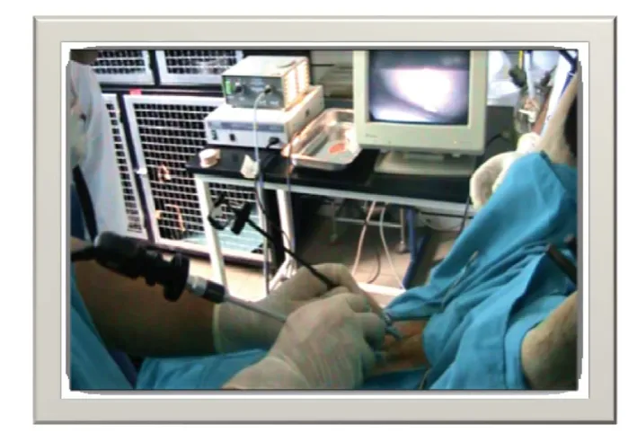
With the introduction of video cameras and other ancillary instruments (Fig. 3.) laparoscopy rapidly advanced from being just a diagnostic
pro-Figure 1. Preovulatory follicle on sheep ovary - laparoscopic view
Figure 2. Laparoscopic view of a sheep uterus in estrus
cedure to one used in numerous surgical procedures in all surgical disciplines for a variety of indications (30).
Moreover, the development of assisted repro-ductive methods brought laparoscopy into the focus as a technique for study of ovulation, follicles and corpora lutea development, as well as for pregnancy diagnosis. Salamon and Maxwell (26) recognized the laparoscopic intrauterine insemination of sheep as the most effective method used to improve the fertility.
Using laparoscopy, the “cervical barrier” prob-lem has been overcome and satisfactory fertility rate has been achieved by signifi cant reduction in the number of spermatozoa per insemination (from 200-300 to 1-10 millions, or less for sex-sorted se-men). Pregnancy rate is commonly over 75% and 50% for goats and sheep respectively (26).
Laparoscopic technique was introduced in MOET programs by McKelvey et al. (19). Primary purpose was to reduce adhesions of the reproductive system after surgical embryo recovery. Laparos-copy enables repeated fl ushing of sheep and goats uterus and satisfactory embryos recovery rate. Also, fresh or frozen-thawed embryos are transferred to the utera or oviducts of recipients by laparoscopy, achieving high pregnancy rates. Laparoscopy is also used for oocytes aspiration (Laparoscopy Ovum Pick Up - LOPU) in small ruminants (13).
The aim of this article was to bring to light the current state (advances and pitfalls) of implementa-tion of laparoscopy in small ruminants, as a valuable tool for assisted reproductive technologies.
Laparoscopic Artifi cial Insemination (LAI)
The last few decades have noticeably changed the management of sheep breeding, through im-proved nutrition and veterinary assistance. Artifi cial insemination is the most commonly applied repro-ductive technology in selection programs (15). Sat-isfactory results of transcervical insemination have not been achieved yet, especially out of the breeding season and using deep frozen-thawed ram semen. Fertilization failure is equally frequent in ewes bred naturally or artifi cially inseminated, and appears to be due to faulty transport of spermatozoa through the cervix (7). This problem can be overcome by intra-uterine deposition of semen through surgi-cal procedure in superovulated ewes or by laparo-scopic insemination (16). Killen and Caffery (17) have developed a laparoscopic artifi cial
insemina-failure of superovulated donors, exhibiting high ovulation rates. In sheep, intra uterine laparoscopic insemination should be carried out in the middle of the estrous period (31). However, some authors (16) reported that when fertile rams are available, natural mating should be used in the superovulation pro-gram, and sheep should be mated every 6 h during standing estrus.
Laparoscopic Artifi cial Insemination
Ewes are fasted and restricted access to water for 16 hours before, and injected with local anes-thetic 15 minutes before the procedure is performed. The ewe is than placed in a laparoscopy cradle. The abdominal region is surgically prepared by shearing the wool and disinfecting the skin. Using the cradle, the ewe is positioned in a supine head-down (Tren-delenburg) position to an approximate angle of 30o (Fig 4).
The t rocars and cannulae for introducing lapa-roscope and insemination pipette are inserted 7 – 10 cm ventral to the udder and 5 – 10 cm on each side of the midline (linea alba) (Fig 5). Usually, a scalpel blade is used to make a small skin incision in orther to facilitate trocar penetration. The 7 mm trocar and cannula that is connected with the CO2 is
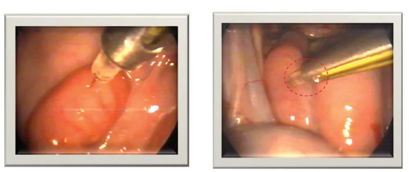
fi rst introduced and the abdomen is slightly infl ated to reduce the chance of injury to organs. Insertions of the fi rst trocar and cannula should be well con-trolled and the sharp trocar is withdrawn as soon as the abdominal wall has been penetrated. The blunt cannula is pushed well into the abdomen, while the second trocar and cannula are inserted after infl ation with CO2. Endoscope and AI instrument go through the cannulae and the uterus is located and fi xed just ventral to the urinary bladder. Semen is deposited in each uterine horn approximately halfway between the uterine bifurcation and the utero-tubal junction (Fig. 6).
sertion of the fi rst trocar. It is possible to pierce a full rumen as well as a full bladder. Operators should be well trained.
Similar procedure, with slight variation, has been described in goat (28). Before laparoscopic AI, animals are deprived of feed for 2 d, and of water for 1 d. Emptiness of the digestive tract and blad-der facilitates fast and easy operation and decreases the pressure exerted by the abdominal organs on the diaphragm while the animals are in a supine head-down position. Laparoscopic insemination, in de-tails is described by Vallet et al. (29). Briefl y, the animals should be tilted into a head-down position at an angle of at least 30o. Approximately 8 cm cra-nial to the udder and 8 cm to the left and right of the midline, the skin should be nicked with the tip of a scalpel blade and, with the aid of a trocar, two 5-mm cannulas have to be inserted through the abdomi-nal wall. The abdomiabdomi-nal cavity is insuffl ated with a moderate amount of sterile air to aid visibility. A laparoscopic telescope and an insemination instru-ment are introduced through the cannulas. The uter-ine horns should be punctured with the fi ne tip of the insemination pipette near the middle of the great curvature, in the least vascularized area, to deposit the semen in the uterine lumen. The sperm could be distributed to one (if preovulatory follicle is found on the ipsilateral ovary) or both uterine horns.
Dovenski et al. (8) have reported high percent-age of fertilization in goats after the insemination by laparoscopic intrauterine and transcervical tech-niques. The lambing rate as well as the fertility rate had higher tendency (80.95% vs. 67.48%) and
(182.35% vs. 176.98%) after intrauterine than after cervical insemination, respectively.
Petkov and Dovenski (21) compared the cervical vs. intrauterine unicornual or bicornual laparoscopic insemination of sheep by frozen-thawed semen out of breeding season. They reported higher fertility rate for I/U insemination (54% and 52% for uni/ bicornual A.I., respectively) than cervical insemina-tion 38%.
More recently Dovenski et al. (10) reported similar results after insemination of Ovchepolean Pramenka sheep by deep frozen/thawed ram semen, based on ultrasound pregnancy diagnosis on day 60. Fertility rate was signifi cantly higher (p<0.05) in sheep inseminated laparoscopically (54.54%) com-paring to the intracervical insemination (42.86%). Laparoscopic insemination in superovulatory estrus induced by eCG (1200 IU) and FSH (200 mg of NIH-FSH-P1 in 6 decreasing doses) have shown very high fertility rate in both groups 83.33% and 71.43% respectively.
Investigating ovarian macrostructures during the periovulatory periods some authors reported (22) that for precise estimation the use of both methods: ultrasonography and laparoscopy is necessary. Tran-srectal ultrasonography enables precise estimation of the ovarian size and diameter of follicles and CL’s. However, ultrasound examination is less ac-curate for determination of number of ovarian struc-tures then laparoscopy: 67±11%, 50±12%, 55±12%, 60±10% for growing follicles, ovulation, CL and Cysts, respectively.
Figure 4. Positioning of the animal in a supine head-down (Trendelenburg) position for laparoscopy
Recovery and transfer of embryos and oocytes
Most MOET programs in sheep and goat consist of a 14 to 17 day progesterone exposure, with mul-tiple FSH injections beginning 2 days before the re-moval of the progesterone releasing device (4, 14). Flushing of the uterus for recovering 6-7 days old embryos (morula or blastocyst stage mostly) usu-ally is done surgicusu-ally. The occurrence of adhesions after this procedure may reduce the number of
flushings obtainable from one donor (20). Laparoscopic techniques for embryo recovery in sheep have been introduced by McKelvey et al. (19), in purpose to to reduce the extent of surgical
Figure 6. Correct laparoscopic semen deposition at the least vascular location of the ewe’s uterus
Figure 7. Incorrect insemination; wrong angle of needle insertion (left horn) and bleeding of right uterine horn
intrusion. Laparoscopic embryo collection is less invasive and allows up to 7 collections per one ani-mal (3).
Procedure for laparoscopic embryo recovery is similar to the LAI, with using of three instruments: endoscope, forceps for holding uterine horn and a three-way balloon catheter for fl ushing of embryos (24). The major problems related to repeated super-ovulation and frequent oocyte or embryo collection procedures are adhesions caused by protrusions of the endometrium at the puncture site. However, similar problem have been recorded during embryo recovery by laparotomy (Fig. 8. and Fig. 9.).
Laparoscopic transfer of embryos in uterine horn (27) or tubal transfer (5) resulted with high-er pregnancy rates than the surgical transfhigh-er. They have concluded that laparoscopic transfer is a safe, minimally invasive surgical procedure and it should be recommended for transfer of embryos in small ruminants.
Successful transcervical embryo transfers in small ruminants also have been reported, but only limited studies have been carried out on this mat-ter (11). The same authors also reported that lapa-roscopic oocyte aspiration technique is relatively simple and effective, as well as being less traumatic than normal embryo recovery procedures (12).
Considering the possible negative consequences of laparoscopy, there is evidence that twofold and threefold applications of laparoscopy for diagnostic purposes or A.I. are not related with severe stress symptoms or any dramatic increase of the plasma cortisol level (23).
Armstrong and Evans (2), concluded that intra-uterine insemination using the technique of laparos-copy is a relatively simple and convenient means of achieving high fertilization rates in superovulated ewes when frozen semen is used. Embryo recovery was not impaired in ewes inseminated by this te-chnique, and with proper care to avoid damage to mammary blood vessels, or deposition of semen within the uterine stroma, there were no detrimental consequences to the ewes.
In conclusion, application of laparoscopy in small ruminant assisted reproduction would be economically reasonable in MOET programs (I/U insemination as well as for uterine fl ushing and embryo transfer), especially for insemination of ge-netically valuable animals with high-quality import-ed ram and goats semen. This technique has factual limitations for wider application in the expensive equipment, and the special staff training.
REFERENCES
1. Alexander, B., Mastromonaco, G., King, WA. (2010). Recent Advances in Reproductive Bio-technologies in Sheep and Goat. J Veterinar Sci Technol 1:101. doi:10.4172/2157-7579.1000101 2. Armstrong, D. T., Evans, G. (1984). Intrauterine
insemination enhances fertility of frozen semen in superovulated ewes. Journals of Reproduction & Fertility 71: 89-94.
3. Baril, G., Pougnard, J., Freitas, V., Leboeuf, B., Saumande, J. (1996). A new method for control-ling the precise time of occurrence of the pre-ovulatory gonadotropin surge in superovulated goats. Theriogenology 45: 697–706.
4. Barry, D.M., Van Niekerk, C.H., Rust, J., van der Walt, T. (1990). Cervical embryo collec-tion in sheep after ripening of the cervix with prostaglandin E2 and estradiol. Theriogenology 33:190.
5. Besenfelder, U., Zinovieva, N., Dietrich, E., Sohnrey, B., Holtz, W. & Brem, G. (1994). Tubal transfer of goat embryos using endoscopy. Vet. Rec. 135, 480-481.
6. Betteridge KJ. (2003). A history of farm animal embryo transfer and some associated techniques. Anim Reprod Sci. 15;79(3-4):203-244.
7. Boland, M.P., Crosby, T.F., Gordon, I. (1983). Factors infl uencing the superovulatory response in sheep. Theriogenology 19:114.
8. Dovenski, T., Popovski, K., Petkov, V., Mickovs-ki, G., KocosMickovs-ki, Lj., Trojacanec, P., StojanovsMickovs-ki, B. (1997). Comparison of intrauterine and cervi-cal insemination in goats with deep-frozen se-men. Croatian veterinary congress, Proceedings pp. 279-285, Cavtat, Croatia
9. Dovenski, T., Trojacanec, P., Petkov, V., Pop-ovska-Percinic, F., Kocoski, Lj. (2011). Using of ultrasound guided Ovum Pick-Up (OPU) in Bovine Embryo Industry as an alternative to superovulatory treatment. Proceedings of the 2nd Conference of Balkan Network for Animal Reproduction Biotechnology, pp. 36-39, Sofi a, Bulgaria
10. Dovenski, T., Trojacanec, P., Petkov, V., Pop-ovska-Percinic, F., Dovenska, M., Atanasov, B., Ilievska, K., Mickov, Lj., Nikolovski, M. (2012). Ovine Laparoscopic Intrauterine Artifi -cial Insemination by Deep-Frozen/Thawed Ram Semen from Ovchepolean Pramenka Breed. Abstracts from the SEE.ERA.net Project Indi. Sheep.Tradi.Cheese., pp. 12-13, Thessaloniki, Greece
12. Flores-Foxworth, G., Coonrod, S.A., Moreno, J.F., Byrd, S.R., Kraemer, D.C., Westhusin, M. (1995). Interspecifi c transfer of IVM IVF-de-rived Red sheep (Ovis orientalis gmelini) em-bryos to domestic sheep (Ovis areies). Therio-genology 44:681-690.
13. Gibbons, A., Pereyra Bonnet, F., Cueto, MI., Catala, M., Salamone, DF., Gonzalez-Bulnes, A. (2007). Procedure for maximizing oocyte harvest for in vitro embryo production in small ruminants.Reprod Domest Anim 42(4):423-426.
14. Gonzalez-Bulnes, A., Baird, DT., Campbell, BK., Cocero, MJ., Garcia-Garcia, RM., (2004). Multiple factors affecting the effi ciency of mul-tiple ovulation and embryo transfer in sheep and goats. Reprod Fertil Dev 16: 421-435.
15. Grazul-Bilska, A. T. (2004). Assisted reproduc-tive technology in sheep. Western Dakota Sheep and Beef Day Report No. 45, pp. 57-67.
16. Ishwar, A.K., Memon, M.A. (1996). Embryo transfer in sheep and goats: a review. Small Ru-minant Res 19:35-43.
17. Killen, I. D. Caffery, G. J. (1982). Uterine in-semination of ewes with the aid of a laparo-scope. Aust Vet J 59:95.
18. Paramio, M.T. (2010). In vivo and in vitro em-bryo production in goats. Small Ruminant Re-search 89, 2, 144-148.
19. McKelvey, W.A.C., J. Robinson, J., Aitken, R.P., Robertson, I.S. (1986). Repeated recoveries of embryos from ewes by laparoscopy. Therio-genology 25:855-865.
20. Nellenschulte, E, Nieman, H. (1992). Collection and transfer of ovine embryos by laparoscopy. Anim Reprod Sci 27:293-304.
21. Petkov, V., Dovenski, T. (2007). Laparoscopic technique of Artifi cial Insemination with frozen semen in small ruminants. 9. Symposium on Veterinary Clinical Pathology and Therapy “Cli-nica Veterinaria” Palic, Serbia.
22. Ralchev, I., Lalev, I., Dovenski, T.,
Trojaca-possibilities of laparoscopy and ultrasound for morphological diagnocs of ovarian structures in sheep during preovulatory period. Proceedings of the Joint Scientifi c Meeting on Reproduction, Veterinary Medicine and Animal Husbandry, 90-95, Sofi a, Bulgaria
23. Ralchev, I., Vlahov, K., Kanchev, L., Lalev, I., Sapundjiev, E., Popovski, K., Dovenski, T., Tro-jacanec, P., Kocoski, Lj. (1996). Blood cortisol levels in ewes subjected to laparoscopy during the periovulatory period. Macedonian Journal of Reproduction 2,2, 173-176.
24. Ralchev, I., Maslev, Tz., Todorov, M., Hristova, Tz. (2005). Technical aspects of accepting the embryos of sheep-donors using median laparot-omy and laparoscopy. Biotechnology in Animal Husbandry 21, (5-6): 89-92.
25. Roberts, E.M., (1968). Endoscopy of the repro-ductive tract of the ewe. Proc. Austr. Soc. Anim. Prod. 7, 192.
26. Salamon, S., Maxwell, W.M.C. (2000). Storage of ram semen. Animal Reproduction Science 62, 77–111.
27. Stefani, J.S., Pahla, M.D.C., Christmann, L., Rosa, J.M., Silveira, M.C., Rodrigues, J.L. (1990). Laparoscopic versus surgical transfer of ovine embryos. Theriogenology 33:330.
28. Sohnrey, B. Holtz, W. (2005). Technical Note: Transcervical deep cornual insemination of goats. J Anim Sci 83:1543-1548.
29. Vallet, J. C., Baril, G., Leboeuf, B., Perrin, J. (1992). Inse´mination artifi cielle intra-ute´rine sous controˆle laparoscopique chez les petits ruminants domestiques. Ann. Zootech 41:305– 309.
30. Vilos, GA., Ternamian, A., Dempster, J., La-berge, PY. (2007). Laparoscopic entry: a review of techniques, technologies, and complications. J Obstet Gynaecol Can 29(5):433-447.