ISSN 0015–5659 www.fm.viamedica.pl
The variable origin of the lateral circumflex
femoral artery: a meta-analysis and proposal
for a new classification system
K.A. Tomaszewski
1, 2, J. Vikse
1, 2, B.M. Henry
1, 2, J. Roy
1, 2, P.A. Pękala
1, 2,
M. Svensen
2, D. Guay
2, K. Saganiak
2,
J.A. Walocha
1, 21International Evidence-Based Anatomy Working Group, Krakow, Poland 2Department of Anatomy, Jagiellonian University Medical College, Krakow, Poland
[Received: 25 May 2016; Accepted: 1 July 2016]
The lateral circumflex femoral artery (LCFA) is responsible for vascularisation of the head and neck of the femur, greater trochanter, vastus lateralis and the knee. The origin of the LCFA has been reported to vary significantly throughout the literature, with numerous branching patterns described and variable distances to the mid-inguinal point reported. The aim of this study was to determine the estimated population prevalence and pooled means of these anatomical char-acteristics, and review their associated clinical relevance. A search of the major electronic databases was performed to identify all articles reporting data on the origin of the lateral circumflex femoral artery and its distance to the mid-inguinal point. Additionally, an extensive search of the references of all relevant articles was performed. All data on origin, branching, and distance to mid-inguinal point was extracted and pooled into a meta-analysis. A total of 26 articles (n = 3731 lower limbs) were included in the meta-analysis. Lateral circumflex femoral artery most commonly originates from the deep femoral artery with a pooled prevalence of 76.1% (95% confidence interval 69.4–79.3). The deep femoral artery-derived lat -eral circumflex femoral artery was found to originate with a mean pooled distance of 51.06 mm (95% confidence interval 44.61–57.51 mm) from the mid-inguinal point. Subgroup analysis of both gender and limb side data were consistent with these findings. Due to variability in the lateral circumflex femoral artery’s origin and distance to mid-inguinal point, anatomical knowledge is crucial for clinicians to avoid iatrogenic injuries when performing procedures in the femoral region, and thus radiographic assessment prior to surgery is recommended. Lastly, we propose a new classification system for origin of the lateral circumflex femoral artery. (Folia Morphol 2017; 76, 2: 157–167)
Key words: evidence-based anatomy, common femoral artery, lateral circumflex femoral artery
INTRODUCTION
The lateral circumflex femoral artery (LCFA) is
a laterally running branch of the deep femoral artery
(DFA), or less frequently, the common femoral artery (CFA) [1, 30, 31]. It most often arises from the root of
the DFA and passes between divisions of the femoral nerve, posteriorly to the sartorius and rectus femoris
muscles. The LCFA subsequently divides into its as
-cending, descending and transverse branches [32].
The LCFA, along with the medial circumflex femo
-ral artery (MCFA), supplies the proximal femo-ral epi
-physis at birth. The LCFA then regresses at 3 years of
age, leaving only the MCFA and its branches to supply the entire femoral epiphysis and proximal femoral
epiphyseal plate [18]. In adults, the LCFA primarily
supplies blood to the head and neck of the femur, greater trochanter, vastus lateralis, and the knee [32].
The LCFA is used in a diverse number of clinical
procedures, including aortopopliteal bypass [10, 29],
anterolateral thigh flaps [33] and coronary artery
bypass grafting [8], giving its normal and variant
anatomy a high degree of clinical significance. Fur
-thermore, its branches may also be used in vari-ous procedures, for example its ascending branch is often used for vascularised iliac transplant, and its descending branch can be used as a collateral for an
obstructed superficial femoral artery (SFA) [12, 32]. Significant differences in the arterial origins of the LCFA exist in the literature. It has been reported that the LCFA originates from the DFA in 64% [9] to 90% of individuals [21], and from the CFA in 4% [7] to 35%
[28] of studied subjects. Other rarer variations in the
origin of the LCFA have been reported in the literature,
including branches from the external iliac artery [8] or the SFA [6]. Variations also exist in the origin of
the ascending (La) and descending (Ld) branches of the LCFA. These branches commonly originate from the LCFA, but have been reported in numerous cases
to originate from the CFA [7], DFA [20] or SFA [8].
Additionally, variations in the distance of the LCFA
to the mid-inguinal point [25, 32] have also been reported, providing relevant clinical information for
interventional procedures involving the LCFA.
Due to the large reported degree of variation in
the origin of the LCFA, the aim of our study was to de -termine an accurate population prevalence estimate
of the various LCFA branching patterns and formulate a new classification system to provide simplicity to the multitude of reported origins of the LCFA.
MATERIALS AND METHODS
Search strategy
In order to recognise all articles containing rel-evant data, which can be used in the meta-analysis, a broad search through several electronic databases
(PubMed, EMBASE, Scopus, ScienceDirect, Web of Science, SciELO, BIOSIS, and CNKI) was performed
through July 2015. During the search the following search terms were used: femoral head circulation, femoral head blood supply, femoral neck circulation, femoral neck blood supply, superior gluteal artery,
inferior gluteal artery, medial femoral circumflex artery, lateral femoral circumflex artery, superficial
femoral artery, deep femoral artery, retinacular ar-teries, extracapsular arterial ring of femoral neck, intracapsular arterial ring of femoral neck, arteries of the round ligament, posterior superior nutrient artery, posterior inferior nutrient artery, piriformis branch of the inferior gluteal artery, and profunda femoris. No date and language restrictions were applied.
Additionally, a reference search of all included studies was conducted in order to identify any further relevant articles. During the entirety of this meta-analysis the Preferred Reporting Items for
System-atic Reviews and Meta-Analyses (PRISMA) guidelines were strictly followed (Supplement S1. PRISMA 2009 checklist — see journal website, supplementary file).
Eligibility assessment
Eligibility for inclusion into the meta-analysis was
assessed by two independent reviewers. All cadaveric or radiographic studies containing extractable
ana-tomical data concerning the LCFA origin in humans
were included into the analysis. All reviews, case reports, case series, letters to the editor, and confer-ence abstracts were excluded. Additionally, studies with incomplete or non-extractable data, studies concerning limbs with congenital hip and femur pathologies, and studies conducted on animals were excluded from the meta-analysis. All manuscripts in
languages other than those spoken fluently by the
authors were translated by medical professionals
fluent in both the language of the original article and English. Any differences in opinions among the
Data extraction
All relevant anatomical data including prevalence
of the various origins of the LCFA, prevalence of the various types of CFA origins of the LCFA, and the mean distance of the various origins of the LCFA to the mid-inguinal point (MIP) were extracted individually
by two reviewers. In the event of any discrepancies in the data, the authors of the original study were
contacted via email for clarification. Morphometric
data obtained from any foetal studies were excluded from the analysis.
Statistical analysis
To determine the multi-categorical pooled
preva-lence of the LCFA origins, the extracted data was pooled into a meta-analysis using MetaXL analysis version 2.0 EpiGear Pty Ltd (Wilston, Queensland, Australia). For morphometric anatomical data, pooled
means were calculated using Comprehensive
Meta-Analysis version 3.0 by Biostat (Englewood, New Jer
-sey, USA). A random effects model was applied for all
analyses. The c2 test and Higgins I2 statistics were used
to assess heterogeneity between the included stud-ies. For the c2 test, significant heterogeneity among
studies was indicated by a p-value of < 0.10. The I2 statistic was interpreted as follows: 0% to 40%
might not be important; 30% to 60% might indicate moderate heterogeneity; 50% to 90% may indicate substantial heterogeneity; and 75% to 100% may
represent considerable heterogeneity [14].
To probe for sources of heterogeneity, subgroup analyses based on type of study, geography, gender, and side were conducted. Additionally, a sensitivity analysis was performed by restricting inclusion to only studies with ≥ 100 lower limbs. To compare results
between subgroups, confidence intervals were used. Statistically insignificant results were considered in cases of overlapping confidence intervals between
the two or more compared groups [13].
Establishment of a classification system
For the establishment of a simple classification sys
-tem for the origin of LCFA, the authors set an a priori threshold level of a minimum 1% pooled population
prevalence of a variant origin in the overall analysis,
for it to be eligible for inclusion into the classification
system. For any sub-variant origins not represented in
the overall analysis (i.e. various types of CFA origins of the LCFA), eligibility for inclusion was determined
by multiplying the pooled prevalence of the particular
sub-variant by the pooled prevalence of its main vari-ant representative in the overall analysis. If the calcu-lated value was ≥ 1%, the sub-variant origin would
be deemed eligible for inclusion as an independent
variant in the new classification system.
RESULTS
Study identification
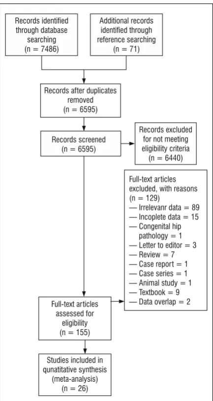
An overview of the process of study identification is summarised in Figure 1. Extensive searching of all
major databases revealed an initial 7486 articles.
A further 71 articles were identified through reference searching. One hundred and fifty-five articles were
assessed by full text for potential eligibility, of which
129 articles were deemed ineligible and 26 articles were included into the meta-analysis. Articles that were not considered eligible included case reports, case series, letters to the editor and reviews.
Characteristics of included studies
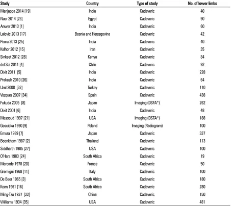
The characteristics of included studies are
pre-sented in Table 1. Twenty-eight studies (n = 3766 lower limbs) were considered eligible and included
in the meta-analysis. The dates of the included stud-ies ranged from 1934 to 2014, and mostly included cadaveric studies, except for the studies by Fukuda
et al. [8] (Digital Subtraction Femoral Arteriography),
Massoud and Fletcher [21] (Digital Subtraction Trans
-femoral Aortogram) and Gościcka et al. [9] (Radio
-gram) which utilised different imaging modalities. The
studies also varied geographically and hailed from
Asia, Europe, North America and Africa.
Origin of the lateral circumflex femoral artery
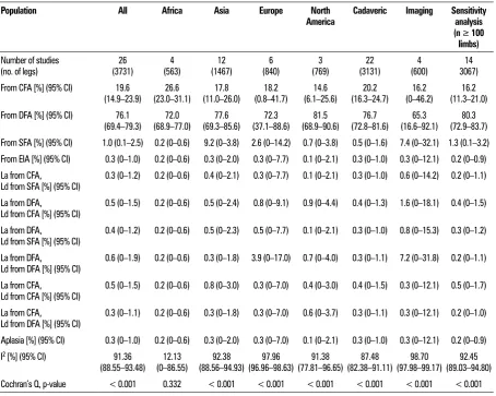
Twenty-six studies (n = 3731 lower limbs) reported the prevalence of the various origins of the LCFA
(Table 2). Our results showed that the LCFA most com
-monly originates from the DFA with a pooled
preva-lence of 76.1% (95% confidence interval 69.4–79.3). The second most common origin of the LCFA was from the CFA in 19.6% of cases, of which 81.8% of these
cases arose as a single branch. Detailed results on the
vario us origins of the LCFA are presented in Tables 2 and 3 and illustrated in Figures 2 and 3 (Supplement S2. For
-rest plots for origins of the lateral circumflex femoral artery — see journal website, supplementary file).
Table 1. Characteristics of included studies
Study Country Type of study No. of lower limbs
Manjappa 2014 [19] India Cadaveric 40
Nasr 2014 [23] Egypt Cadaveric 90
Anwer 2013 [1] India Cadaveric 60
Lalovic 2013 [17] Bosnia and Herzegovina Cadaveric 42
Peera 2013 [25] India Cadaveric 40
Kalhor 2012 [15] Iran Cadaveric 35
Sinkeet 2012 [28] Kenya Cadaveric 84
del Sol 2011 [4] Chile Cadaveric 92
Dixit 2011 [5] India Cadaveric 228
Prakash 2010 [26] India Cadaveric 64
Uzel 2008 [32] Turkey Cadaveric 110
Vazquez 2007 [34] Spain Cadaveric 438
Fukuda 2005 [8] Japan Imaging (DSFA*) 262
Dixit 2001 [6] India Cadaveric 48
Massoud 1997 [21] USA Imaging (DSTA*) 188
Goscicka 1990 [9] Poland Imaging (Radiogram) 100
Emura 1989 [7] Japan Cadaveric 337
Boonkham 1987 [2] Thailand Cadaveric 113
Siddharth 1985 [27] USA Cadaveric 100
O’Hara 1983 [24] South Africa Cadaveric 19
Marcade 1978 [20] France Cadaveric 50
Gremigni 1968 [11] Italy Cadaveric 100
De Beer 1965 [3] South Africa Cadaveric 180
Keen 1961 [16] South Africa Cadaveric 280
Ming-Tzu 1937 [22] China Cadaveric 150
Williams 1934 [35] USA Cadaveric 481
Table 2. Prevalence of the various origins of the LCFA with subgroup and sensitivity analyses
Population All Africa Asia Europe North
America Cadaveric Imaging Sensitivity analysis (n ≥ 100
limbs)
Number of studies
(no. of legs) (3731) 26 (563) 4 (1467) 12 (840) 6 (769) 3 (3131) 22 (600) 4 3067) 14 From CFA [%] (95% CI) 19.6
(14.9–23.9) (23.0–31.1) 26.6 (11.0–26.0) 17.8 (0.8–41.7) 18.2 (6.1–25.6) 14.6 (16.3–24.7) 20.2 (0–46.2) 16.2 (11.3–21.0) 16.2
From DFA [%] (95% CI) 76.1
(69.4–79.3) (68.9–77.0) 72.0 (69.3–85.6) 77.6 (37.1–88.6) 72.3 (68.9–90.6) 81.5 (72.8–81.6) 76.7 (16.6–92.1) 65.3 (72.9–83.7) 80.3
From SFA [%] (95% CI) 1.0 (0.1–2.5) 0.2 (0–0.6) 9.2 (0–3.8) 2.6 (0–14.2) 0.7 (0–3.8) 0.5 (0–1.6) 7.4 (0–32.1) 1.3 (0.1–3.2)
From EIA [%] (95% CI) 0.3 (0–1.0) 0.2 (0–0.6) 0.3 (0–2.0) 0.3 (0–7.7) 0.1 (0–2.1) 0.3 (0–1.0) 0.3 (0–12.1) 0.2 (0–0.9)
La from CFA,
Ld from SFA [%] (95% CI) 0.3 (0–1.2) 0.2 (0–0.6) 0.4 (0–2.1) 0.3 (0–7.7) 0.1 (0–2.1) 0.3 (0–1.0) 0.6 (0–14.2) 0.2 (0–1.1) La from DFA,
Ld from CFA [%] (95% CI) 0.5 (0–1.5) 0.2 (0–0.6) 0.5 (0–2.4) 0.8 (0–9.1) 0.9 (0–4.4) 0.4 (0–1.3) 1.6 (0–18.1) 0.4 (0–1.5) La from DFA,
Ld from SFA [%] (95% CI) 0.4 (0–1.2) 0.2 (0–0.6) 0.5 (0–2.3) 0.5 (0–7.7) 0.1 (0–2.1) 0.3 (0–1.0) 0.8 (0–15.3) 0.3 (0–1.2) La from DFA,
Ld from DFA [%] (95% CI) 0.6 (0–1.9) 0.2 (0–0.6) 0.3 (0–1.8) 3.9 (0–17.0) 0.7 (0–4.0) 0.3 (0–1.1) 7.2 (0–31.8) 0.2 (0–1.1) La from CFA,
Ld from CFA [%] (95% CI) 0.5 (0–1.5) 0.2 (0–0.6) 0.8 (0–3.0) 0.3 (0–7.0) 0.4 (0–3.0) 0.4 (0–1.5) 0.3 (0–12.1) 0.5 (0–1.7) La from CFA,
Ld from DFA [%] (95% CI) 0.3 (0–1.1) 0.2 (0–0.6) 0.3 (0–1.8) 0.3 (0–7.0) 0.6 (0–3.7) 0.3 (0–1.1) 0.3 (0–12.1) 0.2 (0–1.0) Aplasia [%] (95% CI) 0.3 (0–1.0) 0.2 (0–0.6) 0.3 (0–2.0) 0.3 (0–7.0) 0.1 (0–2.1) 0.3 (0–1.0) 0.3 (0–12.1) 0.2 (0–0.9)
I2 [%] (95% CI) 91.36
(88.55–93.48) (0–86.55) 12.13 (88.56–94.93) 92.38 (96.96–98.63) 97.96 (77.81–96.65) 91.38 (82.38–91.11) 87.48 (97.98–99.17) 98.70 (89.03–94.80) 92.45 Cochran’s Q, p-value < 0.001 0.332 < 0.001 < 0.001 < 0.001 < 0.001 < 0.001 < 0.001 CI — confidence interval; CFA — common femoral artery; DFA — deep femoral artery; EIA — external iliac artery; La — ascending branch; LCFA — lateral circumflex femoral artery; Ld — descending branch; SFA — superficial femoral artery
Table 3. Prevalence of the various types of CFA origins of the LCFA with subgroups and sensitivity analyses
Population All Africa Asia Europe North America Cadaveric Imaging
Number of studies
(no. of legs) (614) 23 (151) 4 (197) 10 (116) 5 (126) 3 (548) 19 (66) 4
From CFA (single trunk)
[%] (95% CI) (64.2–92.0) 81.8 (90.7–100) 80.7 (53.0–98.2) 78.0 (33.1–100) 78.8 (60.7–100) 87.6 (60.0–92.3) 80.8 (59.7–100) 86.2 From CFA (with DFA)
[%] (95% CI) (1.9–23.6) 10.7 (0–53.7) 5.7 (1.6–46.2) 18.7 (0–41.3) 7.9 (0–11.9) 0.9 (1.3–26.3) 11.2 (0–32.4) 8.4 From CFA (with MCFA)
[%] (95% CI) 3.2 (0–10.4) 9.3 (0–62.5) 1.5 (0–12.3) 2.1 (0–26.0) 6.1 (0–25.3) 3.0 (0–11.2) 3.9 (0–22.7)
From CFA (with DFA
and MCFA) [%] (95% CI) 4.3 (0–12.4) 4.4 (0–49.9) 1.8 (0–13.4) 11.2 (0–47.9) 5.4 (0–23.7) 5.0 (0–14.9) 1.6 (0–16.1) I2 [%] (95% CI) 94.31
A sensitivity analysis was performed to probe sources of heterogeneity by only including studies
with a sample size of 100 or more lower limbs (Table 2); however, no significant differences were noted from
the results of our overall analysis. Similarly, subgroup analysis with respect to geography was performed
(Table 2), showing no significant differences when
compared with the results of the overall analysis.
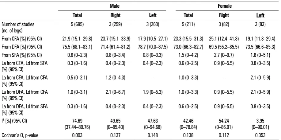
Origins of the lateral circumflex femoral artery with respect to gender and side
Five studies (n = 695 male lower limbs, 211 female lower limbs) reported data on the prevalence of the vari
-ous origins of the LCFA with respect to gender and side (Table 4). Our analysis revealed that the LCFA, consistent
with our overall analysis, most commonly originated
from the DFA. Detailed results on the origins of the LCFA
with respect to gender are presented in Tables 4 and 5.
Nine studies (n = 695 lower limbs) reported data on the prevalence of the various origins of the LCFA with respect to side (Table 6). Our results were consistent with the overall analysis, but the prevalence of the LCFA from the DFA was more common on the left side (77.6%) vs. the right side (73.9%), although these results were not statistically significant. In contrast to LCFA from the
DFA, the LCFA originating from the CFA was more com
-Figure 2. Variants of the origins of the lateral circumflex femoral artery (LCFA); CFA — common femoral artery; DFA — deep femoral artery; EIA — external iliac artery; IL — inguinal ligament; La — ascending branch; Ld — descending branch; MCFA — medial circumflex femoral artery; SFA — superficial femoral artery.
mon on the right side (22.1%) vs. the left side (19.1%);
however, these differences were also not statistically
significant. When the LCFA originated from the CFA, it
most commonly arose as a single trunk, thus consistent
with the results of our overall analysis (Table 7).
Morphometrics of the lateral circumflex femoral artery
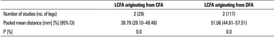
Two studies (n = 29 lower limbs for LCFA origina-ting from CFA, 117 lower limbs for LCFA originaorigina-ting from DFA) reported data on the pooled mean distance
of the various origins of the LCFA to the MIP (Table 8). The pooled mean distance of the LCFA originating from the CFA to the MIP was 38.79 mm (95% confidence interval 28.10–49.48) and the pooled mean distance of the LCFA originating from the DFA to the MIP was 51.06 mm (95% confidence interval 44.61–57.51).
New classification system for origin of the lateral circumflex femoral artery
After a thorough assessment of the results of the
analysis, a new classification system for the origin of
Table 5. Prevalence of the various types of CFA origins of the LCFA according to gender
Male Female
Total Right Left Total Right Left
Number of studies
(no. of legs) 4 (139) 3 (59) 3 (44) 2 (19) 3 (21) 3 (16)
From CFA (single trunk)
[%] (95% CI) (61.6–100) 87.6 (35.9–100) 80.4 (46.0–100) 83.0 (22.5–100) 64.4 (40.5–100) 76.7 (21.5–100) 63.1 From CFA (with DFA)
[%] (95% CI) 7.2 (0–26.1) 11.3 (0–48.3) 12.5 (0–54.0) 30.9 (0–77.5) 13.3 (0–50.4) 24.4 (0–68.4)
From CFA (with MCFA)
[%] (95% CI) 3.2 (0–17.9) 3.9 (0–31.7) 2.2 (0–28.7) 2.3 (0–25.0) 6.6 (0–37.1) 8.4 (0–42.2)
From CFA (with DFA
and MCFA) [%] (95% CI) 1.9 (0–14.5) 4.4 (0–33.2) 2.2 (0–28.7) 2.3 (0–25.0) 3.3 (0–28.3) 4.0 (0–31.4) I2 [%] (95% CI) 89.99
(77.28–95.59) (62.70–95.43) 86.94 (54.39–94.83) 84.65 (0–93.22) 69.85 (4.42–91.67) 71.77 (0–90.63) 67.61
Cochran’s Q, p-value < 0.001 < 0.001 0.001 0.069 0.029 0.046
CI — confidence interval; CFA — common femoral artery; DFA — deep femoral artery; LCFA — lateral circumflex femoral artery; MCFA — medial circumflex femoral artery; SFA — superficial femoral artery
Table 4. Prevalence of the various origins of the LCFA with respect to gender
Male Female
Total Right Left Total Right
Left
Number of studies(no. of legs) 5 (695) 3 (259) 3 (260) 5 (211) 3 (82) 3 (83)
From CFA [%] (95% CI) 21.9 (15.1–29.8) 23.7 (15.1–33.9) 17.9 (10.5–27.1) 23.3 (15.5–31.3) 25.1 (12.4–41.8) 19.1 (11.8–29.4)
From DFA [%] (95% CI) 75.5 (68.1–83.1) 71.4 (61.4–81.2) 78.7 (70.0–87.5) 73.0 (66.3–82.7) 69.5 (55.2–85.5) 73.5 (66.6–85.3)
From SFA [%] (95% CI) 0.6 (0–2.3) 0.8 (0–3.4) 0.8 (0–3.3) 1.5 (0–4.2) 2.7 (0–9.7) 1.6 (0–5.1)
La from CFA, Ld from SFA
[%] (95% CI) 0.3 (0–1.6) 0.4 (0–2.3) 0.4 (0–2.3) 0.6 (0–2.5) 0.9 (0–5.5) 0.8 (0–3.5) La from CFA, Ld from CFA
[%] (95% CI) 0.5 (0–2.1) 1.2 (0–4.3) – 1.0 (0–3.3) – 2.1 (0–5.9) La from DFA, Ld from CFA
[%] (95% CI) 1.0 (0–3.1) 2.1 (0–6.7) 1.9 (0–5.3) 1.0 (0–3.3) 0.9 (0–5.5) 2.1 (0–5.9) La from DFA, Ld from SFA
[%] (95% CI) 0.3 (0–1.6) 0.4 (0–2.3) 0.4 (0–2.3) 0.6 (0–2.5) 0.9 (0–5.5) 0.8 (0–3.5) I2 [%] (95% CI) 74.69
(37.44–89.76) (0–85.40) 49.65 (0–94.68) 47.63 (0–78.84) 42.46 (0–86.91) 54.24 (0–90.01) 3.95
Cochran’s Q, p-value 0.003 0.137 0.148 0.138 0.112 0.353
Table 6. Prevalence of the various origins of the LCFA according to side
Right Left
Number of studies (no. of legs) 9 (695) 9 (695)
From CFA [%] (95% CI) 22.1 (18.1–27.0) 19.1 (15.8–23.3)
From DFA [%] (95% CI) 73.9 (70.1–79.5) 77.6 (74.8–82.5)
From SFA [%] (95% CI) 0.8 (0.1–2.2) 0.7 (0.1–1.8)
From EIA [%] (95% CI) 0.5 (0–1.4) 0.3 (0–1.0)
La from CFA, Ld from SFA [%] (95% CI) 0.4 (0–1.1) 0.3 (0–1.0)
La from CFA, Ld from CFA [%] (95% CI) 0.5 (0–1.6) 0.4 (0–1.2)
La from DFA, Ld from CFA [%] (95% CI) 1.1 (0.2–2.6) 0.9 (0.2–2.1)
La from DFA, Ld from SFA [%] (95% CI) 0.4 (0–1.1) 0.3 (0–1.0)
Aplasia [%] (95% CI) 0.5 (0–1.4) 0.3 (0–1.0)
I2 [%] (95% CI) 43.22 (0–73.79) 28.64 (0–66.86)
Cochran’s Q, p-value 0.079 0.190
CI — confidence interval; CFA — common femoral artery; DFA — deep femoral artery; EIA — external iliac artery; LCFA — lateral circumflex femoral artery; Ld — descending branch; SFA — superficial femoral artery
Table 7. Prevalence of the various types of CFA origins of the LCFA according to side
Right Left
Number of studies (number of legs) 9 (154) 9 (131)
From CFA (single trunk) [%] (95% CI) 78.0 (47.2–98.4) 76.8 (50.2–98.8)
From CFA (with DFA) [%] (95% CI) 15.6 (0–44.9) 18.3 (0.8–48.2)
From CFA (with MCFA) [%] (95% CI) 3.7 (0–19.7) 2.6 (0–16.8)
From CFA (with DFA and MCFA) [%] (95% CI) 2.7 (0–17.2) 2.2 (0–15.6)
I2 [%] (95% CI) 92.13 (87.29–95.13) 90.42 (84.08–94.24)
Cochran’s Q, p-value < 0.001 < 0.001
CI — confidence interval; CFA — common femoral artery; DFA — deep femoral artery; LCFA — lateral circumflex femoral artery; MCFA — medial circumflex femoral artery
Table 8. Pooled mean distance of the various origins of the LCFA to the MIP
LCFA originating from CFA LCFA originating from DFA
Number of studies (no. of legs) 2 (29) 2 (117)
Pooled mean distance [mm] [%] (95% CI) 38.79 (28.10–49.48) 51.06 (44.61–57.51)
I2 [%] 0.0 0.0
CI — confidence interval; CFA — common femoral artery; DFA — deep femoral artery; LCFA — lateral circumflex femoral artery; MIP — mid-inguinal point
the LCFA was established and presented in Figure 4.
Five different types of origin variations that met the
a priori thresholds were included in the classification: Type 1 (normal) — LCFA branching from the DFA; Type 2 — LCFA branching from the CFA as a single trunk; Type 3 — LCFA branching from the CFA with the DFA; Type 4 — LCFA branching from the SFA; and Type 5 — direct branching patterns (from CFA,
DFA, and SFA), where the La and Ld branches of the LCFA originate directly from one of the main femoral
vessels without a common branch. While none of the individual direct branching patterns alone reached a threshold of ≥ 1%, due to the several different
reported patterns in combination reaching a value
well above the threshold, a single category (Type 5)
-Figure 4. New classification system of the origin of the lateral circumflex femoral artery (LCFA); CFA — common femoral artery; DFA — deep femoral artery; La — ascending branch; Ld — descending branch; MCFA — medial circumflex femoral artery; SFA — superficial femoral artery.
ing patterns. All other variants of the LCFA origin not included in the classification system should be considered rare anomalies (Supplement S3. Raw data — see journal website, supplementary file).
DISCUSSION
The LCFA has been shown to supply the head and
neck of the femur, the greater trochanter, the vastus lateralis muscle, and the knee [32]. The overall aim of our study was to gather and analyse all available data
from a comprehensive literature search on LCFA, with
a focus on its branching and morphometric variations, to provide a detailed analysis of the artery, and its clinically important characteristics.
Detailed anatomical knowledge about the LCFA is
useful in a number of clinical procedures, including aortopopliteal bypass [10, 29], anterolateral thigh
flaps [33], and coronary artery bypass grafting [8],
thus giving its normal and variant anatomy a high
de-gree of clinical significance. Furthermore, its branches
may also be used in various procedures. The
ascend-ing branch of the LCFA can be used for vascularised
iliac transplant, and its descending branch may be employed as a collateral for an obstructed SFA, and can also be used in coronary artery bypass grafting [12, 32].
Our results demonstrated that the LCFA most
commonly originated from the DFA with a pooled
prevalence of 76.1%. Thus, we consider a DFA origin to be the normal type of LCFA origin (Type 1). This
trend held true across all subgroups including side, gender, and geographical regions.
The second most common origin of the LCFA was
found to be from the CFA, with a pooled prevalence of
19.6%. A CFA-derived LCFA was also most commonly
found to branch as a single trunk, with a pooled
prevalence of 81.8%. These findings were consistent
when acknowledging gender, side and
geographi-cal location. Though not statistigeographi-cally significant, it is interesting to note that the LCFA originated from the CFA on the right limb more frequently than the
the origin of the LCFA, such as the SFA [8], external
iliac artery [5] and instances where the anterior and
descending limbs of the LCFA branch directly from
the main femoral vessels without a common trunk
[8], have been reported (Table 2).
The highly variable branching pattern of a
CFA--derived LCFA was found to branch alongside the
DFA, the MCFA, or with the DFA and MCFA
concur-rently (Fig. 3). Accurate anatomical details concerning the LCFA origin can help physicians make informed
decisions during interventional procedures and op-erations in the femoral region. Moreover, knowledge
on the existence of these rarer LCFA origin variations
is crucial for surgeons to avoid iatrogenic injuries. In order to supply some organisation to the
multitude of reported origin patterns of the LCFA, we formulated a simple classification system based
on the results of our comprehensive meta-analysis
(Fig. 4). Previous classifications [35] often attempted
to encompass the origins and anatomy of multiple
arteries versus a simple classification system for LCFA origin. Difficulty in adhering to such systems can be
seen in the lack of consistency in the reporting of
LCFA origin between studies in the literate, and thus indicating the need for a new, simple classification
system. Our system is inclusive of all the most com-mon variants with a population prevalence ≥ 1.0%, and organised as most common — Type 1 (DFA origin) to least common — Type 4 (SFA origin). Additionally,
we formulated a Type 5 for direct branching patterns,
inclusive of all variants where the La and Ld branch
directly from one of the main femoral vessels without
a common branch. The proposed classification system
should be further assessed and evaluated in future original anatomical studies.
The distance from LCFA origin to the MIP was also
analysed, with a pooled mean of 51.06 mm when originating from the DFA, the most common origin of
the artery (Table 8). When the LCFA originated from
the CFA, it was found to do so with a pooled mean distance from the MIP of 38.79 mm, substantially shorter than that of a DFA origin. The differences in distance, however, were not found to be statistically
significant. Due to the lack of studies on this param -eter [2], as well as the overall lack of other substantial
morphometric studies on the LCFA, further research
is needed to determine the precise vessel measure-ments throughout populations, as this data may be of value to interventional radiologists and orthopaedic
surgeons. Additionally, future studies should examine
the spatial arrangement of the LCFA, to better under
-stand the exact location of its origin.
Our study was limited by the lack of a quality as -sessment tool and risk of bias as-sessment method for anatomical studies, as well as lack of a proper meas-ure for publication bias in multi-categorical pooled prevalence analysis. Furthermore, our meta-analysis was also limited by the high heterogeneity among the included studies, which persisted despite extensive subgroup analysis. We there attribute the high heterogeneity to be most likely due to the highly variable nature of the vessel itself.
CONCLUSIONS
In conclusion, the most common origin of the
LCFA is from the DFA, branching at an average
distance of 51.06 mm from the MIP. However, the
origin of the LCFA is variable throughout the gen
-eral population and thus a new simple
anatomi-cal classification system was proposed. Accurate
knowledge of the anatomical properties of the
LCFA may convey important information to sur
-geons, especially during aortopopliteal bypass
sur-gery and anterolateral thigh flap procedures. The high degree of variability within this artery requires
physicians to proceed with caution in order to decrease the risk of iatrogenic injuries. Thus, we highly recommended radiographic assessment of the vessel anatomy prior to surgical procedures in the femoral region.
REFERENCES
1. Anwer D, Karmalkar AS, Humbarwadi RSA. study of varia-tions in the profunda femoris artery and its branches. Int J Biomed Adv Res. 2013; 04: 366–368.
2. Boonkham Y, Plakornkul V. Variational anatomy of the profunda femoris artery in Thais. Siriraj Hospital Gazette. 1987; 39: 441–445.
3. De Beer PM. The profunda femoris and circumflex femo-ral arteries in the South African Bantu-speaking Negro. S Afr J Med Sci. 1965; 30(1): 1–10, indexed in Pub -med: 5854977.
4. Sol Md, Galdames IS, Vásquez B. Las Arterias Cir -cunflejas Femorales en el Triángulo Femoral. Int J Morphol. 2011; 29(2): 644–649, doi: 10.4067/s0717-95022011000200057.
5. Dixit D, Kubavat DM, Rathod SP, et al. study of variations in the origin of the profunda femoris artery and its circumflex branches. Int J Biomed Res. 2011; 2: 1084–1089. 6. Dixit DP, Mehta LA, Kothari ML. Variations in the origin
7. Emura S, Shoumura S, Ishizaki N, et al. [The anatomi -cal study on the branches of the femoral artery (II). Comparison with the findings of Adachi’s classification]. Kaibogaku Zasshi. 1989; 64(3): 196–205, indexed in Pubmed: 2801014.
8. Fukuda H, Ashida M, Ishii R, et al. Anatomical variants of the lateral femoral circumflex artery: an angiographic study. Surg Radiol Anat. 2005; 27(3): 260–264, doi: 10.1007/ s00276-004-0312-5, indexed in Pubmed: 15682274. 9. Gościcka D, Gielecki J, Zietek Z. Digital image analysis of
variations in the origin of the deep femoral artery in hu-man foetuses. Acta Morphol Hung. 1990; 38(2): 95–107, indexed in Pubmed: 2099102.
10. Gradman WS. Bypass to the lateral circumflex femoral artery. Ann Vasc Surg. 1992; 6(4): 344–346, doi: 10.1007/ BF02008791, indexed in Pubmed: 1390022.
11. Gremigni D. [Influence of sex on the point of origin of the 2 femoral circumflex arteries and on the methods of subdivision of the lateral femoral circumflex artery]. Boll Soc Ital Biol Sper. 1968; 44(2): 105–107, indexed in Pubmed: 5661659.
12. Hage JJ, Woerdeman LAE. Lower limb necrosis after use of the anterolateral thigh free flap: is preoperative angi -ography indicated? Ann Plast Surg. 2004; 52(3): 315–318, indexed in Pubmed: 15156989.
13. Henry BM, Tomaszewski KA, Walocha JA. Methods of Evidence-Based Anatomy: a guide to conducting systematic reviews and meta-analysis of anatomical studies. Ann Anat. 2016; 205: 16–21, doi: 10.1016/j. aanat.2015.12.002, indexed in Pubmed: 26844627. 14. Higgins J, Green S (2011) Cochrane handbook for system
-atic reviews of interventions. Version 5.1.0. The Cochrane Collaboration. http://cochrane-handbook.org.
15. Kalhor M, Horowitz K, Gharehdaghi J, et al. Anatomic variations in femoral head circulation. Hip Int. 2012; 22(3): 307–312, doi: 10.5301/HIP.2012.9242, indexed in Pubmed: 22740271.
16. Keen JA. A study of the arterial variations in the limbs, with special reference to symmetry of vascular patterns. Am J Anat. 1961; 108: 245–261, doi: 10.1002/aja.1001080303, indexed in Pubmed: 14454801.
17. Lalović N, Cvijanović R, Malis M, et al. [Surgical anatomy of the initial segment of the lateral circumflex femoral artery]. Med Pregl. 2013; 66(7-8): 326–330, indexed in Pubmed: 24069816.
18. Li M, Cole PA. Anatomical considerations in adult femoral neck fractures: how anatomy influences the treatment issues? Injury. 2015; 46(3): 453–458, doi: 10.1016/j.in-jury.2014.11.017, indexed in Pubmed: 25549821. 19. Manjappa T, Prasanna LC. Anatomical variations of the
profunda femoris artery and its branches-a cadaveric study in South Indian population. Indian J Surg. 2014; 76(4): 288–292, doi: 10.1007/s12262-012-0677-3, indexed in Pubmed: 25278652.
20. Marcade E, Leguerrier A, Scarabin JM, et al. L’artere femorale profonde etude anatomo-radiologique. Bulletin de L’Association des Anatomistes (Nancy. 1978; 62: 453–459. 21. Massoud TF, Fletcher EW. Anatomical variants of the
profunda femoris artery: an angiographic study. Surg
Radiol Anat. 1997; 19(2): 99–103, indexed in Pub -med: 9210243.
22. Ming-Tzu P. Origin of deep and circumflex femoral group of arteries in the Chinese. Am J Physical Anthropol. 1937; 22(3): 417–424, doi: 10.1002/ajpa.1330220304. 23. Nasr AY, Badawoud MH, Al-Hayani AA, et al. Origin of
pro-funda femoris artery and its circumflex femoral branches: anatomical variations and clinical significance. Folia Mor -phol. 2014; 73(1): 58–67, doi: 10.5603/FM.2014.0008, indexed in Pubmed: 24590524.
24. O’Hara JP, Dommisse GF. Extraosseous blood supply to the neonatal femoral head. Clin Orthop Relat Res. 1983(174): 293–297, indexed in Pubmed: 6339140.
25. Peera SH, Sugavasi R. Morphological study of branches of femoral artery in the femoral triangle – a human cadaveric study. Int J Health Sci Res. 2013; 3: 14–19.
26. Kumari J, Kumar Bhardwaj A, Jose BA, et al. Variations in the origins of the profunda femoris, medial and lat-eral femoral circumflex arteries: a cadaver study in the Indian population. Rom J Morphol Embryol. 2010; 51(1): 167–170, indexed in Pubmed: 20191139.
27. Siddharth P, Smith NL, Mason RA, et al. Variational anatomy of the deep femoral artery. Anat Rec. 1985; 212(2): 206–209, doi: 10.1002/ar.1092120216, indexed in Pubmed: 3842043.
28. Sinkeet SR, Ogeng’o JA, Elbusaidy H, et al. Variant origin of the lateral circumflex femoral artery in a black Kenyan population. Folia Morphol. 2012; 71(1): 15–18, indexed in Pubmed: 22532179.
29. Sugawara Y, Sato O, Miyata T, et al. Utilization of the lateral circumflex femoral artery as a midway outflow for aorto-popliteal grafting: report of a case. Surg Today. 1998; 28(9): 967–970, doi: 10.1007/s005950050264, indexed in Pubmed: 9744412.
30. Tomaszewski K, Henry B, Vikse J, et al. Variations in the origin of the deep femoral artery: A meta-analysis. Clin Anat. 2016; 30(1): 106–113, doi: 10.1002/ca.22691. 31. Tomaszewski KA, Henry BM, Vikse J, et al. The origin of
the medial circumflex femoral artery: a meta-analysis and proposal of a new classification system. Peer J. 2016; 4: e1726, doi: 10.7717/peerj.1726, indexed in Pubmed: 26966661.
32. Uzel M, Tanyeli E, Yildirim M, et al. An anatomical study of the origins of the medial circumflex femoral artery in the Turkish population. Folia Morphol. 2006; 65(3): 209–212, indexed in Pubmed: 16988917.
33. Valdatta L, Tuinder S, Buoro M, et al. Lateral circumflex femoral arterial system and perforators of the anterolateral thigh flap: an anatomic study. Ann Plast Surg. 2002; 49(2): 145–150, indexed in Pubmed: 12187341.
34. Vazquez MT, Murillo J, Maranillo E, et al. Patterns of the circumflex femoral arteries revisited. Clin Anat. 2007; 20(2): 180–185, doi: 10.1002/ca.20336, indexed in Pub-med: 16617441.