Open Access
Research article
Sequence variation in human succinate dehydrogenase genes:
evidence for long-term balancing selection on
SDHA
Bora E Baysal*
1,2, Elizabeth C Lawrence
2and Robert E Ferrell
2Address: 1Department of Obstetrics, Gynecology and Reproductive Sciences, University of Pittsburgh School of Medicine, Pittsburgh, PA 15213, USA and 2Department of Human Genetics, Graduate School of Public Health, University of Pittsburgh, Pittsburgh, PA 15213, USA
Email: Bora E Baysal* - [email protected]; Elizabeth C Lawrence - [email protected]; Robert E Ferrell - [email protected] * Corresponding author
Abstract
Background: Balancing selection operating for long evolutionary periods at a locus is characterized by the maintenance of distinct alleles because of a heterozygote or rare-allele advantage. The loci under balancing selection are distinguished by their unusually high polymorphism levels. In this report, we provide statistical and comparative genetic evidence suggesting that the SDHA gene is under long-term balancing selection. SDHA encodes the major catalytical subunit (flavoprotein, Fp) of the succinate dehydrogenase enzyme complex (SDH; mitochondrial complex II). The inhibition of Fp by homozygous SDHA mutations or by 3-nitropropionic acid poisoning causes central nervous system pathologies. In contrast, heterozygous mutations in SDHB, SDHC, and SDHD, the other SDH subunit genes, cause hereditary paraganglioma (PGL) tumors, which show constitutive activation of pathways induced by oxygen deprivation (hypoxia).
Results: We sequenced the four SDH subunit genes (10.8 kb) in 24 African American and 24 European American samples. We also sequenced the SDHA gene (2.8 kb) in 18 chimpanzees. Increased nucleotide diversity distinguished the human SDHA gene from its chimpanzee ortholog and from the PGL genes. Sequence analysis uncovered two common SDHA missense variants and refuted the previous suggestions that these variants originate from different genetic loci. Two highly dissimilar SDHA haplotype clusters were present in intermediate frequencies in both racial groups. The SDHA variation pattern showed statistically significant deviations from neutrality by the Tajima, Fu and Li, Hudson-Kreitman-Aguadé, and Depaulis haplotype number tests. Empirically, the elevated values of the nucleotide diversity (% π = 0.231) and the Tajima statistics (D = 1.954) in the
SDHA gene were comparable with the most outstanding cases for balancing selection in the African American population.
Conclusion: The SDHA gene has a strong signature of balancing selection. The SDHA variants that have increased in frequency during human evolution might, by influencing the regulation of cellular oxygen homeostasis, confer protection against certain environmental toxins or pathogens that are prevalent in Africa.
Published: 21 March 2007
BMC Biology 2007, 5:12 doi:10.1186/1741-7007-5-12
Received: 9 September 2006 Accepted: 21 March 2007
This article is available from: http://www.biomedcentral.com/1741-7007/5/12
© 2007 Baysal et al; licensee BioMed Central Ltd.
Background
Succinate dehydrogenase (SDH; mitochondrial complex II) is an essential enzyme complex that has dual roles in the Krebs cycle and the electron transport chain (ETC) in mitochondria [1]. SDH is composed of four subunits encoded by the nuclear genes SDHA, SDHB, SDHC, and
SDHD. SDHA at chromosome band 5p15 and SDHB at
chromosome band 1p35 encode the two catalytical hydrophilic subunits flavoprotein (Fp; 70 kDa) and iron-sulfur (Ip; 35 kDa), respectively. SDHC at chromosome band 1q23 and SDHD at chromosome 11q23 encode the two membrane-spanning hydrophobic subunits, cybL (15 kDa) and cybS (12 kDa), respectively. The SDHA, SDHB, SDHC, and SDHD gene products are encoded by 15, 8, 6, and 4 exons, which span genomic distances of ~38 kb, 35 kb, 50 kb. and 10 kb, respectively [2,3].
The identification of the SDHD subunit gene as the hered-itary paraganglioma type 1 locus (PGL1) has uncovered unexpected links between SDH and tumor susceptibility, and highlighted the role of mitochondria in cancer [4]. Since then, mutations in SDHB, SDHC, and SDHD subu-nit genes (PGL genes) have been established as an impor-tant cause of sporadic and familial paragangliomas [5-10]. The paraganglia specificity of PGL tumors [4] and data from global gene-expression analysis [11], cell biology [12], animal-model studies [13], and gene-environment interaction and population genetics [14] support the hypothesis that constitutive hypoxic stimulation under-lies the pathogenesis of PGL.
The role of SDH in disease pathogenesis has been impli-cated independently through a series of studies on a widely distributed plant and fungal neurotoxin, 3-nitro-propionic acid (3-NPA)[15]. Acute food poisoning with 3-NPA, which can lead to central nervous system defects with lifelong disability and to mortality in ~10% of the cases, have been associated with consumption of moldy sugarcanes in China [16]. The neurodegeneration induced by 3-NPA poisoning often involves the basal ganglia, hip-pocampus, spinal tracts, and peripheral nerves, and the symptoms mimic those of Huntington's disease [17]. 3-NPA irreversibly inhibits SDH, owing to the similarity of the chemical structures of 3-NPA to succinate [18]. It has been suggested that 3-NPA may form a covalent adduct with an arginine residue at amino acid position 345 in the active site of the Fp subunit [19].
Surprisingly, mutations in the major catalytical subunit
SDHA have yet to be associated with PGL. Although
homozygous mutations in SDHA have been found in Leigh syndrome [20], a severe neurodegenerative disorder of childhood, and with neuromusculopathies, no genetic link between SDHA and paraganglioma susceptibility has ever been established. Current biochemical knowledge on
SDH provides very few clues for the phenotypic dichot-omy arising from the germline subunit gene mutations.
SDHA and SDHB subunits encode the two physically-interacting catalytical subunits, so it is surprising that their mutations would have such different phenotypic conse-quences [21]. Recently, after identifying cDNA seconse-quences encoding a missense Fp variant containing the Y629F and V657I polymorphisms, Tomitsuka et al [22,23] proposed that distinct genetic loci encode two Fp variants, namely type I and type II. They reached this conclusion after observing tissue-specific and cell line-specific differential expression of the cDNA variants and PCR amplification from genomic DNA of processed SDHA gene fragments that lacked introns (i.e, a functional SDHA retrogene). However, the genomic location of the retrogene that was proposed to encode the second SDHA gene could not be determined. A retrogene for SDHA is not present in the human genome, according to the March 2006 assembly in The UCSC database. [24] Finally, Briere et al [25] showed the presence of the missense SDHA variants in several dif-ferent cell types and assumed that these variants originate from two different genes, although they provided no experimental or bioinformatic evidence for the genomic presence of a second SDHA locus. Briere et al [25] sug-gested that the presence of two SDHA genes in paraganglia prevents tumorigenesis. If Fp were encoded by two differ-ent loci, this would indeed have provided a simple expla-nation for why SDHA mutations would not be associated with PGL susceptibility.
An alternative approach to gain insights into gene func-tion involves analysis of sequence variafunc-tion in the popu-lation. To date, no study has systematically addressed the variation patterns in the SDH subunit genes in normal subjects from different racial or ethnic groups. To gain fur-ther insights into the multiple roles of SDH in disease pre-disposition and to help to integrate the seemingly disparate phenotypic consequences of SDH subunit defects, we examined sequence variation in the complete coding and partial flanking intronic sequences of the four SDH subunit genes in 24 samples from an African Ameri-can population and 24 samples from a white population. These analyses uncovered an unexpected degree of nucle-otide diversity in the SDHA gene.
Results
Sequence variants in the SDH subunit genes
samples. Except for two non-coding indels in SDHA and one in SDHC, all variants were single nucleotide polymor-phisms (SNPs) involving base replacements. A full list of the identified sequence variants is provided in Additional File 1 and has also been submitted to the SDH mutation database [26]. SDHA variant density was 2.6-fold and 2.3-fold higher in the coding and non-coding regions, respec-tively, than the average of 106 genes [27]. The minor allele frequencies of all variants are shown in Figure 1.
Nucleotide diversity in SDH subunit genes
We calculated the nucleotide diversity in SDH subunit genes using the population genetic parameters π and θs (Table 2). As expected, all diversity indices were higher in the African American samples. The nucleotide diversity (%) in the total sample set was low at the PGL genes:
SDHB (π = 0.008), SDHC (π = 0.065), and SDHD (π = 0.044). In contrast, the nucleotide diversity of the SDHA
gene (π = 0.199%) was 5.1-fold higher than the average of the PGL genes and 3.4-fold higher than the average (π = 0.058%) of 292 autosomal genes [28]. The θs and π esti-mates of nucleotide diversities were similar for the mem-brane-spanning subunits SDHC and SDHD, but differed substantially for the two catalytic subunits. Whereas the π estimate was ~1.6-fold higher than θs for the SDHA gene, consistent with the enrichment of alleles with intermedi-ate frequencies, the θs estimate was ~4-fold higher for the
SDHB gene, indicating the very low frequency of the allelic variants. For comparison, 90% of the genes in a recent survey had θs estimates higher than the π estimates [28], indicating an abundance of rare alleles, which is thought to be a result of recent population expansion in humans. FST statistics provided statistically significant evi-dence of population differentiation between the two racial groups for the SDHA, SDHC, and SDHD genes, but not for the SDHB gene (Table 2). This was attributable to the very low frequencies and the absence of SDHB allelic variants in the African American and European American samples, respectively.
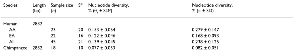
Comparison of the human and chimpanzee SDHA genes for sequence diversity
To test whether high nucleotide diversity also character-izes the chimpanzee SDHA gene, we used the human PCR primers to amplify and sequence 18 unrelated
chimpan-zee samples. We obtained high-quality sequences for exons 3–6, 8, 12, 13, and 15, which together comprise a total genomic sequence size of 2832 bp (Table 1). We identified one silent exonic and seven intronic fixed-nucleotide differences between the human and chimpan-zee SDHA genes (Additional file 2), corresponding to a substitution rate of 0.28%. The nucleotide substitution rate in SDHA is lower than the average of 127 known genes (0.75%) that were recently sequenced in human and chimpanzee [29]. The chimpanzee SDHA gene has 10 polymorphic variants, compared with 21 in the human gene in the same region, and showed ~2.9-fold lower nucleotide diversity (π) than the human gene (Table 3). Furthermore, θs and π estimates of nucleotide diversities were similar in the chimpanzee, consistent with neutral expectations. These findings indicate that the mutation rate in SDHA is not inherently high and that the increased nucleotide diversity in the human gene must have occurred after the split of the two species from their com-mon ancestor 5–6 million years ago.
Tests of neutrality
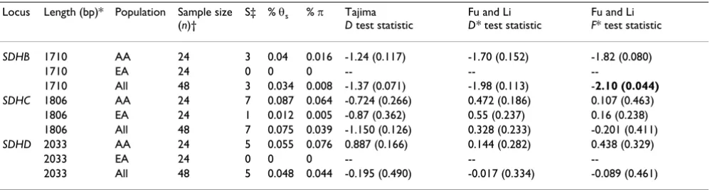
We employed three commonly used tests (the Tajima D
tests and the Fu and Li D* and F* tests) to identify depar-tures of the allelic distributions from neutral expectations. None of the PGL genes showed statistically significant departures from neutrality in samples from either racial group (Table 4). In contrast, the allelic distribution of the
SDHA gene showed positive test values at statistically sig-nificant levels in both racial samples (Table 5). Notably, the neutrality statistics were supportive of balancing selec-tion on SDHA despite the presence of six singleton vari-ants in the African American samples and one singleton variant in the European American samples (Additional file 1). To obtain a clearer picture of the departure of
SDHA allelic distributions from neutral expectations, we analyzed non-coding, coding, synonymous, and non-syn-onymous variants separately (Table 5). Nominally signif-icant departures from neutrality were obtained in seven of the nine test statistics for the non-coding variants, although the SDHA coding region variation was also sug-gestive of an excess of variants in intermediate frequencies in the African American samples.
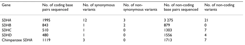
Table 1: Summary of variants in SDH subunit genes
Gene No. of coding base
pairs sequenced
No. of synonymous variants
No. of non-synonymous variants
No. of non-coding base pairs sequenced
No. of non-coding variants
SDHA 1995 12 3 3 275 21
SDHB 843 1 2 879 0
SDHC 510 1 0 1303 7
SDHD 480 1 0 1556 4
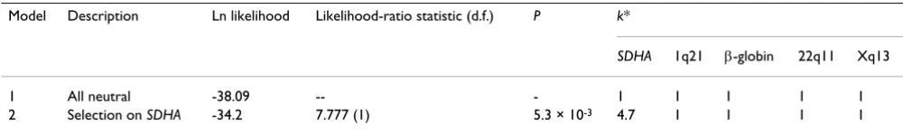
To test whether the level of silent diversity in SDHA corre-lates with level of divergence between human and chim-panzee, as predicted by the neutral theory, we used the Hudson-Kreitman-Aguadé (HKA) test. Sequence data from four loci that were assumed to be evolving neutrally were used for comparison. These loci include non-coding regions on chromosome bands 1q24 [30], 22q11 [31], and Xq13.3 [32] and the promoter region of β-globin at 11p15 [33]. Locus-by-locus comparison provided statisti-cal significance in two of the four tests, suggesting increased diversity in SDHA relative to these two loci (Additional file 3). To further address whether the SDHA
variation pattern is unusual when information from the comparison loci is jointly used, we used a recently devel-oped maximum-likelihood-ratio test [34]. The likelihood of two models were compared; the first assumes that all five loci evolve neutrally, whereas the other assumes that
SDHA is subject to selection while the other four loci
evolve neutrally. The model assuming selection on SDHA
was statistically supported over the model of neutrality (p
= 5.3 × 10⁻³; Table 6). These results further support the
hypothesis that increased nucleotide diversity in SDHA is maintained by balancing selection.
Empirical assessment of neutrality in SDH subunit genes
Because population history plays an important role in shaping the variation patterns in the genome, we sought to assess whether the nucleotide diversity of complex II genes were unusual compared with other genes across the genome. We used the summary statistics for Tajima's D
test and nucleotide diversity of 282 genes listed in the SeattleSNP database for comparison. When compared with the database genes, the statistics for nucleotide diver-sity and Tajima's D were not outstanding for any of the complex II genes in the European American samples or for the SDHC and SDHD genes in the African American sam-Minor allele frequency of each SDH subunit variant (also see Additional Table 1) is shown
Figure 1
Minor allele frequency of each SDH subunit variant (also see Additional Table 1) is shown. Filled vertical bars refer to African American, unfilled vertical bars refer to European American samples. Synonymous and non-synonymous coding var-iants are marked by S and NS, respectively. ID refers to insertion/deletion polymorphisms.
0 0.1 0.2 0.3 0.4 0.5 0.6
1 3 5 7 9 11 13 15 17 19 21 23 25 27 29 31 33 35
0 0.1 0.2 0.3 0.4 0.5
1 2 3 4 5 6 7 8
0 0.1 0.2 0.3
1 2 3 4 5
0 0.1 0.2
1 2 3
SDHA
SDHB
SDHC
SDHD
SNP No.
Minor allele
frequency
S S S
S S
S S
S S
S S N
N N
S
N N
S
S ID
ID
ples. However, the SDHA nucleotide diversity was higher than that of 279 (p < 0.015) of the genes and the Tajima
D statistic was higher than that of 281 (p < 0.0036) of the genes in the African American samples (Figure 2). In con-trast, SDHB had less sequence diversity than 280 of the SeattleSNP genes (p < 0.011) in the African American sam-ples. A recent analysis of 151 loci in the SeattleSNP set has indicated that the D statistic of the ABO locus (D = 1.58) retains its significance in an African American population under several demographic scenarios [35]. Because the magnitude of D in SDHA in our African American sam-ples (D = 1.95) is higher than that in the ABO locus (Fig-ure 2), it is likely that the statistical support for balancing selection on SDHA would be retained by different popu-lation histories. In summary, the departure of SDHA
allelic distribution from neutral expectations is empiri-cally supported in the African American samples, consist-ent with a balancing selection mechanism.
Haplotype structures of the SDH subunit genes
Haplotypes, haplotype-block structures and the tagging SNPs for each block were inferred using the web-based
HAP software (see methods). As expected, the haplotypes were more variable in the African American than in the European American samples. The SDHA haplotype varia-tion could be defined by 6 haplotype blocks and 13 tag-ging SNPs in the African American samples but only by 3 haplotype-blocks and 5 tagging SNPs in the European American samples (Additional file 1). In contrast, haplo-type variation in the PGL genes could be defined by sin-gle-haplotype blocks. The most common haplotype accounted for ~99% of the haplotypes of the PGL genes in the European American samples (Additional file 4). Simi-larly, the most common haplotype and its 1-nucleotide neighbors covered ~98%, 79% and 73% of the variation in the SDHB, SDHC, and SDHD genes, respectively, in the African American samples.
The commonness of a single haplotype and its 1-nucleo-tide neighbors in the PGL genes was in stark contrast to the presence of two common but highly dissimilar haplo-types in SDHA in both racial groups. The two most com-mon SDHA haplotypes, A1 and A2, accounted for ~19% (17/90) and ~9% (8/90) of all haplotype diversity,
respec-Table 2: Sequence diversity in succinate dehydrogenase subunit genes
Locus Length (bp)* Population Sample size (n)† S‡ Gene diversity ± SD
Nucleotide diversity, % (θ ± SD)
Nucleotide diversity, % (π ± SD)
FST (P§)
SDHA 5270 AA 24 35 0.974 ± 0.010 0.141 ± 0.045 0.231 ± 0.118 0.093 (0.012)
EA 24 27 0.903 ± 0.030 0.107 ± 0.036 0.147 ± 0.077
All 48 36 0.964 ± 0.009 0.126 ± 0.037 0.199 ± 0.101
SDHB 1722 AA 24 3 0.231 ± 0.078 0.039 ± 0.024 0.016 ± 0.019 0.061 (0.113)
EA 24 0 0 0 0
All 48 3 0.120 ± 0.045 0.034 ± 0.021 0.008 ± 0.013
SDHC 1813 AA 24 8 0.680 ± 0.060 0.086 ± 0.040 0.103 ± 0.067 0.176 (~0)
EA 24 2 0.082 ± 0.053 0.012 ± 0.012 0.013 ± 0.017
All 48 8 0.441 ± 0.061 0.075 ± 0.033 0.065 ± 0.046
SDHD 2036 AA 24 5 0.609 ± 0.063 0.055 ± 0.028 0.077 ± 0.052 0.234 (~0)
EA 24 0 0 0 0
All 48 5 0.361 ± 0.059 0.048 ± 0.024 0.044 ± 0.034
AA, African American samples; EA, European American samples; SD, standard deviation. *Includes coding and non-coding sequences.
†Number of unrelated subjects. ‡Number of segregating sites.
§p value for population genetic structure between AA and EA.
Table 3: Sequence diversity in the human and chimpanzee SDHA genes
Species Length
(bp)
Sample size (n)
S* Nucleotide diversity, % (θs ± SDc)
Nucleotide diversity, % (π ± SD)
Human 2832
AA 23 20 0.153 ± 0.054 0.279 ± 0.147
EA 22 16 0.122 ± 0.046 0.168 ± 0.093
All 45 21 0.139 ± 0.045 0.238 ± 0.125
Chimpanzee 2832 18 10 0.077 ± 0.033 0.082 ± 0.051
tively, and differed from each other in 22 of the 36 variant positions (Figure 3). Haplotype A1 and A2 encode the missense Fp variants Y629-V657 and F629-657, by the SNPs 27 and 35, respectively, indicating an allelic associ-ation of the missense variants in these two amino-acid sites. Notably, the variant Fp amino acids Y629 and V657 were conserved in mammalian Fp sequences, including orangutan, macaque, mouse, dog, rat, and bovine. How-ever, different amino acids were found in phylogenetically more distant species such as the zebrafish, which had Y629-I657 and Dirofilaria, an infectious nematode, which had E629-I657.
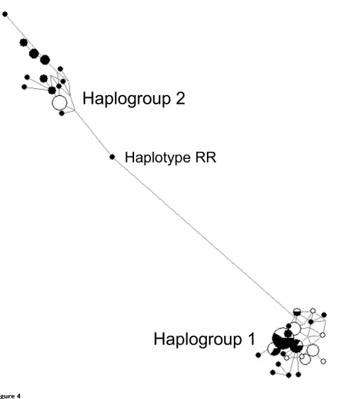
All of the remaining 34 SDHA haplotypes were highly similar to one of the two commonest haplotypes, and formed two distinct haplotype sets, referred to as haplo-group 1 and haplohaplo-group 2. The haplotypes within each group differed from the most common haplotype of the group in up to seven variant positions, with a median number of three differences. The frequencies of haplo-groups 1 and 2 were ~56% and ~44% in the African Amer-ican samples and ~82% and ~18% in the European American samples, respectively. A median-joining net-work of all haplotypes clustered all but one haplotype within two distinct haplogroup clusters (Figure 4). The only haplotype (RR) that mapped outside of the two hap-logroups clusters was probably a recombination product between haplogroup 1 and haplogroup 2.
Haplotype number test
To test whether the number of predicted SDHA haplo-types in the African American samples is compatible with neutral evolution, we employed the Depaulis and Veuille haplotype number test [36]. In total, 35 variants in 46 African American sequences defined 27 different haplo-types (Figure 3). Using Depaulis and Veuille simulations under assumptions of neutrality showed that when there
are 40 variants in 50 sequences, the upper limit of the 95% confidence interval for the expected number of dif-ferent haplotypes is 24. Thus, the number of SDHA hap-lotypes is statistically significantly higher than expected under neutrality, and is consistent with an ancient bal-anced polymorphism in the African American popula-tion.
Estimating age of the SDHA haplogroups
We estimated the age of the two haplogroups by compar-ing the sequence divergence between them with that between the human and chimpanzee genes, assuming a constant evolutionary rate of nucleotide substitutions. Haplogroups 1 and 2 have eight fixed nucleotide differ-ences, at SNPs 8–12, 17, 21, and 22 (Figure 3), within 5255 bp, whereas human and chimpanzee genes have eight fixed nucleotide differences within 2832 bps. On the basis of these fixed nucleotide substitutions, we estimated haplogroups 1 and 2 to be as old as [(8/5255)/(8/2832)] times the divergence time of human and chimpanzees. Thus, SDHA balanced polymorphisms were estimated to be 2.69–3.23 million years old, assuming a divergence time of 5–6 million years for human and chimpanzees. This is probably a conservative estimate, as the fixed dif-ferences between the haplogroups erode in time by recombination and gene conversion.
Discussion
Our results establish a foundation to understand the selective and demographic forces that have shaped the variation patterns in SDH subunit genes, and have impor-tant functional implications. Our findings indicate that the variation pattern in SDHA is characterized by the pres-ence of higher sequpres-ence diversity, two common and highly dissimilar haplogroups, and statistical and empiri-cal support for the operation of a balancing selection mechanism. Our data also refute the previous suggestions
Table 4: Tests of Neutrality in PGL genes
Locus Length (bp)* Population Sample size (n)†
S‡ % θs % π Tajima
D test statistic
Fu and Li D* test statistic
Fu and Li F* test statistic
SDHB 1710 AA 24 3 0.04 0.016 -1.24 (0.117) -1.70 (0.152) -1.82 (0.080)
1710 EA 24 0 0 0 -- --
--1710 All 48 3 0.034 0.008 -1.37 (0.071) -1.98 (0.113) -2.10 (0.044)
SDHC 1806 AA 24 7 0.087 0.064 -0.724 (0.266) 0.472 (0.186) 0.107 (0.463)
1806 EA 24 1 0.012 0.005 -0.87 (0.362) 0.55 (0.237) 0.16 (0.238)
1806 All 48 7 0.075 0.039 -1.150 (0.126) 0.328 (0.233) -0.201 (0.411)
SDHD 2033 AA 24 5 0.055 0.076 0.887 (0.166) 0.144 (0.282) 0.438 (0.329)
2033 EA 24 0 0 0 -- --
--2033 All 48 5 0.048 0.044 -0.195 (0.490) -0.017 (0.334) -0.089 (0.461)
AA, African American samples; EA, European American samples; SD, standard deviation. Significant p values (within parentheses) are in bold.
*Includes coding and non-coding sequences. †Number of unrelated subjects.
that the Y629F and V657I variants originate from two dis-tinct genetic loci because these missense variants are encoded by a single, highly polymorphic SDHA gene.
The PGL genes had much lower nucleotide diversity, which was especially evident in SDHB, suggesting that the
SDHB gene product might be under functional
con-straints that preclude the accumulation of variants. If slightly deleterious variants in PGL genes increase the risk of paraganglioma tumor development, such variants would be eliminated before they reach high frequencies in the population. This potential mechanism might apply especially to SDHB because its mutations are associated with malignancy and early-onset pheochromocytomas that could lead to severe hypertensive crises [37,38]. In contrast, because there is no evidence that heterozygous mutations in SDHA are associated with a pathologic phe-notype, negative selection of deleterious SDHA alleles may operate only when they are in the homozygous state, which often leads to a lethal metabolic syndrome in child-hood.
A major finding of our study is the unexpectedly high nucleotide diversity in the SDHA gene in the African American samples. It has been suggested that high local recombination rates may increase SNP density [39]. How-ever, this mechanism is unlikely to contribute to SDHA
variant density, because a recent high-resolution recombi-nation map indicates a very low recombirecombi-nation rate at the tip of chromosome 5 short arm, where SDHA is located [40]. It is conceivable that the four SDHA pseudogenes, generated by complete or partial gene duplications, may increase the de novo mutation rate in the SDHA gene through illegitimate recombination or gene conversion during meiosis to increase variant density. However, lack of high nucleotide diversity in the chimpanzee SDHA
gene does not suggest that the mutation rate in SDHA is inherently high, even though the chimpanzee genome also contains the duplicated SDHA pseudogenes. Rather, our findings suggest that the high nucleotide diversity of the human SDHA gene is a consequence of persistence of two distinct haplogroups for long periods during human evolution, leading to acquisition of a distinct set of poly-morphisms by each haplogroup.
Table 6: Maximum-likelihood HKA analysis of silent polymorphisms in SDHA relative to four other neutrally evolving loci
Model Description Ln likelihood Likelihood-ratio statistic (d.f.) P k*
SDHA 1q21 β-globin 22q11 Xq13
1 All neutral -38.09 -- - 1 1 1 1 1
2 Selection on SDHA -34.2 7.777 (1) 5.3 × 10⁻³ 4.7 1 1 1 1
*Relative measure of diversity.
Table 5: Tests of neutrality in SDHA
Locus Length (bp) Population Sample size (n)* S† % θs % π Tajima D test statistic Fu and Li D* test
statistic
Fu and Li F* test statistic
Coding 1995 AA 23 14 0.16 0.238 1.515 (0.053) -0.352 (0.428) 0.320 (0.367) 1995 EA 22 10 0.115 0.147 0.797 (0.181) 0.805 (0.098) 0.945 (0.154) 1995 All 45 15 0.148 0.204 1.046 (0.116) -1.065 (0.210) -0.361 (0.361) Synonymous 1995 AA 23 11 0.125 0.191 1.542 (0.051) -0.249 (0.491) 0.399 (0.344)
1995 EA 22 8 0.092 0.116 0.726 (0.192) 0.612 (0.416) 0.761 (0.215) 1995 All 45 12 0.119 0.163 1.003 (0.132) -0.988 (0.235) -0.339 (0.383) Non-synonymous 1995 AA 23 3 0.034 0.047 0.810 (0.197) -0.390 (0.449) -0.036 (0.495) 1995 EA 22 2 0.023 0.031 0.597 (0.231) 0.761 (0.428) 0.827 (0.236) 1995 All 45 3 0.03 0.041 0.687 (0.215) -0.561 (0.499) -0.199 (0.453) Non-coding 3275 AA 23 19 0.133 0.217 2.046 (0.015) 0.908 (0.084) 1.534 (0.036)
3275 EA 22 15 0.106 0.145 1.176 (0.092) 1.565 (0.038) 1.696 (0.020)
3275 All 45 19 0.115 0.195 2.020 (0.019) 1.707 (0.025) 2.174 (0.005)
Coding and non-coding 5255 AA 23 33 0.143 0.225 1.954 (0.017) 0.402 (0.279) 1.128 (0.091) 5255 EA 22 25 0.11 0.17 1.114 (0.106) 1.452 (0.037) 1.584 (0.029)
5255 All 45 34 0.128 0.198 1.721 (0.036) 0.521 (0.228) 1.183 (0.079)
AA, African American samples; EA, European American samples; SD, standard deviation. Significant p values (within parentheses) are in bold.
*Number of unrelated subjects.
The most important finding of our study is the statistical and empirical support for a balancing selection mecha-nism on SDHA. A classic example of balancing selection is found at the major histocompatibility complex (MHC) loci [41], where high levels of polymorphisms in the func-tional MHC genes may confer a selective advantage to the heterozygotes by enabling them to process a wider range of pathogen antigens on T cells. The variation in a few other human genes may also have been shaped by balanc-ing selection. For example, the 5' cis-regulatory region of
CCR5, encoding the principal coreceptor for HIV-1 [42], protocadherin alpha gene cluster promoters [43] and the bitter-taste receptor gene, PTC [44], have two major ancient haplotype groups and positive D test statistics, similar to SDHA. However, in contrast to SDHA, these genes did not show significant Tajima D statistics in the African or African American samples. In general, the aver-age Tajima D value is positive in the European American
population and negative in the African American popula-tion. Positive Tajima D statistics in European Americans are often interpreted to reflect population contraction that occurred during the migration of modern humans out of Africa, whereas negative Tajima D statistics in African Americans may reflect admixture between African and European populations [35]. Thus, evidence of balancing selection on a gene, suggested by statistically significantly positive Tajima D values, is more likely to be confounded by population history in European American samples than in African American samples.
It is conceivable that an environmental factor prevalent in Africa may have contributed to the increased frequency of certain SDHA variants that might have differential roles in the regulation of oxygen homeostasis by the SDH com-plex. A candidate environmental factor is the neurotoxin 3-NPA and its aliphatic nitrocompounds derivatives. In Tajima's D statistic and nucleotide diversity in SDHA and SDHB genes (squares) relative to the 282 genes in SeattleSNP database are shown in the African American population
Figure 2
Tajima's D statistic and nucleotide diversity in SDHA and SDHB genes (squares) relative to the 282 genes in SeattleSNP database are shown in the African American population. Positions of two highly polymorphic loci, ABO
and FUT2, the latter of which encodes the alpha(1,2)fucosyltransferase, are shown.
-2.5 -2 -1.5 -1 -0.5 0 0.5 1 1.5 2 2.5
0 5 10 15 20 25 30 35
Nucleotide diversity (X10
-4
)
Tajima’s D
ABO
SDHA
FUT2
addition to being a product of certain fungi such as
Arthrinium species, 3-NPA and its derivatives are also found in several higher plants. The toxicity of these plants is well established, because their aliphatic nitrocom-pound contents have been linked to acute and chronic diseases in some domestic animals. Major livestock losses were attributed to plant nitrocompounds in the western United States, Canada and Mexico [45]. Thus, although human toxicity involving moldy sugarcane poisoning have to date been reported only in China, human expo-sure to 3-NPA and other nitrocompounds might be more common throughout the world than is indicated by the numbe of clinical cases [18,46]. 3-NPA exposure might be more prevalent in Africa partly because a hot and humid climate promotes the growth of fungi. If certain SDHA
variants confer a selective advantage against 3-NPA poi-soning by affecting gene expression levels, protein transla-tion efficiency, and/or the binding affinity for 3-NPA,
then such variants may provide a survival advantage for their carriers against 3-NPA poisoning. Alternatively, SDH may play a currently unrecognized role against infectious pathogens such as malaria, which are prevalent in Africa. Genetic studies of PGL suggest that inactivation of SDH by subunit mutations inappropriately activates hypoxia-inducible pathways. If the SDHA variants that have increased in frequency during human evolution are hypo-morphs or encode Fps that have slight functional deficits, these variants might promote the activation of hypoxia-inducible pathways and help the immune cells to survive better under sustained hypoxic microenvironments of the infected tissues.
Finally, our findings do not support the previous explana-tions as to why SDHA mutations are not associated with PGL susceptibility because these explanations assume the presence of two SDHA genes in the human genome. Haplotypes of SDHA
Figure 3
Haplotypes of SDHA. Periods denote the identical SNP variant when compared to the most common haplotype of each hap-logroup. @NC = non-coding, S = synonymous coding, NS = non-synonymous coding. I = insertion allele, D = deletion allele. ^AA = African-American samples, EA = European-American samples.
SNPs Haplotype
Chimpanzee sequence ? ? G G G G T A C C G D C G C C C C C C A A T C G G A C A G C C A G/A G G counts^ SNP type@ NC NC NC S NC NC NC S NC NC NC NC S NC S S NC S S NC NC NC S NC NC S NS NC NC NC NC S NS S NS NC
Haplogroup 1 1 2 3 4 5 6 7 8 9 10 11 12 13 14 15 16 17 18 19 20 21 22 23 24 25 26 27 28 29 30 31 32 33 34 35 36 AA EA A1 C g T A G g C A T C A T T g c c T C c c A A t C g G A c G A c c a G G g 7 10 B1 . . . . . . . . . . . . . . . T . . . . . . . . . . . . . . . . . . . . 5 1 C1 . . . . . . T . . . . . . . . . . . . . . . . . . . . . . . . . . . . . 2 4 D1 . A . . . . . . . . . . . . . . . . . . . . . . . . . . . . . . . . . . 0 6 E1 . A . . . . . . . . . . . A . T . . . . . . . . . . . . . . . . . . . . 0 5 F1 . . . . . . . . . . . . . A . T . . . . . . . . . . . . . . . . . . . . 0 4 G1 . . . . . . . . . . . . G . . T . . . . . . . . . . . . . . . . . . . . 2 0 H1 I A . . . . . . . . . . . . . . . . . . . . . . . . . . . . . . . . . . 1 1 I1 I A . . . . . . . . . . . . . T . . . . . . . . . . . . . . . . . . . . 1 0 J1 I A . . . . . . . . . . . . . T . . . A . . . . . . . . . . . . . . . . 1 0 K1 I A . . . . T . . . . . . . . . . . . . . . . . . . . . . . . . . . . . 1 0 L1 I A . . . . . . . . . . . . . . . . . . . . . . . . . T . . . . . . . . 0 1 M1 . . . G . . . . . . . . . . . T . . . . . . . . . . . . . . . . . . . . 1 0 N1 . . . . . . . . . . . . . . . T . . . A . . . . . . . . A G . . . . . . 1 0 O1 . . . . . . . . . . . . . . . T . . . . . . . T . A . . . . . . . . . . 1 0 P1 . . . . . . . . . . . . . . . T . . . . . . . . . . . . A G . . . . . . 1 0 R1 . . . . . . . . . . . . . . . T . . . . . . . . . . T . A G . . . . . . 1 0 S1 . . . . . . . . . . . . . . . . . . . . . . . . . . . . . . . . . A A . 1 0 T1 . . . . . . T . . . . . . . . T . . . . . . . . . . . . . . . . . . . . 0 1 U1 . . . . . . T . . . . . . . . T . . . . . . . . . . . T . . . . . . . . 0 1 V1 . A . . . . . . . . . . . A . T . . . . . . C . . . . . . . . . . . . . 0 1 W1 . A . . . . . . . . . . . . . T . . . . . . . . . . . T . . . . . . . . 0 1
Total 26 36
Haplogroup 2
A2 I g G G T g T C C T G D C g c c C G c c T C t T g A T c A G c c a A A g 0 8 B2 . . . . . A . . . . . . . . . . . . T . . . . . . . . . . . . . . . . . 3 0 C2 . . . . . A . . . . . . . . . . . C T . . . . . . . . . . . . . . . . . 3 0 D2 . . . . . . . . . . . . . . . . . . . . . . . . . . . . . . T . . . G . 2 0 E2 . . . . . A . . . . . . . . . . . . T . . . . . A . . . . . . . . . . . 2 0 F2 . . . . . . . . . . . . . . . . . . . . . . . . . . . . . . . . . . G . 2 0 G2 . A . . . . . . . . . . . . . . . C . . . . . . . . . . . . . . . . G . 1 0 H2 C A . . . . . . . . . . . . . . . . . . . . . . . . . . . . . . . . . . 1 0 I2 . . . . . A . . . . . . . . . . . C T . . . . . . . . . . . T . G . G C 1 0 RR . A T A G . . . . . . . . . . . . . . . . . . . . . . T . . . . . . . . 1 0 J2 . . . . . . . . . . . . . . . . . C . . . . . . . . . . . . . . . . . . 1 0 K2 . . . . . . . . . . . . . A . . . . T . . . . . . . . . . . . . . . G . 1 0 L2 . A . . . . . . . . . . . . . . . C T . . . . . . . . . . . T . . . . . 1 0 M2 . . . . . . . . . . . . . . T . . . . . . . . . . . . . . . . T . . G . 1 0
A median-joining network groups all SDHA haplotypes (Figure 3) on the basis of number of nucleotide differences
Figure 4
A median-joining network groups all SDHA haplotypes (Figure 3) on the basis of number of nucleotide differ-ences. The haplotype RR is probably a recombinant between the two haplogroups. The pie chart for each haplotype depicts the proportional contribution of the African American (filled portion) and the European American (unfilled portion) samples.
Haplogroup 1
Haplogroup 2
Instead, the contrasting patterns of sequence variation between SDHA and the PGL genes suggest the presence of two functionally distinct modules in SDH: one formed by the three closely-associated PGL gene products (PGL mod-ule), and the other a loosely-interacting, highly-variable
SDHA protein product. This model provides an alterna-tive explanation as to why SDHA mutations do not cause PGL and predicts the following two conditions:
(i) The relative concentration of SDHA protein product is much higher (>two-fold) than the PGL module in the par-aganglionic tissues. Thus, even a 50% reduction in SDHA
protein levels, as a result of heterozygous mutations, would not compromise the SDH function in paraganglia to initiate tumor formation.
(ii) The physical interaction between the SDHA protein product and the PGL module is loose and kinetically fast during catalysis, thus a mutant SDHA protein product could not irreversibly trap a PGL module to initiate tumor formation.
Conclusion
Our findings demonstrate that the SDHA gene carries a strong signature of balancing selection in the African American population and that PGL and SDHA gene prod-ucts are subject to distinct selective constraints. Collec-tively, these data provide new insights into SDH biology and may catalyze further research on the causes and the consequences of the unexpectedly high sequence diversity in the SDHA subunit gene.
Methods
Samples
DNA was isolated using standard protocols from samples from 24 unrelated African American and 24 unrelated European American women, which are part of an ano-nymized sample collection in the Department of Human Genetics at The University of Pittsburgh School of Public Health. The samples were collected under research proto-cols approved by the internal review board review com-mittee. One African American and two European American samples that failed to amplify multiple SDHA
exons on repeated attempts were removed from certain analyses, including minor allele frequency calculations, haplotype analysis, and neutrality statistics. We also sequenced the SDHA gene in 18 unrelated common chim-panzees (Pan troglodytes), which are part of the primate DNA collection in the Department of Human Genetics.
PCR and sequencing
PCR amplification for each exon was performed by using oligonucleotide primers that were designed from the flanking intronic or untranslated sequences of the exons. The primer sequences and the amplicon sizes for each
SDH subunit gene exon are provided in Additional file 5. The PCR amplification was performed using Taq polymer-ase under standard conditions. The PCR amplification of
SDHA is potentially confounded by the presence of mul-tiple pseudogenes created by genomic duplications. These pseudogenes contain multiple mutations in their coding regions. BLAST analyses of human expressed sequences database in GenBank reveal no evidence for expression of the SDHA pseudogenes (data not shown). The PCR prim-ers for specific amplification of the SDHA gene were designed so that the 3' ends of the primers were placed at nucleotides that showed divergence from the pseudo-genes. The human genome March 2006 sequence assem-bly at UCSC database indicates that SDHA has two complete and one truncated gene duplications within ~3 Mb at chromosome band 3q29 and one truncated dupli-cation ~100 kb centromeric to the functional gene at chro-mosome band 5p15 [24]. The duplicated SDHA copies have 92.5–98.4% sequence identity with the functional gene within the exons and in the flanking introns. This high degree of sequence identity has erroneously led to the designation of some of the fixed nucleotide differ-ences between the functional SDHA gene and its pseudo-genes as real SNPs in the SDHA gene in the dbSNP database. In our experiments, we confirmed the specific amplification of each SDHA exon by analyzing the nucle-otide positions of the amplicons where there are fixed dif-ferences between the functional and the duplicated gene copies (number of fixed nucleotide differences between
SDHA and its duplicated pseudogenes are indicated in Additional file 5). In addition, we confirmed that all
SDHA exonic variants, except the rare variants of SNPs 15, 33, and 36, which were observed only once in our whole sample set (i.e. were singletons), are represented by mul-tiple expressed sequence tags (ESTs) in the human EST database at NCBI as determined by BLAST analyses [47]. Taken together, these results confirm that our genomic primers have specifically amplified the exons of the func-tional SDHA gene while avoiding the duplicated pseudo-genes.
Computational analyses
estab-lished using Network software (version 4.1) [50]. All soft-ware programs were operated on a PC platform. Haplotype analyses and the prediction of tagging SNPs were performed using HAP, a free web-based haplotype analysis software.
Sequence databases
We used the BLAT function of UCSC genome browser to determine the genomic locations of and sequence similar-ities between SDHA genomic duplications [24]. The Ensembl genome browser was used to determine the intron-exon junction, transcription initiation sites, and start/stop codons of the SDH subunit genes [51]. Gene variation data in the SeattleSNP database (August 2006) derived from 24 African American individuals and 23 Europeans [52] were used to compare with our results.
Population genetics
Nucleotide diversity
Two measures of nucleotide diversity were derived using unphased genotypic data: π, which measures the mean number of differences per nucleotide between two ran-domly chosen sequences and θs, which measures the
pro-portion of segregating sites under the assumption of an infinite site-neutral model. Both measures estimate the mutation rate, θs = 4Neμ, where Ne is the effective popula-tion size and μ is the neutral mutation rate per generation.
In a sample of n chromosomes, π = Σi<j πi, j/nc, where πi, j is the number of nucleotide differences between ith and
jth DNA sequences and nc = n(n - 1)/2 and
θs = S/a, where
Tests of neutrality
θs is strongly affected by the existence of deleterious alle-les, because such alleles are usually present in low fre-quencies, but θs is not affected by the frequency of mutants. Conversely, π is not significantly affected by the presence of rare deleterious alleles because π incorporates the frequency of mutants. If some of the variants in the sample have selective effects, then the estimates of θs and π will be different. Tajima [53] used the difference between these two estimates to detect selection among the sequences.
Tajima's D statistic is calculated as D = (π - θs)/[Var(π -θs]1/2
The value of D is expected to be zero for selectively neutral variants in a constant population. A non-zero D value is a sign of departure from the neutral model caused by a
rel-ative excess (positive D values) or deficiency (negative D
values) of substitutions of various frequencies [54].
Departures from the neutral model of the allelic distribu-tions can also be tested by Fu and Li's D* and F* test sta-tistics [55]. These tests compare the number of mutations between internal and external branches of a sequence genealogy with their expectations under selective neutral-ity. D* and F* tests compare the number of nucleotide variants observed only once in a sample with the total number of nucleotide variants and with the mean pair-wise difference between the sequences, respectively. We assessed the significance of neutrality test statistics by comparing the observed test values to those obtained by 10000 coalescent simulations using sample size and number of segregating sites as variables and assuming a standard neutral model with no recombination. Coales-cent simulations were performed by DnaSp software (ver-sion 4.10).
We used the HKA test for excesses of variation in SDHA
gene. This test compares whether the level of intra-specific polymorphism parallels the level of nucleotide divergence between two species in a given locus relative to neutrally evolving loci. We used the direct HKA mode in the DNAsp software for locus-by-locus comparison. We also used a software testing maximum likelihood ratio of selection on
SDHA in a multilocus framework as described previously [34]. Twice the difference of log likelihoods for two com-peting models is approximately χ2 distributed, with the
degree of freedom (d.f.) equal to the number of selected loci. We seeded 100000 and 200000 cycles of the Markov chain to run two independent tests on a PC. Both chains provided similar results.
Genetic structure of populations
The genetic structure of populations was investigated by the analysis of molecular variance (AMOVA) approach, as implemented in Arlequin software [48]. This approach is based on the analyses of variance of gene frequencies. The proportion of total variation among populations is esti-mated by FST, Wright's fixation index.
Haplotype analyses
We used HAP, a software employing a highly accurate method for common haplotype prediction from genotype data [56] to calculate minor allele frequencies of all vari-ants. The haplotype resolution employs a phasing method that uses imperfect phylogeny. This method partitions the SNPs into haplotype blocks, and for each block, it predicts the common haplotypes and each individual's haplotype. We used Network (version 4.1), a phylogenetic network analysis software, to generate an evolutionary tree net-work that links the predicted haplotypes on the basis of their similarity [50].
a i
i n
= =
−
∑
1 1Authors' contributions
BEB and REF conceived and designed the study. BEB per-formed the statistical analyses and drafted the manuscript. REF and ECL revised the manuscript critically for impor-tant intellectual content. ECL performed the sequence analyses. BEB and REF obtained funding. All authors read and approved the final manuscript.
Additional material
Acknowledgements
We thank Joan W. Willett-Brozick for technical help and three reviewers for helpful suggestions. This research is supported in part by a National Institute of Health grant CA112364 to BEB.
References
1. Scheffler IE: Molecular genetics of succinate:quinone oxidore-ductase in eukaryotes. Prog Nucleic Acid Res Mol Biol 1998,
60:267-315.
2. Yankovskaya V, Horsefield R, Tornroth S, Luna-Chavez C, Miyoshi H, Leger C, Byrne B, Cecchini G, Iwata S: Architecture of succinate dehydrogenase and reactive oxygen species generation. Sci-ence 2003, 299:700-704.
3. Sun F, Huo X, Zhai Y, Wang A, Xu J, Su D, Bartlam M, Rao Z: Crystal structure of mitochondrial respiratory membrane protein complex II. Cell 2005, 121:1043-1057.
4. Baysal BE, Ferrell RE, Wilett-Brozick JE, Lawrence EC, Myssiorek D, Bosch A, van der Mey A, Taschner PEM, Rubinstein WS, Myers EN, Richard CW III, Cornelisse CJ, Devilee P, Devlin B: Mutations in SDHD, a mitochondrial complex II gene, in hereditary para-ganglioma. Science 2000, 287:848-851.
5. Niemann S, Muller U: Mutations in SDHC cause autosomal dominant paraganglioma, type 3. Nat Genet 2000, 26:268-270. 6. Astuti D, Latif F, Dallol A, Dahia PL, Douglas F, George E, Skoldberg
F, Husebye ES, Eng C, Maher ER: Gene mutations in the succi-nate dehydrogenase subunit sdhb cause susceptibility to familial pheochromocytoma and to familial paraganglioma.
Am J Hum Genet 2001, 69:49-54.
7. Baysal BE, Willett-Brozick JE, Lawrence EC, Drovdlic CM, Savul SA, McLeod DR, Yee HA, Brackmann DE, Slattery WH III, Myers EN, Fer-rell RE, Rubinstein WS: Prevalence of SDHB, SDHC, and SDHD germline mutations in clinic patients with head and neck paragangliomas. J Med Genet 2002, 39:178-183.
8. Baysal BE, Willett-Brozick JE, Filho PA, Lawrence EC, Myers EN, Fer-rell RE: An Alu-mediated partial SDHC deletion causes famil-ial and sporadic paraganglioma. J Med Genet 2004, 41:703-709. 9. Bayley JP, Devilee P, Taschner PE: The SDH mutation database: an online resource for succinate dehydrogenase sequence variants involved in pheochromocytoma, paraganglioma and mitochondrial complex II deficiency. BMC Med Genet 2005,
6:39.
10. Schiavi F, Boedeker CC, Bausch B, Peczkowska M, Gomez CF, Strass-burg T, Pawlu C, Buchta M, Salzmann M, Hoffmann MM, Berlis A, Brink I, Cybulla M, Muresan M, Walter MA, Forrer F, Valimaki M, Kawecki A, Szutkowski Z, Schipper J, Walz MK, Pigny P, Bauters C, Willet-Brozick JE, Baysal BE, Januszewicz A, Eng C, Opocher G, Neu-mann HP: Predictors and prevalence of paraganglioma syn-drome associated with mutations of the SDHC gene. JAMA
2005, 294:2057-2063.
11. Dahia PL, Ross KN, Wright ME, Hayashida CY, Santagata S, Barontini M, Kung AL, Sanso G, Powers JF, Tischler AS, Hodin R, Heitritter S, Moore F, Dluhy R, Sosa JA, Ocal IT, Benn DE, Marsh DJ, Robinson BG, Schneider K, Garber J, Arum SM, Korbonits M, Grossman A, Pigny P, Toledo SP, Nose V, Li C, Stiles CD: A HIF1alpha regulatory loop links hypoxia and mitochondrial signals in pheochromocyto-mas. PLoS Genet 2005, 1:72-80.
12. Selak MA, Armour SM, MacKenzie ED, Boulahbel H, Watson DG, Mansfield KD, Pan Y, Simon MC, Thompson CB, Gottlieb E: Succi-nate links TCA cycle dysfunction to oncogenesis by inhibiting HIF-alpha prolyl hydroxylase. Cancer Cell 2005, 7:77-85. 13. Piruat JI, Pintado CO, Ortega-Saenz P, Roche M, Lopez-Barneo J: The
mitochondrial SDHD gene is required for early embryogen-esis, and its partial deficiency results in persistent carotid body glomus cell activation with full responsiveness to hypoxia. Mol Cell Biol 2004, 24:10933-10940.
14. Astrom K, Cohen JE, Willett-Brozick JE, Aston CE, Baysal BE: Alti-tude is a phenotypic modifier in hereditary paraganglioma type 1: evidence for an oxygen-sensing defect. Hum Genet
2003, 113:228-237.
15. Alexi T, Hughes PE, Faull RL, Williams CE: 3-Nitropropionic acid's lethal triplet: cooperative pathways of neurodegeneration.
Neuroreport 1998, 9:R57-R64.
16. Ming L: Moldy sugarcane poisoning – a case report with a brief review. J Toxicol Clin Toxicol 1995, 33:363-367.
17. Brouillet E, Jacquard C, Bizat N, Blum D: 3-Nitropropionic acid: a mitochondrial toxin to uncover physiopathological mecha-nisms underlying striatal degeneration in Huntington's dis-ease. J Neurochem 2005, 95:1521-1540.
18. Ludolph AC, He F, Spencer PS, Hammerstad J, Sabri M: 3-Nitropro-pionic acid-exogenous animal neurotoxin and possible human striatal toxin. Can J Neurol Sci 1991, 18:492-498. 19. Huang LS, Sun G, Cobessi D, Wang AC, Shen JT, Tung EY, Anderson
VE, Berry EA: 3-nitropropionic acid is a suicide inhibitor of mitochondrial respiration that, upon oxidation by complex II, forms a covalent adduct with a catalytic base arginine in the active site of the enzyme. J Biol Chem 2006, 281:5965-5972. 20. Bourgeron T, Rustin P, Chretien D, Birch-Machin M, Bourgeois M, Viegas-Pequignot E, Munnich A, Rotig A: Mutation of a nuclear succinate dehydrogenase gene results in mitochondrial res-piratory chain deficiency. Nat Genet 1995, 11:144-149.
Additional file 1
Additional Table 1 – S DH subunit gene variants Click here for file
[http://www.biomedcentral.com/content/supplementary/1741-7007-5-12-S1.doc]
Additional file 2
Additional Table 2 – Sequence variants and fixed differences in chimpan-zee SDHA
Click here for file
[http://www.biomedcentral.com/content/supplementary/1741-7007-5-12-S2.doc]
Additional file 3
Additional Table 3 – Locus-by-locus HKA tests of SDHA versus neutrally evolving loci
Click here for file
[http://www.biomedcentral.com/content/supplementary/1741-7007-5-12-S3.doc]
Additional file 4
Additional Table 4 – Haplotype structures of SDHB, SDHC, and SDHD Click here for file
[http://www.biomedcentral.com/content/supplementary/1741-7007-5-12-S4.xls]
Additional file 5
Additional Table 5 – PCR oligonucleotide primers and the amplicon sizes for SDHA, SDHB, SDHC, and SDHD exons
Click here for file
Publish with BioMed Central and every scientist can read your work free of charge "BioMed Central will be the most significant development for disseminating the results of biomedical researc h in our lifetime."
Sir Paul Nurse, Cancer Research UK
Your research papers will be:
available free of charge to the entire biomedical community
peer reviewed and published immediately upon acceptance
cited in PubMed and archived on PubMed Central
yours — you keep the copyright
Submit your manuscript here:
http://www.biomedcentral.com/info/publishing_adv.asp
BioMedcentral
21. Baysal BE, Rubinstein WS, Taschner PE: Phenotypic dichotomy in mitochondrial complex II genetic disorders. J Mol Med 2001,
79:495-503.
22. Tomitsuka E, Goto Y, Taniwaki M, Kita K: Direct evidence for expression of type II flavoprotein subunit in human complex II (succinate-ubiquinone reductase). Biochem Biophys Res Com-mun 2003, 311:774-779.
23. Tomitsuka E, Hirawake H, Goto Y, Taniwaki M, Harada S, Kita K:
Direct evidence for two distinct forms of the flavoprotein subunit of human mitochondrial complex II (succinate-ubiq-uinone reductase). J Biochem (Tokyo) 2003, 134:191-195. 24. UCSC genome browser 2006 [http://genome.ucsc.edu]. 25. Briere JJ, Favier J, Benit P, El GV, Lorenzato A, Rabier D, Di Renzo
MF, Gimenez-Roqueplo AP, Rustin P: Mitochondrial succinate is instrumental for HIF1alpha nuclear translocation in SDHA-mutant fibroblasts under normoxic conditions. Hum Mol Genet 2005, 14:3263-3269.
26. SDH mutation database 2006 [http://chromium.liacs.nl/ lovd_sdh].
27. Cargill M, Altshuler D, Ireland J, Sklar P, Ardlie K, Patil N, Shaw N, Lane CR, Lim EP, Kalyanaraman N, Nemesh J, Ziaugra L, Friedland L, Rolfe A, Warrington J, Lipshutz R, Daley GQ, Lander ES: Character-ization of single-nucleotide polymorphisms in coding regions of human genes. Nat Genet 1999, 22:231-238.
28. Stephens JC, Schneider JA, Tanguay DA, Choi J, Acharya T, Stanley SE, Jiang R, Messer CJ, Chew A, Han JH, Duan J, Carr JL, Lee MS, Koshy B, Kumar AM, Zhang G, Newell WR, Windemuth A, Xu C, Kalbfleisch TS, Shaner SL, Arnold K, Schulz V, Drysdale CM, Nandabalan K, Jud-son RS, Ruano G, Vovis GF: Haplotype variation and linkage dis-equilibrium in 313 human genes. Science 2001, 293:489-493. 29. Shi J, Xi H, Wang Y, Zhang C, Jiang Z, Zhang K, Shen Y, Jin L, Zhang
K, Yuan W, Wang Y, Lin J, Hua Q, Wang F, Xu S, Ren S, Xu S, Zhao G, Chen Z, Jin L, Huang W: Divergence of the genes on human chromosome 21 between human and other hominoids and variation of substitution rates among transcription units.
Proc Natl Acad Sci USA 2003, 100:8331-8336.
30. Yu N, Zhao Z, Fu YX, Sambuughin N, Ramsay M, Jenkins T, Leskinen E, Patthy L, Jorde LB, Kuromori T, Li WH: Global patterns of human DNA sequence variation in a 10-kb region on chro-mosome 1. Mol Biol Evol 2001, 18:214-222.
31. Zhao Z, Jin L, Fu YX, Ramsay M, Jenkins T, Leskinen E, Pamilo P, Trex-ler M, Patthy L, Jorde LB, Ramos-Onsins S, Yu N, Li WH: World-wide DNA sequence variation in a 10-kilobase noncoding region on human chromosome 22. Proc Natl Acad Sci USA 2000,
97:11354-11358.
32. Kaessmann H, Heissig F, von Haeseler A, Paabo S: DNA sequence variation in a non-coding region of low recombination on the human X chromosome. Nat Genet 1999, 22:78-81.
33. Fullerton SM, Bond J, Schneider JA, Hamilton B, Harding RM, Boyce AJ, Clegg JB: Polymorphism and divergence in the beta-globin replication origin initiation region. Mol Biol Evol 2000,
17:179-188.
34. Wright SI, Charlesworth B: The HKA test revisited: a maxi-mum-likelihood-ratio test of the standard neutral model.
Genetics 2004, 168:1071-1076.
35. Stajich JE, Hahn MW: Disentangling the effects of demography and selection in human history. Mol Biol Evol 2005, 22:63-73. 36. Depaulis F, Veuille M: Neutrality tests based on the distribution
of haplotypes under an infinite-site model. Mol Biol Evol 1998,
15:1788-1790.
37. Young AL, Baysal BE, Deb A, Young WF Jr: Familial malignant cat-echolamine-secreting paraganglioma with prolonged sur-vival associated with mutation in the succinate dehydrogenase B gene. J Clin Endocrinol Metab 2002,
87:4101-4105.
38. Gimenez-Roqueplo AP, Favier J, Rustin P, Rieubland C, Crespin M, Nau V, Van Kien PK, Corvol P, Plouin PF, Jeunemaitre X: Mutations in the SDHB gene are associated with extra-adrenal and/or malignant phaeochromocytomas. Cancer Res 2003,
63:5615-5621.
39. Lercher MJ, Hurst LD: Human SNP variability and mutation rate are higher in regions of high recombination. Trends Genet
2002, 18:337-340.
40. Myers S, Bottolo L, Freeman C, McVean G, Donnelly P: A fine-scale map of recombination rates and hotspots across the human genome. Science 2005, 310:321-324.
41. Hughes AL, Nei M: Pattern of nucleotide substitution at major histocompatibility complex class I loci reveals overdominant selection. Nature 1988, 335:167-170.
42. Bamshad MJ, Mummidi S, Gonzalez E, Ahuja SS, Dunn DM, Watkins WS, Wooding S, Stone AC, Jorde LB, Weiss RB, Ahuja SK: A strong signature of balancing selection in the 5' cis-regulatory region of CCR5. Proc Natl Acad Sci USA 2002, 99:10539-10544. 43. Noonan JP, Li J, Nguyen L, Caoile C, Dickson M, Grimwood J,
Sch-mutz J, Feldman MW, Myers RM: Extensive linkage disequilib-rium, a common 16.7-kilobase deletion, and evidence of balancing selection in the human protocadherin alpha clus-ter. Am J Hum Genet 2003, 72:621-635.
44. Wooding S, Kim UK, Bamshad MJ, Larsen J, Jorde LB, Drayna D: Nat-ural selection and molecular evolution in PTC, a bitter-taste receptor gene. Am J Hum Genet 2004, 74:637-646.
45. Anderson RC, Majak W, Rassmussen MA, Callaway TR, Beier RC, Nisbet DJ, Allison MJ: Toxicity and metabolism of the conju-gates of 3-nitropropanol and 3-nitropropionic acid in forages poisonous to livestock. J Agric Food Chem 2005, 53:2344-2350. 46. Peraica M, Domijan AM: Contamination of food with
mycotox-ins and human health. Arh Hig Rada Toksikol 2001, 52:23-35. 47. Basic Local Alignment Search Tool (BLAST) 2006 [http://
www.ncbi.nlm.nih.gov/BLAST/].
48. Schneider S, Roessli D, Excoffier L: Arlequin: A software for pop-ulation genetics data analysis. Ver 2.000 Geneva 2000. 49. Rozas J, Sanchez-DelBarrio JC, Messeguer X, Rozas R: DnaSP, DNA
polymorphism analyses by the coalescent and other meth-ods. Bioinformatics 2003, 19:2496-2497.
50. Bandelt HJ, Forster P, Rohl A: Median-joining networks for infer-ring intraspecific phylogenies. Mol Biol Evol 1999, 16:37-48. 51. Ensembl genome browser 2006 [http://www.ensembl.org/
index.html].
52. SeattleSNP database 2006 [http://pga.gs.washington.edu/ summary_stats.html].
53. Tajima F: Statistical method for testing the neutral mutation hypothesis by DNA polymorphism. Genetics 1989, 123:585-595. 54. Bamshad M, Wooding SP: Signatures of natural selection in the
human genome. Nat Rev Genet 2003, 4:99-111.
55. Fu YX, Li WH: Statistical tests of neutrality of mutations.
Genetics 1993, 133:693-709.
56. Halperin E, Eskin E: Haplotype reconstruction from genotype data using Imperfect Phylogeny. Bioinformatics 2004,