Neuroprotective effects of
Abelmoschus
moschatus
seed extract on
fluoride-induced myelin degeneration in
developing brain of rats
Kurmeti Sudhakar, K. Pratap Reddy
Department of Zoology, Osmania University, Hyderabad, Telangana, IndiaAbstract
Aim: Fluoride at higher concentrations affects various soft tissues, including brain, heart, kidney, and other tissues in addition to dental and skeletal systems. Particularly, in brain it induces various complications such as oxidative stress, alters in the levels of neurotransmitters, and histological changes. The aim of the present study is to report the protective effects of Abelmoschus moschatus seed extract against sodium fluoride-induced neurohistological changes with particular emphasis on myelin degeneration, cell shape, size, and Gamma-aminobutyric acid (GABA) as well as aspartate alterations. In addition, antioxidants (glutathione peroxidase [GSH-Px] and superoxide dismutase [SOD]), spatial navigation, and learning ability were observed. Materials and Methods: The pregnancy confirmed Wistar rats were segregated into six groups, five subjects for each and doses started from 1st day of pregnancy. Control group
received normal tap water, fluoride group fed on 20 ppm fluoridated water, 3rd group treated with NaF (20 ppm)
+ A. moschatus aqueous extract (AMAE) (300 mg/kg b. wt.), 4th group received NaF (20 ppm) + A. moschatus
ethanolic extract (AMEE) (300 mg/kg b. wt.), and 5th and 6th groups treated with AMAE and AMEE alone. Treatment
continued for 51 days (21 gestational and 30 postnatal days [PND]).On PND 1, 7, 14, 21, and 30 rat pups were sacrificed, dissected out the brain and used to assess antioxidants, GABA, aspartate and also used for histological studies. Days 21 and 30, rats were used to behavioral studies before they sacrificed. Results and Discussion: The decreased learning ability is observed in NaF exposed rats compared to control and protective groups of rats. GSH-Px activity is increased and SOD activity is decreased in fluoride received rats. Moreover, GABA and aspartate levels are increased (P < 0.001). The GABA, aspartate, and myelin have a crucial role in the maturation of brain. Decreased neural connections, networks, dendritic branches, and degenerating myelin sheath are observed in NaF intoxicated rats through H and E stain and luxol fast blue stain. These all are reverted on the administration of AMAE and AMEE toward NaF toxicity. AMEE showed good results over AMAE. Conclusion: It is concluded that the seed extract of A. moschatus possesses neuroprotective effects against fluoride toxicity.
Key words: Abelmoschus, aspartate, fluoride, Gamma-aminobutyric acid, histology, myelin
Address for correspondence:
Dr. K. Pratap Reddy, Department of Zoology, Osmania University, Hyderabad - 500 007, Telangana, India. E-mail: [email protected]
Received: 10-12-2018 Revised: 04-06-2019 Accepted: 14-06-2019
INTRODUCTION
F
luoride is widely distributed in nature, readily available in various sources and enters into the body through drinking water, food, toothpaste, mouth rinses, and other dental products such as drugs. In addition, fluoride dust and fumes from industries using fluoride containing salt and hydrofluoric acid also are sources for fluoride. Fluorosis is caused in human beings predominantly through fluoride in drinking water and also burning coal, drinking tea, and supplementing food with additives such as calcium monohydrogen phosphate containing high levels of fluoride contribute to fluorosis.[1] High levels of fluorideconsumption are known to cause structural changes, altered activities of enzymes, and metabolic lesions in the brain and influence the metabolism of lipids.[2]
The fluoride-induced alterations in the central nervous system (CNS) include morphological and functional
ORIGINAL
AR
changes. Dong et al.[3] reported the decreased nAChRs’ expression in fluoride-exposed rats which received fluoride chronically; thus, they are resulted in reduced learning and memory ability. Fluoride produces excess free radicals which act as an excitotoxin, stimulates overexcitation of aspartate and Gamma-aminobutyric acid (GABA) receptors which, in turn, leads to the production of free radicals and consequently damage CNS. Thus, fluoride exposure induces oxidative stress which has an immense effect on superoxide dismutase (SOD) and Glutathione peroxidase (GSH-Px) enzymes activity, alters neurotransmitters levels and morphological changes in the cerebral cortex and as well as hippocampal regions.[3] Exposure to chronic fluorosis also induced alterations in behavioral changes, depression, deterioration of short-term spatial memory[4] and neural damage in rodents as well as in humans.[3] Inhibited SOD, glutathione reductase, catalase, GSH-Px activity, and glutathione content were reported in fluoride-exposed rats by Blaylock.[5]
High levels of fluoride exposure during gestational periods have an adverse effect on the levels of fetal neurotransmitters.[6] GABA is principal excitatory neurotransmitter in developing brain (in contrast to the mature brain in which it is inhibitory NT),[7] influences the processes of cell proliferation, migration, and differentiation,[8] regulate the development of excitatory synapses at early stages of cortical circuit formation.[9]Altered NTs levels and their abnormal functioning on NaF administration is resulted in decreased learning and memory ability.[10] Increased free radicals from fluoride initiate oxidative stress pathway which leads to damage in the neuronal cell membranes, destruct them and in turn leads to the release and subsequent extracellular accumulation of glutamate, contributing to excitotoxicity[11] and further promoting reactive oxygen species (ROS) generation.[12] In our earlier report, increased glutamate levels were observed in NaF treated rats[13] and thus, the altered levels of GABA, glutamate, and aspartate leads to the excitotoxic mechanism and alter the brain maturation process.
Myelin sheath is a plasma membrane of neural cells enriched with phospholipids. It is formed by oligodendrocytes in CNS and by Schwann cells in PNS and acts as an insulator and increase the speed of propagation in conducting action potentials. Myelin sheath, in addition to rapid signal conduction, is important for axon maintenance and function.[14] Myelin degeneration is associated with many CNS pathological conditions such as congenital, autoimmune, and metabolic disorders.[15] Structural perturbations also result in axonal degeneration and it occurs due to disruption in axo-oligodendrocytic signaling. Reddy et al.[16] reported that the destruction of the myelin sheath in fluoride (20 ppm) exposed rats.
Most recent researchers are now looking at natural antioxidants as persuasive therapeutic agents against several neurological disorders, as they have the proficiency to combat by neutralizing free radicals. The primary source of antioxidants is our diet. However, the medicinal herbs are catching attention to be a commercial source of antioxidants at present.
In earlier studies, researchers attempted to treat a variety of diseases with antioxidants, which acts through scavenging free radicals. In previous reports, for example, Ginkgo biloba extract,[17] quercetin,[18] resveratrol,[19] Vitamin E,[20] Vitamin C,[17] curcumin,[21] tamarind fruit pulp,[22,23] and silymarin[24] were used to scavenge free radicals. All these observations suggesting that the involvement of ROS in the pathogenesis of several diseases including neurodegenerative disorders and a possibility of the therapeutic use of free radical scavengers and antioxidants in the prevention of free radical-mediated neurological disorders. Many research studies evidenced that free radical scavenging substances inhibit the toxic effect of β-amyloid or hydrogen superoxide on cell cultures and organotypic hippocampal cultures.[25,26] Three free radical scavenging drugs used for therapeutic purposes in different fields were also scrutinized in clinical studies of AD and produced beneficial results: Vitamin E (α-tocopherol), selegiline (also a monoamine oxidase B inhibitor), and G. biloba extract EGb 761[26].
The earlier studied natural compounds provided positive results against fluoride toxicity, but, no compound is suitable for treating fluorosis completely. In view of this, the present study focused on Abelmoschus moschatus plant, which belongs to Malvaceae family and possesses a number of pharmacologically important chemicals. Lai et al.[27] isolated and identified a number of flavonoids, namely myricetin, myricetin 3-O-beta– D-glucopyranoside, and quercetin from the plant Abelmoschus manihot. Lai et al.[28] isolated a glucuronide from the flowers of A. manihot along with traces of hibifolin. Jain and Bari[29] isolated of Stigmasterol and γ-Sitosterol from the petroleum ether extract of the woody stem of A. manihot.[30] The various parts of the plant, A. moschatus (seeds, leaves, flowers, and some extent roots) is used in Bangladesh by traditional healers;[31] used in the tribal and traditional medicine of India;[32] for stomach pain and disorder in Trinidad and Tobago.[33] From the seeds of Ambrette, there are four natural polyphenolic compounds, namely 1-(6-ethyl-3-hydroxypyridin-2-yl) ethanone, 1-(3-hydroxy-5,6-dimethylpyridin-2-yl) ethanone, 1-(3-hydroxy-6-methylpyridin-2-yl) ethanone, and 1-(3-hydroxy-5-methylpyridin-2-yl) ethanone were isolated by Du et al.[34] In our earlier reports, Okra seed extract showed the protective results toward NaF toxicity by reducing ROS production,[35,36] maintaining NT system,[13] and reversed altered pain and alterations in cell shape, size, Nissl granules, and amyloid plaque formation.[37]
Based on these observations, the present study reports the protective role of A. moschatus seed extract against fluoride induced neuronal disturbances in terms of GABA, aspartate alterations, myelin destruction, and learning abilities.
MATERIALS AND METHODS
in laboratory conditions as per the guidelines of the CPCSEA (CPCSEA No: 383/01/a/CPCSEA) and Institutional Animal Ethical Committee approval was taken for experimentation. Animals were acclimatized for 7 days to light from 6:00 AM to 6:00 PM alternating with 12 h dark and after that doses were started. The animals are housed in stainless steel cages and which were maintained in an air-conditioned room with a temperature at 25 ± 2°C. Rats were allowed to feed on standard chow diet and water ad libitum throughout the experiment. Female and male rats of the same strain are categorized in 2:1 ratio into separate cages for breeding and after confirmed the pregnancy, females were randomized into six groups five subjects for each, and they were treated as:
1. Group I – Control rats (untreated) received normal tap water
2. Group II – Sodium fluoride (NaF) received (20 ppm) rats in their drinking water
3. Group III – NaF (20 ppm) + A. moschatus aqueous seed extract (AMAE) at the rate of 300 mg/kg body weight/rat/day
4. Group IV – NaF (20 ppm) + A. moschatus ethanolic seed extract (AMEE) at the rate of 300 mg/kg body weight/rat/day
5. Group V – AMAE at the rate of 300 mg/kg body weight/rat/day
6. Group VI – AMEE at the rate of 300 mg/kg body weight/rat/day.
The treatment continued for 51 days (prenatal or gestational – 21 days and postnatal 30 days), and the pups from all experimental groups were sacrificed at different age groups such as postnatal day (PND) 1, 7, 14, 21, and 30 and used the brain for all experiments.
Methods
Behavioral parameter
Maze learning test was performed on rats’ pups with age 21 and 30 days. PND 21 and day 30 young rats from all experimental groups were used to perform the maze task.
Maze learning
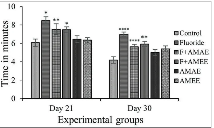
The maze test was conducted according to the method of Bromley-Brits et al.[38] Maze apparatus (60” × 30” × 15”) is used to study the ability of spatial navigation of animal, in which rats explore novel situations and make decisions based on reward (food) that produce desirable outcome. During the training period, all rats were under starvation for a period of 8–12 h and trials were conducted around 10 A.M.–2 P.M. At the beginning of each trial, animals were placed at the start point of the maze and allowed to explore the maze for 10 min. The training was given to rats for 3 days. On the day of experimentation, rats were allowed into maze task to locate the food which present at the end of the set up and noted the latency time to reach the goal. The data were exposed to statistical analysis and results (i.e., goal reaching time) were expressed in minutes.
Antioxidant markers
PND 1, 7, 14, 21, and 30 rats from all experimental groups were used for assess oxidative stress markers.
GSH-Px
Glutathione peroxidase was estimated by the method of Rotruck et al.[39]
SOD
SOD assay was carried out by the method of Marklund and Marklund.[40] This method is based on the ability of the enzyme to inhibit oxygen dependent auto-oxidation of pyrogallol. The rate of auto-oxidation is measured by noting the increase in absorbance at 420 nm.
Neurotransmitters
Neurotransmitters are assessed from PND 1, 7, 14, 21, and 30 rats of all experimental groups.
Aspartate
Aspartate was assessed by the modified method of Murai et al.[41]
GABA
GABA was assessed by the modified method of Ippolito and Piwnica.[42]
Histological studies
Histological studies were carried out from PND 1, 7, 14, 21, and 30 rats of all experimental groups.
H and E stain
H and E staining was performed by the method of Leeson et al.[43] and observed under Lawrence digital microscope.
Luxol fast blue (LFB) stain
The LFB stain is preferably used for staining myelin sheath and Nissl substance and observed under Lawrence digital microscope.[44]
Statistical Analysis
One-way analysis of variance (one-way ANOVA) to compare the means between the groups and t-test was used to determine the statistical differences between groups. Results were represented as the mean ± standard error of the mean. Significant of the data is P < 0.001.
RESULTS
Maze Learning
compound-treated rats [Figure 1]. The percent of change in time latencies in NaF treated rats as compared to control rats is 39.70% and 34.74% after 21 and 30 days. The time latencies reduced in NaF+AMAE about 24.05% and 16.40% after 21 and 30, respectively, and in NaF+AMEE about 22.38% and 14.28% in after 21 and 30 days, respectively. The percent of change in AMAE and AMEE from control is 7.13% and 5.14% in PND 21 and −3.28% and 4.05% in PND 30 (P < 0.01), respectively.
GSH-Px
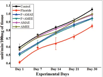
The GSH-Px activity in NaF treated rats was increased progressively with age, and its percent of change from control was about 23.28%, 23.28%, 26.92%, 24.05%, and 30.12% in PND 1, 7, 14, 21, and 30, respectively. GSH-Px activity significantly (P < 0.001) reversed in NaF+AMAE and NaF+AMEE treated groups and it was about 4.22% and 7.04% in PND 1, 8.21% and 6.84% in PND 7, 6.41% and 5.12% in PND 14, 7.59% and 6.32% in PND 21, and 4.81% and 3.61% in PND 30 rats. AMAE and AMEE treated rats showed the normal enzyme activity as compared to the control group [Figure 2].
SOD
In NaF intoxicated rats, the decreased activity of SOD was found with respect to control rats. The percentage of decrease in its activity in NaF is about −30.50%, −20.27%, −23.80%, −21.73%, and −19.04% from PND 1–30, respectively. Its activity was reverted in AMAE and AMEE administered rats toward fluoride received rats. The reverted activity of SOD in NaF+AMAE and NaF+AMEE from day 1 to day 30 by −11.86% and −15.25%, −12.16% and −13.51%, −7.14% and −9.52%, −5.43% and −7.60%, and −9.52% and −10.47% correspondingly with compared to control. The results showed that the enzyme activity was more prominently decreased in PND 1, 7, and 14 [Figure 3].
Aspartate
Aspartate levels were increased [Figure 4] in the brain tissue of NaF intoxicated rats, and it was about −42.85% in day 1, −37.50% in day 7, −44.44% in day 14, −45.45% in day 21, and −41.66% in day 30 when compared to respective age control rats. On administration of AMAE and AMEE against NaF toxicity, its levels were reversed. The percent of reversal observed in NaF+AMAE treated rats is about −28.57%, −12.50%, −11.11%, −18.18%, and −16.16% in PND 1, 7, 14, 21, and 30, respectively, and in NaF+AMEE treated rats is by −28.57%, −12.50%, −11.11%, −18.18%, and −16.66% in PND 1, 7, 14, 21, and 30 correspondingly. Ethanolic extract of seeds provides better results over aqueous extract.
GABA
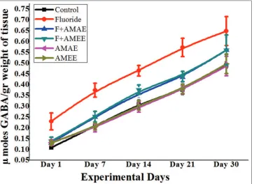
γ-aminobutyric acid levels were increased [Figure 5] in the brain tissue of NaF intoxicated rats compared to the control group. The percent of the increase in GABA levels is 53.84%, 43.47%, 22.58%, 18.42%, and 12.24% from PND 1 to PND 30 correspondingly. A. moschatus extract treatment along with NaF reverted the GABA levels. In NaF+AMAE received rats, it was about 15.38%, 12.90%, 8.16%, 10.00%, and 7.89% from PND 1 to 30, respectively. NaF+AMEE treated rats showed that the percent of reversal is about 10.00%, 10.52%, 7.69%, 9.67%, and 6.12% from day 1 to day 30, respectively. AMEE showed more protection than AMAE.
H and E stain
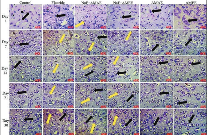
The cells of control rats’ brain tissue are with round regular shape and size and are represented by the yellow color arrow
mark. In NaF alone fed rats, the cell with irregular shape and size was observed and they were represented by black color arrow mark. In NaF+AMAE and NaF+AMEE treated some of the cells with irregular shape and size. AMAE and
AMEE treated groups showed the normal cells comparable to control. PND 14–30 day rats treated with NaF showed obvious alterations in their shape and size than PND 1 and PND 7 rats of fluoride exposed [Figure 6].
Figure 2: Protective effects of Abelmoschus moschatus extract on glutathione peroxidase activity in brain tissue of rats exposed to NaF. Results were presented as the mean ± standard error of the mean (n = 5). Significant of the data is P < 0.001.Units: Glutathione peroxidase activity was expressed as µg/mg protein
LFB Stain
Rats exposed to NaF during pre- and early post-natal periods showed the destructing myelin sheath and cells undergoing
apoptosis. The NaF+AMAE, NaF+AMEE treated rats showed the reverted myelin degeneration, and only AMAE and AMEE exposed rats displayed normal myelin sheath as compared with control rats. The myelin sheath degeneration Figure 4: Protective effects of Abelmoschus moschatus extract on aspartate levels in brain tissue of rats exposed to NaF. Results were presented as the mean ± standard error of the mean (n = 5). Significant of the data is P < 0.001. Units: Aspartate levels were expressed as µ mole of aspartate/g weight of tissue
Figure 5: Protective effects of Abelmoschus moschatus extract on Gamma-aminobutyric acid (GABA) levels in brain tissue of rats exposed to NaF. Results were presented as the mean ± standard error of the mean (n = 5). Significant of the data is
was clearly seen in PND 14–30 rats. The reason for this may be a long duration of exposure to fluoride [Figure 7] .
DISCUSSION
The present study is undertaken to evaluate the consequences of high levels of fluoride exposure during pregnancy and
lactation on neurodegeneration in young rats’ developing brain whose mothers exposed to fluoride (20 ppm) during pregnancy and on different PNDs (lactation periods) and simultaneous protective treatment of A. moschatus seed extract. The fetus is with poorly formed protective mechanisms including blood-brain barrier against xenobiotics that circulate in the maternal blood and the placenta also does not block the entry of numerous environmental toxicants Figure 6: Cerebral cortex region of rat brain stained with H and E stain. Protective effect of Abelmoschus moschatus seed extract on rat brain exposed to NaF. Normal cells with regular round shape and size were found in control rat brain and which are represented by the yellow color arrow mark. Black color arrow mark showing the cells which were with altered shape and size, swelling and undergoing necrosis were seen in NaF received a group of rats. NaF+AMAE and F+AMEE treated rat brain sections were found with normal cells (×40, Lawrence digital microscope)
from the maternal circulation to fetal circulation.[45] Mullenix et al.[46] observed that the various behavioral alterations and which were common to weanling and adult exposure and were different from those after prenatal exposures. In the present study, the NaF fed rats showed decreased learning ability and spatial navigation compared to control rats. The similar results were observed by Gao et al.,[47] they found the decreased capacity of learning and memory of rats which exposed to high fluoride levels. AMAE and AMEE treated rats showed increased learning ability than fluoride alone treated rats.
The fluoride-exposed rats resulted in reduced activity of SOD when compared to control and NaF+AMAE and NaF+AMEE treated groups. Fluoride has been reported as one of the well-known inhibitors of SOD activity.[48] Fluoride exposure during prenatal and early postpartum periods in rats showed significant decrease in SOD activity in their neural tissues with respect to the control group and the similar results were observed by Nabavi et al.,[24] in which they exposed the rats to 600 ppm of fluoride for a week through their drinking water. The decreased SOD activity in sodium fluoride administered rats result in an extra superoxide onion accumulation in rat’s neural tissue. SOD activity was reversed in NaF+AMAE and NaF+AMEE treated rats when compared to fluoride-exposed rats. Present results reported that the increased GSH-Px activity in 20 ppm fluoride received rats when compared to control group of rats. AMAE and AMEE treated rats along with sodium fluoride showed the reverted GSH-Px activity. AMAE and AMEE alone treated rats do not showed any significant differences in the activity of GSH-Px, and results were similar to control rats.
Increased free radicals from fluoride initiate oxidative stress pathway which leads to damage in the neuronal cell membranes, destruct them and in turn leads to the release and subsequent extracellular accumulation of glutamate, contributing to excitotoxicity[11] and further promoting ROS generation.[12] These effects of oxidative stress on amino acid accumulation may be due to membrane lipid peroxidation, which may impair the GABA transporter, leading to a decrease of GABA accumulation,[49] and/or it may increase the release and extracellular accumulation of aspartate and glutamate.[49] Other mechanisms that could be involved in the inhibition of glutamate uptake by ROS may be the direct oxidation of the transporter sulfhydryl groups or the impairment of Na+-K+-ATPase activity.[49] In agreement with these results, the present data showed increased levels of aspartate in NaF treated rats with compared to control rats, and its levels were reverted on the administration of AMAE and AMEE toward fluoride toxicity. In contrary to the Duarte et al.,[49] GABA levels were increased in NaF treated rats, and it was reverted on AMAE and AMEE treatment. Glutamate, aspartate is excitatory amino acid NTs and GABA also acts as an excitatory NT in developing the brain. In our earlier report, NaF received rats showed the increased levels of glutamate[13] and in this report, the increased levels of GABA and aspartate were observed in NaF treated rats. These
three NTs distributed throughout the CNS and play a very crucial role in cognition, synaptogenesis, memory, etc. Free radicals from fluoride lead to excitotoxicity which, in turn, triggered the release of glutamate, GABA, and aspartate from presynaptic terminals into extracellular space with consequent overstimulation their respective receptors. As a consequence, disturbance in GABAergic, aspartatergic and glutamatergic systems may lead to many psychological and neurodegenerative diseases.
The myelin sheath is a specialized multilayered structure and surrounds selected axons in CNS. Biochemically, myelin is formed of roughly 70–85% lipid, with a high content of cholesterol and only 15–30% of protein, especially with myelin-specific proteins.[50] Myelin degeneration occurs in CNS on chemical toxicity, traumatic brain injury, and demyelinating diseases.[51] Demyelination may induce undesirable inflammation[52] and cell death.[53] Myelin-associated proteins are released after the destruction of the intact myelin sheath.[54] After destruction of the intact myelin sheath, myelin basic protein (MBP) also disassociates from the plasma membrane and acts in a free, membrane-unbound manner in the extracellular matrix.[55]
In the present study, the NaF treated rats showed myelin destruction as compared to control rats. In NaF+AMAE and NaF+AMEE rats, the normal myelin sheath was found. Histological studies indicate myelin destruction, which is correlated with changes in MBP secondary to membrane damage and axonal degeneration on the exposure of fluoride.[56] The histopathological investigations in rat exposure to fluoride seemed thickening and disappearance of dendrites, swelling of mitochondria, dilation of the endoplasmic reticulum in neurons,[57] and diminished hippocampal synaptic interface structure[58] and the changes in the structure of synaptic interface could necessarily affect the neuronal transmission. These findings indicate that high consumption of fluoride resulted in structural and functional damages of the CNS and is associated with CNS dysfunction. In summary, developing brain is more prone to fluoride toxicity due to poorly formed protective mechanisms such as blood-brain barrier and antioxidant defense. F mediates the generation of superoxide anion (O2–) and the supplementary
production of hydrogen peroxide, peroxynitrite, and hydroxyl radicals. Exposure to NaF during pregnancy and lactation has resulted in a wide range of changes, including histopathological changes. This resulted in alterations in the levels of neurotransmitters and as consequence of behavioral alterations. A. moschatus treatment resulted in a reversal of the alterations resulted in fluoride exposure.
CONCLUSION
membrane lipids and thus destruct the myelin sheath of neural cells. These changes lead to neuronal loss, alters in the levels of neurotransmitters, and finally, behavioral alterations. A. moschatus possess antioxidants principally quercetin, rutin, catechin, epicatechin, and procyanidin and quercetin derivatives which quench free radicals and maintained lipid membranes of the cell. Thus, the normal myelin sheath was maintained in AMAE and AMEE treated rats against fluoride. The ethanol extract has shown better efficacy than aqueous extract. Yet, further studies are needed to know the exact mechanism of components neuroprotective effects of A. moschatus seed extract.
ACKNOWLEDGMENT
K. Sudhakar acknowledges to CSIR, New Delhi, India for SRF fellowship. K. Pratap Reddy acknowledges for partial funding from UGC-DSA-II and BSR mid-career one time grant.
REFERENCES
1. Hassan HA, Yousef MI. Mitigating effects of antioxidant properties of black berry juice on sodium fluoride induced hepatotoxicity and oxidative stress in rats. Food Chem Toxicol 2009;47:2332-7.
2. Shivarajashankara YM, Shivashankara AR, Bhat PG, Rao SH. Brain lipid peroxidation and antioxidant systems of young rats in chronic fluoride intoxication. Fluoride 2002;35:197-203.
3. Dong YT, Wei N, Qi XL, Liu XH, Chen D, Zeng XX, et al. Attenuating effect of Vitamin E on the deficit of learning and memory of rats with chronic fluorosis: The mechanism may involve muscarinic acetylcholine receptors. Fluoride 2017;50:354-64.
4. Wang AG, Xia T, Chu QL, Zhang M, Liu F, Chen XM, et al. Effects of fluoride on lipid peroxidation, DNA damage and apoptosis in human embryo hepatocytes. Biomed Environ Sci 2004;17:217-22.
5. Blaylock RL. Excitotoxicity: A possible central mechanism in fluoride neurotoxicity. Fluoride 2004;37:301-14.
6. Yu Y, Yang W, Dong Z, Wan C, Zhang J, Liu J, et al. Neurotransmitter and receptor changes in the brains of fetuses from areas of endemic fluorosis. Fluoride 2008;41:134-8.
7. van den Pol AN, Gao XB, Patrylo PR, Ghosh PK, Obrietan K. Glutamate inhibits GABA excitatory activity in developing neurons. J Neurosci 1998;18:10749-61. 8. Heck N, Kilb W, Reiprich P, Kubota H, Furukawa T,
Fukuda A, et al. GABA-A receptors regulate neocortical neuronal migration in vitro and in vivo. Cereb Cortex 2007;17:138-48.
9. Wang DD, Kriegstein AR. GABA regulates excitatory synapse formation in the neocortex via NMDA receptor
activation. J Neurosci 2008;28:5547-58.
10. Gao QJ, Zhang SY. The impact of fluoride on the central neurotransmitter and the relationship with impaired learning-memory. Chin J Endemiol 2001;20:315-6. 11. Saransaari P, Oja SS. Alanine release from the adult
and developing hippocampus is enhanced by ionotropic glutamate receptor agonists and cell-damaging conditions. Neurochem Res 1991;24:407-14.
12. Atlante A, Calissano P, Bobba A, Giannattasio S, Marra E, Passarella S, et al. Glutamate neurotoxicity, oxidative stress and mitochondria. FEBS Lett 2001;497:1-5. 13. Sudhakar K, Reddy KP. Protective effects of Abelmoschus
moschatus seed extract on neurotransmitter system of developingbrain of Wistar rats with gestational and post-natal exposure of sodium fluoride. Int J Green Pharm 2017;11:S1-9.
14. Nave KA, Trapp BD. Axon-glial signaling and the glial support of axon function. Annu Rev Neurosci 2008;31:535-61.
15. Love S. Demyelinating diseases. J Clin Pathol 2006;59:1151-9.
16. Reddy PY, Reddy KP, Kumar KP. Neurodegenerative changes in different regions of brain, spinal cord and sciatic nerve of rats treated with sodium fluoride. J Med Allied Sci 2011;1:30-5.
17. Raghu J, Raghuveer VC, Rao MC, Somayaji NS, Babu PB. The ameliorative effect of ascorbic acid and Ginkgo biloba on learning and memory deficits associated with fluoride exposure. Interdiscip Toxicol 2013;6:217-21.
18. Nabavi SF, Nabavi SM, Mirzaei M, Moghaddam AH. Protective effect of quercetin against sodium fluoride induced oxidative stress in rat’s heart. Food Funct 2012;3:437-41.
19. Nalagoni CS, Karnati PR. Protective effect of resveratrol against neuronal damage through oxidative stress in cerebral hemisphere of aluminum and fluoride treated rats. Interdiscip Toxicol 2016;9:78-82.
20. Al-Hayani A, Elshal EB, Aal IH, Al-Shammer E. Does Vitamin E protect against sodium fluoride toxicity on the cerebellar cortex of albino rats? Middle East J Sci Res 2013;16:1019-26.
21. Nabavi SF, Moghaddam AH, Eslami S, Nabavi SM. Protective effects of curcumin against sodium fluoride-induced toxicity in rat kidneys. Biol Trace Elem Res 2012;145:369-74.
22. Khandare AL, Kumar PU, Lakshmaiah N. Beneficial effect of tamarind ingestion on fluoride toxicity in dogs. Fluoride 2000;33:33-8.
23. Reddy MM, Reddy KP. Protective effects of aqueous extract of fruit pulp of Tamarindus indica on motor activity and metabolism of the gastrocnemius muscle of rats treated with fluoride. Int J Toxicol Pharmcol Res 2015;7:241-8.
Chem 2012;142:79-82.
25. Bruce AJ, Malfroy B, Baudry M. Beta-amyloid toxicity in organotypic hippocampal cultures: Protection by EUK-8, a synthetic catalytic free radical scavenger. Proc Natl Acad Sci U S A 1996;93:2312-6.
26. Bastianetto S, Ramassamy C, Christen Y, Poirier J, Quirion E. Ginkgo biloba extract (EGb 761) protects in vitro rat hippocampal cells against toxicity induced by b-amyloid peptides. Soc Neurosci 1998;24:1456. 27. Lai XY, Zhao YY, Liang H. Studies on chemical
constituents in flower of Abelmoschus manihot. Zhongguo Zhong Yao Za Zhi 2006;31:1597-600. 28. Lai X, Liang H, Zhao Y, Wang B. Simultaneous
determination of seven active flavonols in the flowers of Abelmoschus manihot by HPLC. J Chromatogr Sci 2009;47:206-10.
29. Jain PS, Bari SB. Isolation of stigmasterol and γ-sitosterol from petroleum ether extract of woody stem of Abelmoschus manihot. Asian J Biol Sci 2009;2:112-7. 30. Xian YL, Zhao YY, Liang H. A flavonoid glucuronide
from Abelmoschus Manihot (L.) medik. Biochem Syst Ecol 2007;35:891-3.
31. Mollik MA, Hossan MS, Paul AK, Taufiq-Ur-Rahman M, Jahan R, Rahmatullah M. A comparative analysis of medicinal plants used by folk medicinal healers in three districts of Bangladesh and inquiry as to mode of selection of medicinal plants. Ethnobotany Res Appl 2010;8:195-218.
32. Jadeja BA, Nakar RN. Study on ethno-medico botany of weeds from saurashtra region, Gujarat, India. Plant Arch 2010;10:761-5.
33. Lans C. Comparison of plants used for skin and stomach problems in Trinidad and Tobago with Asian ethnomedicine. J Ethnobiol Ethnomed 2007;3:3. 34. Du Z, Clery RA, Hammond CJ. Volatile organic
nitrogen-containing constituents in ambrette seed Abelmoschus moschatus Medik (Malvaceae). J Agric Food Chem 2008;56:7388-92.
35. Sudhakar K, Nageshwar M, Reddy KP. Seed extract of Abelmoschus moschatus Medik reverses NaF-induced behavioral changes through neurodegeneration and oxidative stress in brain of rat. Asian J Pharm Clin Res 2017;10:165-71.
36. Sudhakar K, Nageshwar M, Reddy KP. Protective effect of okra, Abelmoschus moschatus seed extract on developing brain of rats during pre- and post-natal fluoride exposure. Int J Pharm Sci Res 2018;9:1519-28. 37. Sudhakar K, Nageshwar M, Reddy KP. Abelmoschus
moschatus extract reverses altered pain and neurohistology of a rat with developmental exposure of fluoride. J Appl Pharm Sci 2018;8:94-104.
38. Bromley-Brits K, Deng Y, Song W. Morris water maze test for learning and memory deficits in Alzheimer’s disease model mice. J Vis Exp 2011;53:e2920.
39. Rotruck JT, Pope AL, Ganther HE, Swanson AB, Hafeman DG, Hoekstra WG, et al. Selenium: Biochemical role as a component of glutathione peroxidase. Science
1973;179:588-90.
40. Marklund S, Marklund G. Involvement of the superoxide anion radical in the autoxidation of pyrogallol and a convenient assay for superoxide dismutase. Eur J Biochem 1974;47:469-74.
41. Murai S, Saito H, Abe E, Masuda Y, Itoh T. A rapid assay for neurotransmitter amino acids, aspartate, glutamate, glycine, taurine and gamma-aminobutyric acid in the brain by high-performance liquid chromatography with electrochemical detection. J Neural Transm Gen Sect 1992;87:145-53.
42. Ippolito JE, Piwnica-Worms D. A fluorescence-coupled assay for gamma aminobutyric acid (GABA) reveals metabolic stress-induced modulation of GABA content in neuroendocrine cancer. PLoS One 2014;9:e88667. 43. Leeson CR, Leeson TS, Paparo AA. Text Book of
Histology. 5th ed. Philadelphia, PA: WB. Saunders
Company; 1985.
44. Bancroft JD, Marilyn G. Theory and Practice of Histological Techniques. 6th ed. Oxford: Churchill
Livingstone Elsevier; 2008.
45. Needham LL, Grandjean P, Heinzow B, Jørgensen PJ, Nielsen F, Patterson DG Jr, et al. Partition of environmental chemicals between maternal and fetal blood and tissues. Environ Sci Technol 2011;45:1121-6.
46. Mullenix PJ, Kernan WJ, Tassinari MS, Schunior A. Generation of dose response data using activity measures. J Am Coll Toxicol 1989;8:185-97.
47. Gao Q, Liu Y, Guan Z. Decreased learning and memory ability in rats with fluorosis: Increased oxidative stress and reduced cholinesterase activity in the brain. Fluoride 2009;42:277-85.
48. Guo X, Sun G, Sun Y. Oxidative stress from fluoride-induced hepatotoxicity in rats. Fluoride 2003;36:25-9. 49. Duarte AI, Santos MS, Seiça R, Oliveira CR. Oxidative
stress affects synaptosomal gamma-aminobutyric acid and glutamate transport in diabetic rats: The role of insulin. Diabetes 2004;53:2110-6.
50. Boggs JM. Myelin basic protein: A multifunctional protein. Cell Mol Life Sci 2006;63:1945-61.
51. Vallières N, Berard JL, David S, Lacroix S. Systemic injections of lipopolysaccharide accelerates myelin phagocytosis during wallerian degeneration in the injured mouse spinal cord. Glia 2006;53:103-13.
52. Jeon SB, Yoon HJ, Park SH, Kim IH, Park EJ. Sulfatide, a major lipid component of myelin sheath, activates inflammatory responses as an endogenous stimulator in brain-resident immune cells. J Immunol 2008;181:8077-87.
53. Hoffmann K, Lindner M, Gröticke I, Stangel M, Löscher W. Epileptic seizures and hippocampal damage after cuprizone-induced demyelination in C57BL/6 mice. Exp Neurol 2008;210:308-21.
diseases. Ann Neurol 1985;18:324-8.
55. Liu MC, Akle V, Zheng W, Kitlen J, O’Steen B, Larner SF, et al. Extensive degradation of myelin basic protein isoforms by calpain following traumatic brain injury. J Neurochem 2006;98:700-12.
56. Kamel OA. Effect of sodium fluoride on the cerebellar cortex of adult albino rats and the possible protective role of Vitamin B6: A light and electron microscopic study. Egypt J Histol 2009;32:358-67.
57. Shashi A. Histopathological investigation of fluoride induced neurotoxicity in rabbits. Fluoride 2003;36:95-105.
58. Zhang Z, Xu X, Shen X. Effect of fluoride exposure on synaptic structure of brain areas related to learning-memory in mice. Fluoride 2008;41:139-43.