IJPSR (2018), Volume 9, Issue 4 (Research Article)
Received on 12 July, 2017; received in revised form, 13 September, 2017; accepted, 17 September, 2017; published 01 April, 2018
PROTECTIVE EFFECT OF OKRA, ABELMOSCHUS MOSCHATUS SEED EXTRACT ON DEVELOPING BRAIN OF RATS DURING PRE- AND POST-NATAL FLUORIDE EXPOSURE
Kurmeti Sudhakar, Mesram Nageshwar and K. Pratap Reddy*
Department of Zoology, Osmania University, Hyderabad - 500007, Telangana, India.
ABSTRACT: This study reports protective effect of Abelmoschus moschatus seed extract against sodium fluoride (NaF) induced alterations in behavior correlated with neurochemical changes in developing brain of rats. Excessive intake of fluoride during pregnancy cross the blood brain barrier (BBB) and cause adverse effects on neonatal development. As the BBB in fetuses, neonates and infants is immature, it cannot provide protection against the entries. The pregnant wistar rats were randomly categorized into six groups of five animals each. Group I is of control rats received normal tap water. Group II is NaF exposed group with 20 ppm (or 20 mgkg-1 body wt.) in their drinking water. Group III rats were treated with A. moschatus aqueous extract (AMAE) (300 mgkg-1 body wt./day/rat) along with NaF water (20 ppm). Group IV rats were treated with A. moschatus ethanolic extract (AMEE) (300 mgkg-1 body wt./day/rat) along with NaF water (20 ppm).Group V and VI rats were treated with AMAE (300 mgkg-1 body wt./day/rat), and AMEE (300 mgkg-1 body wt./day/rat) respectively. On 1st, 7th, 14th, 21st and 30th day (postpartum days), the pups were sacrificed to assess oxidative stress markers (LPO, CAT), and measured body weight, brain weight, BSI, and estimated protein content of brain tissue of all experimental groups. On post-natal day 21 and day 30 pups behavioral activity (rota rod, hot plate test) was measured. Fluoride exposure significantly increased lipid peroxidation and decreased the activity of catalase, and decreased body and brain weight, BSI and also protein content of the brain of pups indicating oxidative stress and inhibited antioxidant system and protein synthesis which were reverted on administration of AMAE and AMEE against NaF intoxication. The altered behavioral responses on NaF exposure were also reversed to that of control. Hence, this study proves the vulnerability of developing brain to fluoride toxicity during development and growth and protection offered by AMAE and AMEE towards neurotoxicity of NaF.
INTRODUCTION: Prolonged ingestion of fluoride through various sources, mainly drinking water caused fluorosis 1. Fluorosis, as a global public health problem, has been receiving wide and deserving attention in recent years.
QUICK RESPONSE CODE
DOI:
10.13040/IJPSR.0975-8232.9(4).1519-28
Article can be accessed online on:
www.ijpsr.com
DOI link: http://dx.doi.org/10.13040/IJPSR.0975-8232.9(4).1519-28
Prolonged consumption of fluoride in excess during gestation cause adverse effects on neonatal development 2. The mechanisms of maternal fetal transmission of fluoride are poorly understood.
Therefore, the present study focused on maternal exposure of fluoride and it’s effects on developing CNS and it’s amelioration with extract of okra seeds. Fluoride is known to cross the placenta 3, 4 and the blood brain barrier 5 and ultimately increase fluoride levels in fetal brain tissue. The blood brain barrier in the embryo, fetus, and newborn is “immature” that means it is poorly developed,
Keywords:
Sodium fluoride, Oxidative stress, Neurotoxicant,
Abelmoschus, Behavior
Correspondence to Author: Prof. K. Pratap Reddy
Professor,
Department of Zoology,
Osmania University, Hyderabad - 500007, Telangana, India.
leaky, or even absent, rendering the developing brain more vulnerable to fluoride entering the fetal circulation from the mother 6. Once it enter into the brain it can cause adverse effects on the brain cell ultra structure, metabolism, enzymes, the oxidant-antioxidant status and on neurotransmitters and thus overall metabolism of brain 7, 8. The maternal exposure to fluoride and fluoride insobriety at the early periods of life is shown to cause more prominent effects on the oxidant-antioxidant status in the brain than the fluoride exposure at the later stages of life 9, 10. Fluoride increases production of reactive oxygen species (ROS), lipid peroxidation, and decreased catalase activity which have been considered in pathogenesis and recent studies suggest that oxidative stress is a possible pathologic mediating factor in fluoride toxicity 11, 12.
Exposure to fluoride during early developmental stages of life results in long-term irreversible consequences with their structure and function and account for qualitative differences in age related susceptibility 13. The neuronal growth-spurting in rats occurs in the first 3 weeks of postpartum 14. During this period the brain undergoes many profound structural and functional transformations, which makes vulnerable to fluoride 15.
The high intake of plant products is associated with a reduced risk of a number of chronic diseases 16. Earlier reports on natural products which aid in protection against NaF induced behavioral alterations are: Curcumin 17, Quercetin 18, Rutin (extracted compound of Okra) 19, Vitamin A 20, and
Spirulina platensis 21. These natural products reverse behavioral alterations which are induced on NaF exposure. These beneficial effects have been moderately attributed to the compounds which possess antioxidant activity.
Abelmoschus moschatus L., (Okra) is one of the most commonly known and utilized species of the family Malvaceae. The major antioxidants of vegetables are vitamins C and E, carotenoids, and phenolic compounds, especially flavonoids 22. Nutritionally, the richest part of the okra plant is the dried seed. Okra seed is rich in high quality protein especially with regard to its essential amino acids 22 and unsaturated fatty acids such as linoleic acid 22. Seeds are rich in phenolic compounds with derivatives, catechin oligomers and
hydroxyl-cinnamic derivatives. The nutrients content of Okra seed showed that 21% protein, 14% lipids and 5% ash. Proteins play a particularly important role in human nutrition 22. Okra is a popular health food due to its high fiber, Vitamin C, and folate content and also a good source of calcium and potassium. High fiber, “helps to stabilize blood sugar by regulating the rate at which sugar is absorbed from the intestinal tract” 23
. Okra seeds will make available the essential energy to the body and important antioxidants that could boost immune system and prevent diseases 24. The neuroprotectant effects of extract of Okra against fluoride induced behavior and neurodegenerative changes in brain of adult rat were reported in our earlier studies 25. In view of this, the efficacy of AMAE and AMEE in reducing fluoride induced neurobehavioral, and neurochemical alterations in rats of pre and post-natal fluoride exposed is reported in this paper.
MATERIAL AND METHODS: Healthy albino wistar rats (Rattus norvegicus) weighted between 180 to 200 gm were assigned randomly to experimental groups and housed 2 females and 1 male for cage (polypropylene cages) to allow breed. Light cycles were maintained as 12 Light: 12 Dark hours (6.00 AM to 6.00 PM), and temperature between 22 ± 2oC. The rats were maintained on standard diet and water was supplied
ad libitum. After their pregnancy was confirmed, from the first day pregnancy, the females made into six groups and allowed to drink on fluoridated water at the rate of 20 ppm NaF and also
Abelmoschus moshatus seed extract was given to the rats at the rate of 300 mgkg-1 body weight as follows;
Experimental Groups:
Group I: Received normal tap water
Group II: Fed on fluoridated drinking water (20ppm)
Group III: NaF (20 ppm) + Abelmoschus
moschatus seed aqueous extract (AMAE)
(300mgkg-1 b. wt.)
Group IV: NaF (20 ppm) + Abelmoschus moschatus seed ethanolic extract (AMEE) (300mgkg-1 b. wt.)
Group VI: Abelmoschus moschatus seed ethanolic extract (AMEE) (300 mgkg-1 b. wt.) only.
Chemicals: Potassium dichromate, glacial acetic acid, Thiobarbituric acid (TBA), H2O2, sodium carbonate, sodium potassium tartrate, Folin reagent
etc. were purchased from local laboratory chemical suppliers.
Physical Parameters:
Body Weight: The rat pups were weighted at the age of post-natal Day 1, Day 7, Day 14, Day 21 and Day 30 and noted down the readings.
Brain Weight: The same pups were killed by decapitation and their brains were dissected out. These brains weighted and noted down the values.
Brain Somatic Index (BSI): For calculating the brain - somatic index, the weight of the individual rat was noted and the brain was removed carefully and weighed on an electronic weigh balance after removing the blood with tissue paper. BSI was calculated by using the value of brain weight of control rat divided by the same rat body weight value and multiplied with 100 (brain weight / body weight × 100). This formula was applied to all experimental groups to calculate BSI. The Brain - Somatic Index of the rat pups were calculated by the use of equation cited by Ashwini 26.
Brain Somatic Index = Brain weight Body weight
Behavioral Assessments:
Rota Rod: The rota rod test, in which rat pups must balance on a rotating rod, is widely used to assess motor deficit in neurodegenerative
disease models (in rodents). Performance is measured
by the duration that an animal stays on the rod as a function of rotating rod speed. In the present study, the instrument (DolphinTM instruments) had incremental fixed-speed (about 30 rpm) rotating rod. Before experimental testing, rat pups were trained to run on the rotating rod in 3 training trials per day for three consecutive days with a constant speed of 30 rpm. For this purpose, animals were kept on the rotating rod for 2 min, and time of their first falling off and the frequency of falling off the rod are recorded for each rat. After the training period, on the day of testing, the performance of
the rats was measured as maximal time spent on the rod at 30 rpm before falling off and endurance time noted in min 27.
Hot Plate Test: The hot plate test is used for evaluating thermal pain sensitivity. The hot plate test evaluates thermal pain reflexes due to footpad contact with a heated surface. An apparatus consisting of an aluminium plate that is heated and cooled by Peltier elements in contact with its lower surface. During the experiment, the rat was introduced into Remi hot plate kept at 52 ± 0.5 °C. Hot-plate latencies are determined as described by placing each rat pup on a hot plate and observing the occurrence of a nociceptive response (licking of a hind paw, jumping). The time of exhibiting these behaviors was noted as response time and recorded in seconds. To observe the rat behavior, observation area covered with a colourless plastic cylinder was placed on the hot-plate. In order to avoid possible tissue injury, a cut-off time of 12 seconds was used to avoid skin damage. One hot plate test was carried out for each rat 28.
Oxidative Stress Markers: Lipid per-oxidation and catalase were assessed.
Lipid per-oxidation (LPO): The LPO was assessed by the method of Bhuyan 29. This assay is based on the reactivity of an end product of lipid peroxidation, malondialdehyde (MDA) with TBA to produce a pink adduct, which is measured at 533 nm on spectrophotometer. The Levels of lipid peroxidation products expressed as µmol of MDA/gr. weight of tissue.
Catalase: Catalase activity was estimated by measuring the rate of decomposition of H2O2. Disappearance of peroxide in the presence of enzyme source is the basis for assay which is followed spectrophotometrically at 505 nm. One unit decomposes one micromole of H2O2 per minute 30.
Protein Estimation of Brain: Brain tissue total protein content was estimated by the method of Lowry 31. The principle of Lowry method is the peptide nitrogen(s) with the copper [II] ions under alkaline conditions and the subsequent reduction of
the Folin - Ciocalteau phosphomolybdic
acids. The color was read at 540 nm. Protein content in the brain tissue expressed as mg of protein / gm weight of tissue.
Per cent of change between groups was calculated by the formula of;
% change = Experimental – Control Control
Data Analysis: Statistical significance was determined by one way analysis of variance. Differences between means were determined by t-test.
RESULTS:
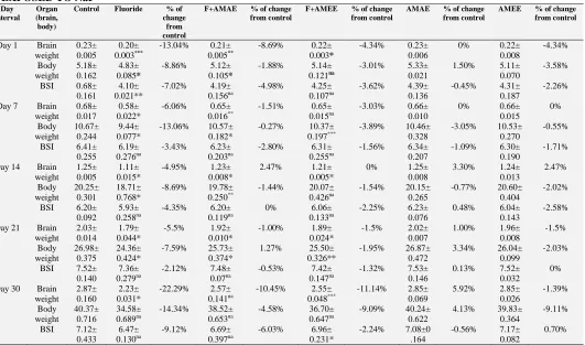
Body Weight: The body weight was progressively decreased (Table 1) in NaF treated rats from post-natal day 1 to post-post-natal day 30 rats as compared to control. Among these experimental days the maximum per cent (-14.34%) of decreased body weight was recorded in post-natal day 30 NaF treated one.
[image:4.612.47.578.280.593.2]Furthermore, the reverted body weight was noticed in F+AMAE and F+AMEE compared to fluoride alone treated group of respective age and it was similar to control group.
TABLE 1: EFFECT OF ABELMOSCHUS MOSCHATUS SEED EXTRACT ON BODY WEIGHT, BRAIN WEIGHT,
BRAIN – SOMATIC INDEX (BSI) OF RAT PUPS (FROM POST-NATAL DAY 1 TO POST - NATAL DAY 30) EXPOSED TO NaF
Day interval
Organ (brain, body)
Control Fluoride % of change
from control
F+AMAE % of change from control
F+AMEE % of change from control
AMAE % of change from control
AMEE % of change from control
Day 1 Brain weight
0.23± 0.005
0.20± 0.003***
-13.04% 0.21± 0.005**
-8.69% 0.22± 0.003*
-4.34% 0.23± 0.006
0% 0.22± 0.008 -4.34% Body weight 5.18± 0.162 4.83± 0.085*
-8.86% 5.12± 0.105*
-1.88% 5.14± 0.121na
-3.01% 5.33± 0.021
1.50% 5.11± 0.070
-3.58% BSI 0.68±
0.161
4.10± 0.021**
-7.02% 4.19± 0.156na
-4.98% 4.25± 0.107na
-3.62% 4.39± 0.136
-0.45% 4.31± 0.187
-2.26% Day 7 Brain
weight
0.68± 0.017
0.58± 0.022*
-6.06% 0.65± 0.016**
-1.51% 0.65± 0.015na
-3.03% 0.66± 0.010
0% 0.66± 0.015 0% Body weight 10.67± 0.244 9.44± 0.077*
-13.06% 10.57± 0.182*
-0.27% 10.37± 0.197***
-3.89% 10.46± 0.328
-3.05% 10.53± 0.270
-0.55% BSI 6.41±
0.255
6.19± 0.276na
-3.43% 6.23± 0.203na
-2.80% 6.31± 0.255na
-1.56% 6.34± 0.207
-1.09% 6.30± 0.190
-1.71% Day 14 Brain
weight
1.25± 0.005
1.11± 0.015*
-4.95% 1.23± 0.008*
2.47% 1.21± 0.005*
0% 1.25± 0.008
3.30% 1.24± 0.013 2.47% Body weight 20.25± 0.301 18.71± 0.768*
-8.69% 19.78± 0.250**
-1.44% 20.07± 0.426na
-1.54% 20.15± 0.265
-0.77% 20.60± 0.404
-2.02% BSI 6.20±
0.092
5.93± 0.258na
-4.35% 6.20± 0.119na
0% 6.06± 0.133na
-2.25% 6.23± 0.076
0.48% 6.04± 0.143
-2.58% Day 21 Brain
weight
2.03± 0.014
1.79± 0.044*
-5.5% 1.92± 0.010*
-1.00% 1.89± 0.024*
-1.5% 2.02± 0.007
1.00% 1.96± 0.008 -1.5% Body weight 26.98± 0.375 24.36± 0.424*
-7.59% 25.73± 0.374*
1.27% 25.50± 0.326**
-1.95% 26.87± 0.472
3.34% 26.04± 0.099
-2.03% BSI 7.52±
0.140
7.36±
0.279na -2.12% 7.48± 0.07na -0.53% 0.1477.42± na -1.32% 7.53± 0.146 0.13% 7.52± 0.032 0% Day 30 Brain
weight
2.87± 0.160
2.23± 0.031*
-22.29% 2.57± 0.141na
-10.45% 2.55± 0.048***
-11.14% 2.85± 0.069
5.92% 2.85± 0.026 -1.39% Body weight 40.37± 0.716 34.58± 0.689na
-14.34% 38.52± 0.653na
-4.58% 36.70± 0.647na
-9.09% 40.24± 0.622
4.13% 39.83± 0.364
-9.11% BSI 7.12±
0.433
6.47±
0.130na -9.12% 0.3976.69± na -6.03% 0.231* 6.96± -2.24% 7.08±0.164 -0.56% 7.17± 0.082 0.70% Percentage of decreased body weight, brain weight and BSI of all experimental groups compared with control was presented. There is no significant change in case of mean value of control group and experimental group. Data presented as mean ± S.E.M. (n = 5). Data exposed to one-way ANOVA and t-test to determine the statistical differences between groups. t-test was conducted in all possible combinations to compare significance of data. Superscript symbols indicated significant differences observed from experimental groups; * denotes the p-value of P < 0.01, ** for the p-value of P < 0.05, *** for the p-value of P < 0.001, and na represent non-significant difference between groups.
Brain Weight: The brain weight was decreased in NaF intoxicated rats when compared to control as shown in the Table 1. But there is no uniform decrease in the brain weight of experimentally treated rats. The reverted brain weight was reported in F+AMAE and F+AMEE compared to fluoride alone treated group of respective age. AMAE and
AMEE received rats brain weight is similar to control.
when compared to control group of respective aged pups. Among these day 30 post-natal rats showed the highest per cent of decreased BSI. AMAE and AMEE treated groups against NaF exposed pups showed the reversed BSI as compared to NaF group.
Furthermore, AMAE and AMEE alone treated groups BSI is same as that of control.
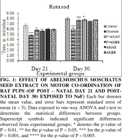
Rota Rod Test: Decreased latencies and repeated fall offs (Fig. 1) from the rotating rod of rota rod apparatus were noted down in NaF intoxicated pups than control. In day 21 experimental groups the per cent of decreased retention time is -21.69% in NaF and similarly, in day 30 it is decreased by -25.52% when compared to respective control group.
It was increased in AMAE and AMEE treated protective groups against NaF as compared to NaF alone treated. AMEE treated group exhibited superior efficacy than AMAE treated group.
FIG. 1: EFFECT OF ABELMOSCHUS MOSCHATUS SEED EXTRACT ON MOTOR CO-ORDINATION OF RAT PUPS (OF POST – NATAL DAY 21 AND POST-
NATAL DAY 30) EXPOSED TO NaF) Each bar denotes
the mean value, and error bars represent standard error of mean (n = 5). Data exposed to one-way ANOVA and t-test to determine the statistical differences between groups. Superscript symbols indicated significant differences observed from experimental groups; * denotes the p-value of P < 0.01, ** for the p-value of P < 0.05, *** for the p-value of P < 0.001, and **** for the p-value of P < 0.005.
Hot Plate Test: The threshold intensity of stimulus for thermal sensitivity was conspicuously increased (P < 0.001) in NaF administered group (Fig. 2) by 44.38% in Day 21 pups with compared to control pups of respective age group and it was 48.45% on Day 30 rats. The response to thermo nociceptive
pain in AMAE and AMEE treated groups towards NaF toxicity was found to be reversed. AMEE treated group showed better efficacy than AMAE treated group.
FIG. 2: EFFECT OF ABELMOSCHUS MOSCHATUS
SEED EXTRACT ON THERMOCEPTION OF RAT PUPS (OF POST-NATAL DAY 21 AND POST-NATAL DAY 30) EXPOSED TO NaF
Each bar denotes the mean value, and error bars represent standard error of mean (n = 5). Data exposed to one-way ANOVA and t-test to determine the statistical differences between groups. Superscript symbols indicated significant differences observed from experimental groups; * denotes the p-value of P < 0.01, *** for the p-value of P < 0.001 and **** for the p-value of P < 0.005.
Oxidative Stress Markers: Lipid per-oxidation and catalase were assessed.
Lipid Per-oxidation (LPO): Significant increased LPO levels were found in brain tissues of NaF exposed pups of all age groups when compared to control group of respective age. Noticeably diminished levels of LPO were found in AMAE and AMEE towards NaF fed rats. AMAE exhibited better protective effect than AMEE. (Table 2)
Catalase: Reduced catalase activity was noticed in the NaF received rats with compared to control. Simultaneous treatment of AMAE and AMEE restored the catalase activity. AMAE presented better result than AMEE. (Table 3)
[image:5.612.314.565.121.292.2] [image:5.612.48.299.371.655.2]TABLE 2: EFFECT OF ABELMOSCHUS MOSCHATUS SEED EXTRACT ON LPO LEVELS IN BRAIN TISSUE OF RAT PUPS (FROM POST-NATAL DAY 1 TO POST-NATAL DAY 30 WITH ONE WEEK INTERVAL) EXPOSED TO NaF
Control Fluoride % of change from control
F+AMAE % of change from control
F+AMEE % of change
from control
AMAE % of change
from control
AMEE % of change from control
Day 1 0.84± 0.023
1.19± 0.022***
41.66% 0.95± 0.036***
13.09% 1.07± 0.01***
27.38% 0.85± 0.026
1.19% 0.85± 0.03
1.19% Day 7 0.86±
0.022
1.14± 0.045****
33.73% 1.07± 0.051***
24.41% 1.08± 0.059*
25.58% 0.84± 0.016
1.20% 0.85± 0.029
2.40% Day 14 1.05±
0.057
1.32± 0.038*
25.71% 1.20± 0.054*
14.28% 1.20± 0.041*
14.28% 1.00± 0.03
-4.76% 1.01± 0.02
-3.80% Day 21 1.21±
0.065
1.57±
0.062*** 29.75% 0.0241.34± * 10.74% 0.0241.32± * 9.09% 1.21± 0.05 0% 1.19± 0.048 -1.65% Day 30 1.26±
0.03
1.60± 0.062*
26.98% 1.38± 0.031*
9.52% 1.40± 0.014***
11.11% 1.31± 0.034
3.96% 1.32± 0.042
4.76% Data presented as mean ± S.E.M. (n = 5). Data exposed to one-way ANOVA and t-test to determine the statistical differences between groups. Superscript symbols indicated significant differences observed from experimental groups; * denotes the value of P < 0.01, *** for the p-value of P < 0.001, **** for the p-p-value of P < 0.005. LPO levels expressed as Nano-mole MDA/gm weight of tissue.
TABLE 3: EFFECTS OF ABELMOSCHUS MOSCHATUS SEED EXTRACT ON CATALASE ACTIVITY IN BRAIN TISSUE OF RAT PUPS (FROM POST-NATAL DAY 1 TO POST-NATAL DAY 30 WITH ONE WEEK INTERVAL) EXPOSED TO NaF
Control Fluoride % of change
from control
F+AMAE % of change from control
F+AMEE % of change
from control
AMAE % of change from control
AMEE % of change
from control
Day 1 0.37± 0.019
0.27± 0.023*
-27.02% 0.34± 0.019***
-8.10% 0.30± 0.017*
-18.91% 0.36± 0.01
-2.70% 0.35± 0.007
-5.40% Day 7 0.45±
0.023
0.35± 0.012***
-22.22% 0.39± 0.026**
-13.33% 0.41± 0.025*
-8.88% 0.43± 0.023
-4.44% 0.42± 0.025
-6.66% Day 14 0.48±
0.026
0.36± 0.028***
-25.00% 0.43± 0.026*
-10.41% 0.41± 0.03*
-14.58% 0.47± 0.029
-2.08% 0.46± 0.029
-4.16% Day 21 0.57±
0.027
0.44± 0.022***
-22.80% 0.49± 0.025*
-14.03% 0.49± 0.026*
-14.03% 0.57± 0.037
0% 0.55± 0.027
-3.50% Day 30 0.61±
0.029
0.48± 0.04***
-21.31% 0.57± 0.026*
-6.55% 0.55± 0.028*
-9.83% 0.59± 0.03
-3.27% 0.59± 0.029
-3.27% Data presented as mean ± S.E.M. (n = 5). Data exposed to one-way ANOVA and t-test to determine the statistical differences between groups. Superscript symbols indicated significant differences observed from experimental groups; * denotes the p-value of P < 0.01, ** for the p-value of P < 0.05, *** for the p-value of P < 0.001. CAT activity expressed as µ mole/min/mg of tissue.
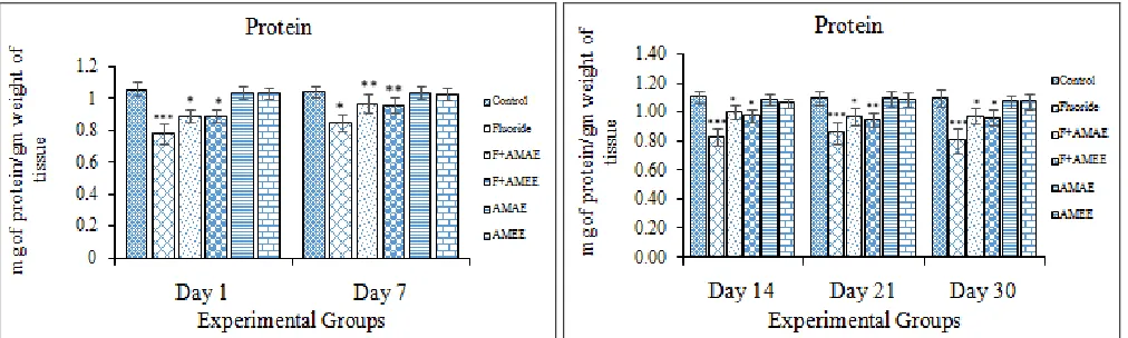
FIG. 3: EFFECT OF ABELMOSCHUS MOSCHATUS SEED EXTRACT ON PROTEIN CONTENT OF BRAIN TISSUES OF RAT PUPS (FROM POST-NATAL DAY 1 TO POST-NATAL DAY 30 WITH ONE WEEK INTERVAL) EXPOSED TO NaF
Each bar denotes the mean value, and error bars represent standard error of mean (n = 5). Data exposed to one-way ANOVA and t-test to determine the statistical differences between groups. Superscript symbols indicated significant differences observed from experimental groups; * denotes the p-value of P < 0.01, ** for the p-value of P < 0.05, *** for the p-value of P < 0.001.Units: protein content in the brain tissue expressed as mg of protein/gm weight of tissue.
DISCUSSION: The present study demonstrated
that the protective effects of Abelmoschus moschatus seed extract against pre- and post-natal NaF fluoride induced behavioral and neurochemical milieu in albino wistar rat. Fetus is not well protected against fluoride that circulates in the
[image:6.612.40.574.252.398.2] [image:6.612.55.561.438.590.2]Further, the research reports also demonstrated that the fluoride also transferred to the infant through human breast milk 32. That means during foetal life and early infancy, the blood brain barrier provides only partial protection against the entry of chemicals into the central nervous system 33, 34. Thus the fetus and new born pups are much vulnerable to fluoride. In the present research, the results have shown that exposure to 20 ppm of NaF during pre- and post-natal and lactation period affect the body weight, brain weight, and brain somatic index, and decreased behavioral response measured on rota rod and hot plate apparatus. And also found that the increased levels of LPO levels and decreased activity of catalase and protein content of brain tissues of fluoride intoxicated rat pups. These all were reversed on treatment of AMAE and AMEE along with NaF.
Excessive fluoride ingestion significantly decreased animal growth evidenced by the depressed appetite, decrease in the rate of feed and water consumption that eventually lead to the poor growth rate in pups, witnessed by decrease in the body weight, brain
weight and BSI 8. The ingested fluoride not only
disturbed nutrient digestibility (absorption) but also attributed to weight loss, degeneration of structure of organs, and decreased protein and lipid levels, which may be the prime factor in causing fluoride
neuro-toxicity. Decreased organ and body weight
was noticed on NaF treatment in group of animals 35
. And also decreased organo-somatic index (OSI) with loss of body weight and decreased protein content was found in brain tissue.
Shashi 36 found significant decreased in acidic, basic, and total proteins in rabbits treated with NaF for 100 days. A decline in protein content occurred in various soft tissues of rodents treated with different doses of NaF after 30 to 70 days 37. The results showed that the administration of AMAE and AMEE along with NaF showed the increased
body weight, brain weight and BSI. Treatment with
the 60 mg/kg of quercetin, rutin or okra showed a significant reversal in their body weights when compared with the dexamethasone treated group 38. The phyto-chemical studies showed that the okra pods contains high ash content which would provide essential valuable and useful minerals needed for body growth 39. A. esculentus peel and seed powder increases body weight 40.
Behavioral assays are important in toxicology, teratology, phenotype screening, and in other applications that require animals’ functional status to be measured. When assessment involves infant animals, it can be especially challenging because an immature animal is often more fragile, prone to fatigue, has limited sensory and motor capabilities, and may be differentially responsive to standard challenges such as water or food deprivation. Fluoride exposed rat pups showed the decreased latencies and frequent falls from the rod with compared to control group pups. On the contrary, F+AMAE, and F+AMEE treated group of pups were showed increased latencies and infrequent falls from the rod. And AMAE and AMEE alone treated pups were displayed similar latency and fall off times of controlled pups.
These results demonstrating that the higher centers of brain were protected from fluoride-induced damage. The significant decline in endurance time of fluoride-exposed animals compared with the control. This is presumably due to its negative impact of fluoride on the cerebellum 21. An inhibition of spontaneous motor activity and rota-rod performance in adult rats 21, 41 and mice 42 treated with NaF for 60 days indicates that motivation of these animals has been impaired by fluoride.
A deficiency in these behaviors may lead to suppression of eating process. Thus a behavioral impairment has been accounted by these investigators for diminished food ingestion in rats and mice treated chronically with NaF. Thus NaF exposed pups showed decreased body weight, brain weight and also BSI. F-induced inhibition of motivated behavior has been accompanying with the locomotor behavioral deficit that occurred in workers who have chronic exposure to high level of industrial F 43. Moreover, increased time to respond thermoceptive pain on hot plate was noted in NaF intoxicated pups which was reverted in AMAE and AMEE treated rats. Fluoride exposure during gestational period significantly exacerbated the levels of LPO along with the decreased activities of antioxidant enzyme like catalase.
decreased activities of antioxidant enzymes 45. The brain is susceptible to oxidative stress. This is because of the presence of high levels of polyunsaturated fatty acids, relatively low antioxidant capacity, the presence of redox metal ions and high oxygen utilization 2, 46. Further, the oxidative stress not only increases free radical damage but also boosts excito-toxicity, since ROS, reactive nitrogen species and lipid peroxidation products can trigger the excito-toxic process 47, 48. Administration of natural plant extracts to fluoride intoxicated pups alleviated the altered cascade of free radical damage and / or oxidative stress 2. AMAE and AMEE along with NaF treated pups showed the reversal levels of LPO and catalase activity. Free radical scavenging activity is the most important mechanism by which antioxidants inhibit lipid peroxidation 49. The present results stand evident for fluoride activity was decreased with increase in the age. This is because of either increasing complexity of BBB with respect to the age of rats or simultaneous administration of AMAE and AMEE, nullifying production of free radicals from fluoride. Inhibition in the docking phenomenon of free radical and excitotoxicity of NaF by antioxidant properties of AMAE and AMEE produces a decline in NaF toxicity.
The antioxidant activity of phenolics (AMAE and AMEE) is mainly due to their redox properties, which allow them to act as reducing agents, hydrogen donors and singlet oxygen quenchers 50. Natural polyphenols exert their advantageous health properties by their antioxidant activity, these compounds are capable of eliminating free radicals, chelate metal catalysts, activate antioxidant enzymes, reduce α- tocopherol radicals and inhibit oxidases 51. Okra is abundant with several vitamins, minerals and nutrients that handles the health advantages the plant provides 52. Occurrence of hyperoside, quercetin, coumarin scopoletin, uridine, and phenylalanine is reported by several authors 53 - 54. Shui and Peng 55 have reported that quercetin derivatives and (−)-epigallocatechin as chief antioxidant compounds in okra. 70% of the total antioxidant activity comes due to the quercetin derivatives (quercetin 3- O- xylosyl (1”→2”) glucoside, quercetin 3- O- glucosyl (1”→6”) glucoside, quercetin O-glucoside and quercetin 3-O- (6”-3-O-malonyl) - glucoside). Liao 52 reported a
new flavonol glycoside named 5, 7, 3’, 4’-tetrahydroxy- 4”- O- methyl flavonol- 3- O- β- D- glucopyranoside. The dietary supplementation of above mentioned quercetin derivatives have an important role in alleviating fluoride induced toxic effects in brain of developing rat pups.
Decreased protein content in brain of fluoride intoxicated rats is due to either increased breakdown of proteins or decreased synthesis of it. Moreover, inhibition of oxidative decarboxylation of branched chain amino acids is supposed to be involved in protein breakdown in fluorosis 56. Additionally, decreased brain protein contents in fluoride exposed animals may result from decreased ability of brain tissue to synthesize amino acids as a result of suppressed activity of certain metabolic enzymes like glutamine synthetase and methionine activating enzymes 57. Ge et al., 58 observed down-regulation of the G protein in the brains of rats exposed to high F concentration (100 mg/LF) in attempt to avoid overstimulation by this ion. The fluoride ions act as enzymatic poisons, interrupting protein synthesis, increasing glycolysis, activating anti-oxidative pathways, these changes may decrease brain weight and body weight.
Emblica officinalis fruit as a food supplement is beneficial in reducing fluoride induced alterations in body and brain weight and recovery of health of the animal 59. E. officinalis is a good antioxidant, prevent the production of F ions thus the normal role of enzymes involved in protein synthesis helps in normal body and organs’ mass. Natural antioxidants play an important protective role against the fluoride, indicating that the dietary intake of antioxidant rich foods protect from neurodegeneration of fluoride induced 60.
Thus the neuroprotective effect of seed extract may be because of afore said mediatory mechanisms.
growth rate in pups, witnessed by decrease in the body weight, brain weight and BSI. Ultimately, these changes led to neurobehavioral alterations. AMAE and AMEE possess quercetin derivatives which reduce the production of free radicals and provide rich nutritional value to the diet, thus reversing the neurotoxic effect of NaF.
ACKNOWLEDGEMENT: K. Sudhakar, would
like to acknowledge to CSIR, New Delhi, India for JRF fellowship and K. Pratap Reddy acknowledges to UGC-DSA-II for partial funding and BSR mid-career award.
CONFLICT OF INTEREST: The authors state no conflict of interest.
REFERENCES:
1. Bhattacharya P, Banerjee S, Pyne J, Samal AC and Santra SC: Assessment of potential health risk of fluoride consumption through rice, pulses, and vegetables in addition to consumption of fluoride-contaminated drinking water of West Bengal, India. Environ Sci. Pollut Res. 2017; 1-15.
2. Madhusudhan N, Basha PM, Puja Rai, Ahmed F and Prasad RG: Effect of maternal fluoride exposure on developing CNS of rats: Protective role of Aloe vera,
Curcuma longa and Ocimum sanctum. Indian Journal of Experimental Biology. 2010; (48): 830-836.
3. Sudhir M, Kavita M, Vasundhara P and Akhil S: Prenatal Fluoride - Necessity or A Myth. Indian Journal of Dental Science 2015; 2(7): 1-3.
4. Malhotra A, Tewari A, Chawla HS, Gauba K and Dhall K: Placental transfer of fluoride in pregnant women consuming optimum fluoride in drinking water. J. Indian Soc Pedod Prev Dent. 1993; 11(1): 1-3.
5. Geeraerts F, Gijis G, Finne E and Crokaert R: The kinetics of fluoride penetration in the liver and the brain. Fluoride.1986; 19: 108-112.
6. Saunders NR, Liddelow SA and Dziegielewska KM:
Barrier mechanisms in the developing brain. Frontiers in Pharmacology 2012; 3: 46.
7. Shivarajashankara YM, Shivashankara AR, Bhat PG and Rao SH: Brain lipid peroxidation and the antioxidant systems of young rats in chronic fluoride intoxication. Fluoride 2002; 35: 197-203.
8. Vani ML and Reddy KP: Effects of fluoride accumulation on some enzymes of the brain and on the gastrocnemius muscle of mice. Fluoride 2000; 33: 17-26.
9. Bhattacharya P: Analysis of fluoride distribution and community health risk in Purulia district of West Bengal, India. In Proceedings of the 9th National Level Science Symposium organized by the Christ college 2016; 3: 88−92.
10. Basha PM, Rai P and Begum S: Evaluation of fluoride-induced oxidative stress in rat brain: a multigeneration study. Biol Trace Elem Res. 2011; 142: 623-37.
11. Sarkar C, Das N, Pal S and Dinda B: Oxidative stress induced alteration of protein and nucleic acid metabolism in fluoride-intoxicated rat brain: protection by 3α-hydroxy
olean-12-en-27-oic acid isolated from Neanotis wightiana.
IJPSR 2014; 5(7): 3047-3066.
12. Madhusudhan N and Basha PM: Effect of maternal exposure of fluoride on oxidative stress markers and amelioration by selected antioxidants in developing central nervous system of rats. Biologia 2011; 66(1): 187-193. 13. Fu Y and Ji LL: Chronic ginseng consumption attenuates
age-associated oxidative stress in rats. J. Nutr. 2003; 133: 3603– 3609.
14. Dobbing J and Sands J: Comparative aspects of the brain growth spurt. Early Hum Dev. 1979; 3: 79.
15. Tilson HA: Developmental neurotoxicology of endocrine disruptors and pesticides: Identification of information gaps and research needs. Environ Health Perspect 1998; 106(3): 807.
16. Gosslau A and Chen KY: Nutraceuticals, apoptosis, and disease prevention. Nutrition 2004; 20: 95-102.
17. Nagilla B and Reddy PK: Neuroprotective and antinociceptive effect of Curcumin in diabetic neuropathy in rats. Int J Pharm Pharm Sci. 2014; 6(5): 131-138. 18. Mesram N, Nagapuri K, Banala RR, Nalagoni CR and
Karnati PR: Quercetin treatment against NaF induced oxidative stress related neuronal and learning changes in developing rats. Journal of King Saud University-Science 2017; 29: 221–229.
19. Al-Enazi MM: Protective Effects of Combined Therapy of Rutin with Silymarin on Experimentally Induced Diabetic Neuropathy in Rats. Pharmacology and Pharmacy 2014; 5: 876-889.
20. Banala RR and Karnati PR: Vitamin A deficiency: An oxidative stress marker in sodium fluoride (NaF) induced oxidative damage in developing rat brain. Int. J. Devl. Neuroscience 2015; 47: 298-303.
21. Banji D, Otilia JF, Banji N, Pratusha G and Annamalai AR: Investigation on the role of Spirulina platensis in ameliorating behavioural changes, thyroid dysfunction and oxidative stress in offspring of pregnant rats exposed to fluoride. Food Chemistry 2013; 140: 321–331.
22. Hassan MAM and Ali HM: The Nutritional Composition of Three Cultivars ofOkra (Abelmoschus esculentus L.) Seeds Flour. World Journal of Dairy and Food Sciences 2015; 10 (2): 122-131.
23. Gemede HF, Ratta N, Haki GD, Woldegiorgis AZ and Beyene F: Nutritional Quality and Health Benefits of Okra (Abelmoschus esculentus): A Review. Food Science and Quality Management 2014; 33.
24. Dhruve JJ, Shukla YM, Shah R, Patel J and Talati JG: Contribution of Okra (Abelmoschus esculentus L.) seeds towards the nutritional characterization. World Journal of Pharmacy and Pharmaceutical Sciences 2015; 4(7): 1009-1023.
25. Sudhakar K, Nageshwar M and Reddy PK: Seed extract of
Abelmoschus moschatus medik reverses NaF-induced behavioral changes through neurodegeneration and oxidative stress in brain of rat. Asian J Pharm Clin Res. 2017; 10(10): 1-7.
26. Ashwini L, Benakappa S, Anjanayappa HN and Akshay: Observation on the Gonado - Somatic Index - GSI and Hepato-Somatic Index - HSI of Decapterus russelli
Mangaluru coast. IJESC 2016; 6(6).
28. Gunn A, Bobeck EN, Weber C and Morgan MM: The Influence of Non-Nociceptive Factors on Hot Plate Latency in Rats. J Pain 2011; 12(2): 222–227.
29. Bhuyan KC, Bhuyan DK and Johansen N: Estimation of Malondialdehyde. IRCS Med. Sci. 1981; 9: 126-127. 30. Brannan TS, Maker HO and Raess IP: Regional
distribution of catalase in adult rat brain. J. Neurochem. 1981; 86: 307- 309.
31. Lowry OH, Rosebrough NJ, Farr AL and Randall RJ: Protein measurement with the Folin phenol reagent. J. Biol. Chem.1951; 193: 265.
32. Needham LL, Grandjean P, Heinzow B, Jørgensen PJ, Nielsen F, Patterson DG Jr, Sjödin A, Turner WE and Weihe P: Partition of environmental chemicals between maternal and fetal blood and tissues. Environ Sci Technol. 2011; 45: 1121–1126.
33. Zheng W, Aschner M and Ghersi-Egea JF: Brain barrier systems: A new frontier in metal neurotoxicological research. Toxicol. Appl. Pharmacol. 2003; 192: 1-11. 34. Bartos M, Gumilar F, Bras C, Gallegos CE, Giannuzzi L,
Cancela LM and Minetti A: Neurobehavioural effects of exposure to fluoride in the earliest stages of rat development. Physiology and Behavior 2015; 147: 205– 212.
35. Niloufer JC and Dhruva P: Ameliorative role of amino acids on fluoride induced alterations in uterine carbohydrate metabolism in mice. Fluoride 1996; 29(4): 217-226.
36. Shashi A, Thapal SP and Singh JP: Effect of fluoride administration on organs of gastrointestinal tract. An experimental study on rabbits: effect on tissue proteins. Fluoride 1987; 20(3): 183-188.
37. Chinoy NJ, Narayana MV and Dalai V: Amelioration of fluoride toxicity in some accessory reproductive glands and spermatozoa of rat. Fluoride. 1995; 28(2): 75-86. 38. Tongjaroenbuangam W, Ruksee N, Chantiratikul P,
Pakdeenarong N, Kongbuntad W and Govitrapong P: Neuroprotective effects of quercetin, rutin and okra (Abelmoschus esculentus Linn.) in dexamethasone-treated mice. Neurochemistry International 2011; 59: 677–685. 39. Gemede HF, Haki GD, Beyene F, Woldegiorgis AZ and
Rakshit SK: Proximate, mineral, and antinutrient compositions of indigenous Okra (Abelmoschus esculentus) pod accessions: implications for mineral bioavailability Food Science and Nutrition 2016; 4(2): 223–233.
40. Sabitha V, Ramachandran S, Naveen KR and Panneerselvam K: Antidiabetic and antihyperlipidemic potential of Abelmoschus esculentus (L.) Moench. In streptozotocin-induced diabetic rats. J Pharm Bioallied Sci. 2011; 3(3): 397-402.
41. Ekambaram P and Paul V: Calcium preventing locomotor behavioral and dental toxicities of fluoride by decreasing serum fluoride level in rats. Environmental Toxicology and Pharmacology 2001; 9: 141–146.
42. Bhatnagar M, Rao P, Sushma J and Bhatnagar R: Neurotoxicity of fluoride neurodegeneration in hippocampus of female mice. Indian Journal of Experimental Biology 2002; 40: 546-554.
43. Spittle B: Psychopharmacology of fluoride, a review. International Clinical Psychophrmacology 1994; 9: 79–82. 44. Gupta S, Seth AK, Gupta A and Gavane AG:
Transplacental passage of fluorides. J Pediatr. 1993; 123-139.
45. Venkataraman P, Muthuvel R, Krishnamoorthy G, Arunkumar A, Sridhar M, Srinivasan N, Balasubramanian K, Aruldhas MM and Arunakaran J: PCB (Aroclor 1254) enhances oxidative damage in rat brain regions: protective role of ascorbic acid. Neurotoxicology 2007; 28: 490–498. 46. Stadtman ER and Berlett BS: Reactive oxygen-mediated
protein oxidation in aging and disease. Chem Res Toxicol. 1997; 10: 485.
47. Chlubek D: Fluoride and oxidative stress. Fluoride 2003; 36: 217.
48. Li J, Yao L, Shao QL and Wu CY: Translated research report Effects of high fluoride level on neonatal neurobehavioral development Fluoride. 2008; 4: 165. 49. Blokhina O, Virolainen E and Fagerstedt KV:
Antioxidants, oxidative damage and oxygen deprivation stress: a review. Annals of Botany 2003; 91(2): 179–194. 50. Evans RCA, Miller NJ, Bolwell PG, Bramley PM and
Pridham JB: The relative antioxidant activities of plant derived polyphenolic flavonoids. Free Rad. Res. 1995; 22: 375-383.
51. Oboh G: Antioxidant properties of some commonly consumed and underutilized tropical legumes. European Fd. Resr. Technl. 2006; 224: 61–65.
52. Liao H, Dong W, Shi X, Liu H and Yuan K: Analysis and comparison of the active components and antioxidant activities of extracts from Abelmoschus esculentus L. Pharmagnosy Magazine. 2012; 8(30): 156–161.
53. Bandyukova VA and Ligai LV: A chemical investigation of the fruit of Abelmoschus esculentus. Chemistry of natural compounds 1987; 23: 376-7.
54. Lu J, Huanfen L and Linlin J: Chemical constituents in n-butanol extract of Abelmoschus esculentus. Chinese Traditional and Herbal Drug 2011; 41: 1771-3.
55. Shui G and Peng LL: An improved method for the analysis of major antioxidants of Hibiscus esculentus Linn. Journal of Chromatography A 2004; 1048(1): 17–24.
56. Zhavoronkov AA: Non-skeletal forms of fluorosis. Arch Toxicol. 1977; 39: 83-91.
57. Verma RJ, Trivedi MH and Chinoy NJ: Black tea amelioration of sodium fluoride induced alterations of DNA, RNA and protein contents in the cerebral hemisphere, cerebellum, and medulla oblongata regions of mouse brain. Fluoride 2007; 40: 7-12.
58. Ge Y, Niu R, Zhang J and Wang J: Proteomic analysis of brain proteins of rats exposed to high fluoride and low iodine. Arch Toxicol. 2011; 85: 27-33.
59. Shalini B and Sharma JD: Beneficial effects of Emblica officinalis on fluoride induced toxicity on brain biochemical indexes and learning-memory in rats. Toxicol Int. 2015; 22(1): 35–39. doi: 10.4103/0971-6580.172254. 60. Nabavi SF, Nabavi SM, Mirzaei M and Moghaddam AH:
Protective effect of quercetin against sodium fluoride induced oxidative stress in rat's heart. Food Funct. 2012b; 3(4): 437-441.
All © 2013 are reserved by International Journal of Pharmaceutical Sciences and Research. This Journal licensed under a Creative Commons Attribution-NonCommercial-ShareAlike 3.0 Unported License.
This article can be downloaded to ANDROID OS based mobile. Scan QR Code using Code/Bar Scanner from your mobile. (Scanners are available on Google Playstore)
How to cite this article: