IRANIAN JOURNAL OF
VETERINARY SURGERY
(IJVS)
WWW.IVSA.IR
Clinical Report
Immunohistochemical study of giant cell osteosarcoma in a dog
Hamidreza Fattahian∗1, DVSc
Saeed Hesaraki 2, PhD
Omid Fazeli 3, DVM
Farrokhreza Kabir 4, DVSc
Seyed Reza Rohani Hajiagha 5, BVSc
1
Department of Surgery, 2Department of Pathology, 4 Department of Radiology and 5 BVSc degree graduated, Faculty of Specialized Veterinary Sciences,
Science and Research Branch, Islamic Azad University, Tehran, Iran.
3
Small Animal Practitioner, Tehran, Iran.
Abstract
Case Description and Clinical Findings- A seven-year-old male German shepherd dog was referred to Small Animal Clinic with severe lameness (non-weight-bearing) and considerable localized proximal right humerus swelling. Radiographic study revealed severe soft tissue swelling and pathologic fracture of proximal right humerus. Immediately, the right forelimb was removed surgically and once sample was sent to pathologic laboratory for staining and immunohistochemical studies. Seven antibodies were used for immunohistochemical study of sample removed.
Treatment and Outcome- Dog tolerated operation and recovered in 12 hours after surgery with no post-operative complication. The sample removed was giant cell osteosarcoma in H and E staining study. This tumor was with low mitotic and metastatic activity in immunohistochemical study.
Clinical Relevance- Immunohistochemical study showed that antibodies used for histologic study could determine origin of tumor and being metastatic character of tumor for predicting of survival time and how to manage (medical and surgical treatment) this type of bone neoplasia.
Key words: Osteosarcoma, Immunohistochemistry, Humerus, Dog.
∗Corresponding author:
Hamidreza Fattahian, DVSc
Case Description
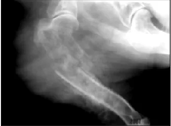
A seven-year-old male German shepherd dog was brought in Small Animal Clinic with lameness and severe localized proximal right humerous swelling, anorexia and weight loss. Firm swelling confirmed in right arm region (Fig. 1). Radiograph survey of affected bones showed pathologic fracture of proximal right humerous, cortical and trabecular bone lysis, periosteal bone proliferation, and severe soft tissue swelling (Fig. 2). Dorsoventral, right and left lateral views of chest were taken for careful evaluation for evidence of tumor metastasis. No abnormalities found in CBC and total serum alkaline phosphatase was 490 u/l.
Treatment and Outcome
Dog received dextrose-saline solution (20 ml/kg/hr, IV) and preoperative medication (atropine sulfate 0.03 mg/kg, SC) prior to anesthesia. Cefazolin (22 mg/kg, IV) was administrated as a prophylactic antibiotic before operation. Anesthesia was induced with diazepam, (0.27 mg /kg, IV) combined to ketamine hydrochloride (0.05 mg/kg, IV). Propofol (7.5 mg/kg, IV) was administrated for maintenance of anesthesia. The patient was positioned in left lateral recumbency. Skin incision from the dorsal border of the scapula, over the scapular spine, to the proximal third of the humerous was made, and then the skin incision was continued around the right forelimb at this level. The trapezius and omotransversarous muscles at the insertion level on the scapula spine were transected. Rhomboideus muscle from its attachment on the dorsal border of the scapula was transected as well. For exposing of the medial surface of scapula, scapula was resect laterally with elevation of the serratus ventralis muscle from the medial surface of scapula, brachial plexus and related muscles were transected. The right forelimb was removed and once was sent to pathologic laboratory. The brachial plexus and vessels were covered with approximately of the muscle bellies. Subcutaneous tissue and skin were sutured using 3-0 polyglactin 910 and 2-0 nylon suture materials, respectively.
All the samples were fixed in 10% formalin, embedded in paraffin wax, and sectioned (6 µm). The sections were stained with Haematoxylin and Eosin processed for immunohistochemistry. Seven antibodies were used such as MoAb anti-human Vimentin, PCNA, CD99, Desmin, CD34, Ki67 and Cyclin D1 all from Dako kit. Post-operative care was including antibiotic-therapy (cefazolin, 22 mg/kg, IM, every 8 hrs for 3 days).
Figure 1. Extensive swelling of right fore-limb.
Dog tolerated operation and recovered in 12 hours after surgery. He started to do weight-bearing second day operatively. Unfortunately, owner euthanized the dog in week 2 post-operatively because of reluctancy to accept chemotherapy and dog appearance.
The cut surface showed a firm white encapsulated mass, measuring about 7.5×5 cm, located in the right humerus area typically involved the epiphysiometaphyseal region of humerus. An invasive mass infiltrated the adjacent tissue.
The regional lymph nodes appeared normal. The tumor extended up to the adjacent articular cartilage, which remained intact. The tumor was eccentric to the long axis of the humerus. The overlying cortex had undergone resorption and the contour of the bone was expanded by the tumor which is covered by a thin shell of subperiosteal new bone. There was no cystification of the tumor, which may be so prominent as to mimic aneurysmal bone cyst.
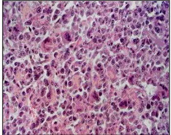
Histologically the lesion is composed of osteoclast-like multinucleated giant cells (MGCs) in a moderately vascularized network of proliferating polygonal or in some areas spindle-shaped stromal cells (Fig. 3).
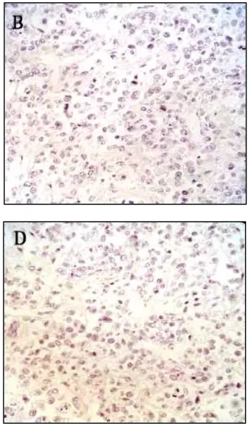
Osteoid was sparse and arranged in thin strands mixed with spindlecells. In the periphery of the mass,irregular trabeculae of woven bone were also present. Immunohistochemical stains for vimentin, PCNA (25%) exhibited diffuse and highly cytoplasmic labeling in MGC and polygonal cells within the stroma (Fig. 4). No cellular staining against Desmin, CD34, Ki67, Cyclin D1 and CD99 was observed (Fig. 5 A-E). Finally, diagnosis was giant cell osteosarcoma.
Figure 4. Immunohistochemical staining for Vimentin (A), PCNA (B). Note cytoplasmic staining of Vimentin and nuclear staining of PCNA ×750.
Discussion
The life span of dogs with osteosarcoma to a median of 300 days with a 40% 1-year survival was extended by multiple modality treatment such as amputation and cisplatin chemotherapy.2,3,7 Treatment of appendicular bone tumors involves limb amputation or tumor resection combined with limb salvage techniques and chemotherapy. Improved changes for
survival are possible with amputation or limb-sparing procedures combined with chemotherapy such as cisplatin.1,4,5,6 Dernell et al, showed that dogs treated for osteosarcoma with amputation alone have a median survival time of 3 to 4 months.3 Dernell and co-workers also showed median survival times of 260 to 400 days and a 385 to 625 1-year survival rate with amputation and cisplatin.3 Recently, biphosphonates have also been used to help diminish bone pain associated with osteosarcoma.3
Histopathologically, osteosarcoma is composed of anaplastic mesenchymal cells that produce osteoid.3 Histologic subgroups include osteoblastic, fibroblastic, osteoclastic, poorly differentiated, and telangiectatic osteosarcoma.3,8 Some pathologists believe that an inflammatory component is often present in fracture-associated sarcoma as sarcoma that arises in the diaphysis of a long bone at the site of a previous fracture.8
Figure 5. Immunohistochemical staining for
CD99: A, Desmin: B, CD34: C, Ki67: D and Cyclin D1: E
were negative × 250.
osteosarcoma is associated with fractures metallic implants, radiation fields following radiation treatment of soft-tissue sarcomas.9 Recent studies were investigating potential molecular and genetic causes of osteosarcoma3. Histological pattern of this tumor is similar to those former authors' findings.8,10 Vimentin positivity implicated that this tumor originated from mesanchymal cells.8 Histological finding and negativity for Desmin (intermediate filament in the muscle cells) identified that this tumor is not originated from myoblasts.11 Also this tumor didn’t originate from hematopoietic precursors and angioblasts, because The CD34 protein that is selectively expressed on lymphoid and myeloid hemopoietic progenitor cells and on vascular endothelium was negative.12 CD99 antigen is reported to be strongly expressed on PNET/EW (Primitive Neuroectodermal Tumor/Ewing’s sarcoma) in human was negative in this tumor then this tumor didn’t have neuroectodermal origin.13 Ki67 is a nuclear cell cycle associated protein, which is expressed in all active parts of the cell cycle (G1, S, G2 and mitosis) but not in resting cells.14 Maximum expression of cyclin D1 occurs at a critical point in mid to late G1 phase of the cell cycle.15 Proliferating cell nuclear antigen (PCNA) is a Cofactor for DNA polymerase delta in S phase and also during DNA synthesis associated whit DNA damage repair mechanisms.16 Negativity for these three later markers means that low mitotic activity of this tumor and endorses low metastatic activity that we observed. In a recent study, dogs with appendicular osteosarcoma that had strong COX-2 expression had significantly decreased overall survival time. Mullins and co-workers in above study showed that the median survival times for dogs with negative (n=10), poor (n=19), moderate (n=11), and strong (n=4) expression were 423, 399, 370, 86 days, respectively.
References
1. Garzotto C, berg J. Musculoskeletal system. In; Slatter D, eds. Textbook of small animal surgery, 3rd ed. Philadelphia: WB Saunders Co, 2003; 2460-2474.
2. Waters DJ. Musculoskeletal system. In; Slatter D, eds. Textbook of small animal surgery, 2nd ed. Philadelphia: WB Saunders Co, 1993; 2213.
3. Fossum TW, Hedlund CS, Hulse DA, et al. Bone neoplasia. In: Fossum TW, Hedlund CS, Hulse DA, Schulz KS, Seim HB, Willard MD, Bahr A, Carroll GL, eds. Small animal surgery. 3rd ed. St. Louis: Mosby, 2006; 1338-1352.
4. Denny HR, Butterworth SJ. Bone neoplasia. In: Denny HR. Butterworth SJ. eds. A guide to canine and feline orthopaedic surgery. 4th ed. London: Blackwell Science, 2000; 618-625.
5. Daly WR. Amputation of the forelimb. In: Bojrab MJ, Ellison GW eds. Current techniques in small animal surgery. 4th ed. Baltimore: Williams and Wilkins, 1998; 1286-1291.
6. Goldschmidt H, Thrall DF. Malignant bone tumors in the dog. In: Newton CD, Nunamaker DM, eds. Textbook of small animal orthopaedics. 1st ed. London: Lippincott, 1985; 887-898.
7. Ferrell EA, Berry CR, Thrall DE. Interpretation paradigms for the appendicular skeleton. Aggressive versus nonaggressive bone lesions. In: Thrall DE, eds. Textbook of veterinary diagnostic radiology. 5th ed. Philadelphia: WB Saunders Co, 2007; 222-230.
8. Negrin A, Bernardini M, Diana A, et al. Giant Cell Osteosarcoma in the Calvarium of a Cat. Vet Pathol 2006;43: 179-182.
9. Boudrieau RJ, McCarthy RJ, Sisson RD. Sarcoma of the proximal portion of the tibia in a dog 5.5 years after tibial plateau leveling osteotomy. J Am Vet Med Asso
10.Thompson JG, Pool RR. Tumors in domestic animal. 4th edn. London: Blackwell Publishing Company, 2002; 281-283.
11.Kenneth DK, Tuyethoa N, Vinh DE. Small cell osteosarcoma of bone: An immunohistochemical study with differential diagnostic considerations. Human Pathology 1993;24: 1211-1225.
12.Kaya M, Wada T, Akatsuka T, et al. Vascular Endothelial Growth Factor Expression in Untreated Osteosarcoma Is Predictive of Pulmonary Metastasis and Poor Prognosis.
Clinical Cancer Research 2000;6: 572–577.
13.De Alava E. Diagnosis of small round cell tumours of bone. Current Diagnostic Pathology 2001;7: 251-261.
14.Jong R, Davis AM, Mendes MG, e al. Proliferative Activity (Ki-67 Expression) and Outcome in High Grade Osteosarcoma: A Study of 27 Cases. Sarcoma 2000; 4(1-2): 47-55.
15.Xiao-hui S, Zheng L. Expression of cyclin D1 and CDK4 in osteosarcoma of the jaws.
Chinese Journal of Cancer Research 2001;13: 140-143.
16.Wenji W, Huiying L, Aiqin W. Expression of survivin and correlation with PCNA in osteosarcoma. Journal of Surgical Oncology 2006;93: 578-584.
هﺪﯿﮑﭼ
ﻮﯾد
يﺎﻣﻮﮐرﺎﺳﻮﺌﺘﺳا
ﯽﻤﯿﺷﻮﺘﺴﯿﻫﻮﻧﻮﻤﯾا
ﻪﻌﻟﺎﻄﻣ
ﮓﺳ
رد
لﻮﻠﺳ
نﺎﯿﺣﺎﺘﻓﺎﺿرﺪﯿﻤﺣ 1 ، ﯽﮐرﺎﺼﺣﺪﯿﻌﺳ 2 ، ﯽﻠﺿﺎﻓﺪﯿﻣا 3 ، ﺮﯿﺒﮐﺎﺿرخﺮﻓ 4
، ﺎﻗآﯽﺟﺎﺣﯽﻧﺎﺣورﺎﺿرﺪﯿﺳ 5 1 هوﺮﮔ آ ﯽﺣاﺮﺟﯽﺷزﻮﻣ ، 2
يژﻮﻟﻮﺗﺎﭘﯽﺷزﻮﻣآهوﺮﮔ ،
4
يژﻮﻟﻮﯾدارﯽﺷزﻮﻣآهوﺮﮔ
و
5
ﯽﮑﺷﺰﭙﻣادﯽﻫﺎﮕﺸﯾﺎﻣزآمﻮﻠﻋﯽﺳﺎﻨﺷرﺎﮐﻪﺘﺧﻮﻣآﺶﻧاد ،
،ﯽﮑﺷﺰﭙﻣادﯽﺼﺼﺨﺗمﻮﻠﻋهﺪﮑﺸﻧاد
ﺪﺣاو اد،تﺎﻘﯿﻘﺤﺗومﻮﻠﻋ ،ناﺮﯾا،ناﺮﻬﺗ،ﯽﻣﻼﺳادازآهﺎﮕﺸﻧ
3
ناﺮﯾا،ناﺮﻬﺗ،ﯽﻠﺿﺎﻓﺮﺘﮐدﮏﭼﻮﮐيﺎﻫمادهﺎﮕﻧﺎﻣرد
.
ﯽﻨﯿﻟﺎﺑيﺎﻫﻪﺘﻓﺎﯾونارﺎﻤﯿﺑﻒﯿﺻﻮﺗ
-ﮓﺳهدﻼﻗﮏﯾ
7 ﯽﯾﻻﺎـﺑيﺎـﻬﺘﻧاﻊﯿﺳومرﻮﺗوﺪﯾﺪﺷﺶﮕﻨﻟﻢﺋﻼﻋﺎﺑدﺮﭙﺷﻦﻣرژﺮﻧﻪﻟﺎﺳ
ﺎﺟراﮏﭼﻮﮐمادهﺎﮕﻧﺎﻣردﻪﺑﺖﺳاروزﺎﺑناﻮﺨﺘﺳا ﺪﺷع
.
نآﻪﺠﯿﺘﻧونﺎﻣرد
- لﺎﺳراﯽﺳﺎﻨﺷﺐﯿﺳآهﺎﮕﺸﯾﺎﻣزآﻪﺑﻪﻠﺻﺎﻓﻼﺑﯽﺘﻓﺎﺑﻪﻧﻮﻤﻧوﺪﺷﻪﺘﺷادﺮﺑﯽﺣاﺮﺟشورﻪﺑﺖﺳارﯽﺘﮐﺮﺣماﺪﻧا
ﺪﯾدﺮﮔ
.
ﻮﻤﻧﻞﻤﺤﺗارﯽﺣاﺮﺟﻞﻤﻋﮓﺳ ﺖﺷﺬﮔزاﺲﭘود
12
ﺪﯿﺳرﻞﻣﺎﮐيرﺎﯿﺷﻮﻫﻪﺑﯽﺣاﺮﺟزاﺖﻋﺎﺳ
. ﺎـﺑهﺪﺷﻪﺘﺷادﺮﺑﻪﻧﻮﻤﻧ
ﺪﺷهدادﺺﯿﺨﺸﺗلﻮﻠﺳﻮﯾدﺎﻣﻮﮐرﺎﺳﻮﺌﺘﺳاﻦﯾزﻮﺋاوﻦﯿﻠﯿﺴﮐﻮﺗﺎﻤﻫيزﺮﯿﻣآﮓﻧرزاهدﺎﻔﺘﺳا
. نﺎـﺸﻧﯽﻤﯿـﺷﻮﺘﺴﯿﻫﻮﻧﻮﻤﯾاﻪـﻌﻟﺎﻄﻣ
ﺪﺷﺎﺑﯽﻣارادارﻻﺎﺑيزﺎﺘﺳﺎﺘﻣﺖﯿﻟﺎﻌﻓوﻦﯿﯾﺎﭘيزﻮﺘﯿﻣﻪﺟردرﻮﻣﻮﺗﻦﯾاداد
.
ﮐ ﯽﻨﯿﻟﺎﺑدﺮﺑرﺎ
رﻮـﻣﻮﺗئﺎﺸﻨﻣﻪﮐﺪﻨﺘﺴﻫردﺎﻗﯽﺳﺎﻨﺷﺖﻓﺎﺑﻪﻌﻟﺎﻄﻣياﺮﺑهﺪﺷهدﺎﻔﺘﺳاﺎﻫيدﺎﺑﯽﺘﻧآﻪﮐدادنﺎﺸﻧﺮﺿﺎﺣﻪﻌﻟﺎﻄﻣ
رﺎﻤﯿﺑﺎﺑدرﻮﺧﺮﺑﯽﮕﻧﻮﮕﭼونﺪﻧﺎﻣهﺪﻧزنﺎﻣزﯽﯾﻮﮔﺶﯿﭘرﻮﻈﻨﻣﻪﺑارنآندﻮﺑيزﺎﺘﺳﺎﺘﻣتﺎﯿﺻﻮﺼﺧو ﻼﺘﺒﻣ
) وﯽـﯾورادنﺎـﻣرد
ﯽﺣاﺮﺟ
(
ﺘﺳايزﻼﭘﻮﺌﻧزاعﻮﻧﻦﯾاﻪﺑ ﺪﻨﻫدنﺎﺸﻧارناﻮﺨ
.
نﺎﮔژاوﺪﯿﻠﮐ
-
ﮓﺳ،وزﺎﺑ،ﯽﻤﯿﺷﻮﺘﺴﯿﻫﻮﻧﻮﻤﯾا،ﺎﻣﻮﮐرﺎﺳﻮﺌﺘﺳا