IBBJ
Spring 2019, Vol 5, No 2
Advancing Chimeric Antigen Receptor-Engineered T-Cell
Immunotherapy Using Genome Editing Technologies:
Challenges and Future Prospects
Yousef Jafari Abarghan
1, Arash Salmaninejad
1, 2, Atieh Eslahi
1, 2, Farzaneh
Alizadeh
1, Majid Mojarrad
1, 3*1.Department of Medical Genetics, Faculty of Medicine, Mashhad University of Medical Sciences, Mashhad, Iran.
2.Student Research Committee, Department of Medical Genetics, Faculty of Medicine, Mashhad University of Medical Sciences, Mashhad, Iran.
3.Medical Genetics Research Center, School of Medicine, Mashhad University of Medical Sciences, Mashhad, Iran.
Submitted 15 Jun 2019; Accepted 28 Jun 2019; Published 26 Jul 2019
Chimeric antigen receptor engineered-T (CAR-T) cells also named as living drugs, have been recently known as
a breakthrough technology and were applied as an adoptive immunotherapy against different types of cancer.
They also attracted widespread interest because of the success of B-cell malignancy therapy achieved by
anti-CD19 CAR-T cells. Current genetic toolbox enabled the synthesis of CARs receptors which are targeted against
tumor-specific antigens, and enabled to arbitrarily T-cells function reprogramming. Approximately all of CAR-T
cell based studies apply autologous CAR-T cells in which, modified T-cells are engineered using patient’s own T
cells. Currently, four different generation of CAR-T cells have been developed, and the evolution of this kind of
therapy illustrates an excellent example of the application of basic research into the clinical trial stage. However,
development of allogenic CAR-T cells can be a turning point for CAR-T cells therapy. Appearance of the reliable
gene editing approach, CRISPR/Cas9 system, provided a new hope for designing universal CAR-T cells which
are off-the-shelf, and enable to use for treatment of any patient with any kind of tumor. This review outlined four
different generations of CAR-T cells. Also, we discussed different types of genome editing systems especially
CRISPR/Cas9 system, and their capabilities for generating engineered T-cells. Additionally, we tried to explain
challenges faced in improving universally generated T-cells.
Keywords: CAR-T cells, cancer, therapy, CRISPR/Cas9 system
espite the fast progress in the appearance
of helpful medical technologies and
therapeutic approaches, cancer is still an intrac-
table problem of public health (1). Most commonly
used conventional therapeutic strategies such as
chemotherapy, surgery, and radiotherapy probably
give short-term advantages but have irritating side
effects because of their invasiveness and highly
biotoxicity (2). Moreover, multidrug resistance
remains a major challenge in the chemotherapy, and
several side effects of radiotherapy depend on the
type of cancer and the radiation therapy dose,
limiting therefore their curative efficacies (3, 4).
Correspondingly, novel therapeutic strategies that
D
Review Article
are highly specified for a targeted tumor and have a
long-term beneficial are required to be developed.
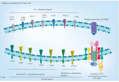
According to the naturally occurring immune
responses against tumor cells (figure 1), currently,
several immune system based therapies such
as usage of T cell receptor (TCR)-engineered T
cells, tumor-infiltrating lymphocytes (TILs), and
chimeric antigen receptor (CAR) –engineered T
cells, have been developed for different human
malignancies (5-7).
TILs are isolated and cultured from resected
tumors, and sent back to patients. Infusion of these
cells in patients with metastatic melanoma have
provided encouraging results (8) , but this method
showed some limitations in other solid tumors
owing to the low efficiency of isolation and in vitro
expansion (9).
However, re-engineering of the immune
system, especially T cells, for targeting and killing
tumor cells has been a primary aim of cancer biology
for many years. Thereby, the idea that adoptively
transferred T cells can act as a harness to cancer
treatment was demonstrated after the seminal study
of Medawar et al. (10). Along with, progresses in
genetic engineering technology, for example
retroviral transduction, has allowed researchers
to insert artificial transgenes into primary T cells,
and their subsequent expression caused the
redirection of T cells against tumor-associated
antigens (TAAs) (11).
Clinical trials of modified T cells in cancer
therapy are based on the genetically modified T cells
which are synthetic receptors that preserve the
primary TCR structure, but are designed for specific
antigens on tumor cells.
Rapoport et al. used New York esophageal
squamous cell carcinoma 1 (NYESO 1) (a
cancer-testis antigen) specific TCR engineered T-cells for
multiple myeloma in phase I/II trial, and showed that
in vivo–expanded genetically altered antigen-specific T cells can be well tolerated without
remarkable safety concerns (12). However, like
native TCRs, these kinds of engineered TCRs need
MHC presentation, but many tumors down-regulate
MHC class I and potentially disappear from these
TCR-modified T cells.
Until now, CAR-modified T cell therapy has
been best effective interesting research field in
cancer therapy. So far, a high number of clinical
trials have been directed to improve the efficiency
and safety of CAR-T cells for cancer treatment (11).
CARs are chimeric molecules that are typically
composed of antigen recognition domain of the
B-cell receptor, such as extraB-cellular antibody
single-chain variable fragment (scFv), a TCR-derived CD3ζ domain, and the co-stimulatory domains of the T cell receptor (13). Unlike early engineered T
cells, CARs allow enhancing the specific targeting
of antigen in an MHC-independent manner.
Therefore, CARs enable the redirection of cytotoxic
T cells toward any various kinds of protein or
non-protein targets that are expressed on tumor cell
surfaces. Accordingly, there is no requirement for
antigen processing and presentation by MHC on
cell surface, and is appropriate to non-classical
targets such as carbohydrates and glycolipids
structures (14).
Eliminating the MHC restriction limitations
afford a promising opportunity to broaden the
applicability of CAR-T cell-dependent
immune-therapy in cancer. Additionally, progresses in ex
vivo expansion enable to rapid development, and
improve the capability of CAR-T cells. Therefore,
construction of clinically appropriate doses of these
Figure 1. Natural occurring immune responses against tumor cells.
“living drugs” becomes possible. Nevertheless, CAR-T cell targets were restricted to cell surface
antigens.
With the proof of concept established, CAR-T
cells therapy have emerged as a probable approach
to treat different kind of malignancies. For example,
adoptive delivery of CD19 directed CAR-T
(CART19) cells have created promising and
resilient therapy in patients with relapsed B cell
malignancies and refractory (15-17). Nevertheless,
there are several challenges in adoptive T cell
immunotherapy for cancer treatment, notably usage
of CAR-T for solid tumors treatments, for example:
target specificity, T cell exhaustion, and suppressive
situation of tumor microenvironments. Mentioned
challenges and limitation thoroughly will be
discussed in this review. In addition, we will explain
some of the latest and promising developments in
adoptive T cell immunotherapy, with a specific
attention to the latest gene-editing systems.
Likewise, we will describe how they are being used
to modified-T cell immunotherapy.
Chimeric antigen receptor structure
CARs are typically composed of four main
regions: extracellular binding region (ectodomain),
hinge or spacer domain, transmembrane domain
and intracellular signaling domain (endodomain)
(figure 2) (18). The ectodomain of CARs has
conventionally provided the antigen binding domain
of receptor, and single-chain variable fragments
(scFvs) are the largest group of extracellular domain
for CARs. ScFv affinity is the major determinant of
CARs function and specificity (19, 20). As a result
of this, Hudecek et al. used receptor tyrosine kinase–
like orphan receptor 1(ROR1), as a tumor-associated
molecule, to create ROR1-CAR-T cells with high
affinity to ROR1 positive tumor cells. Thereby,
ROR1-specific scFvs were able to enhance CAR-T
cell effector function (19). While the ectodomain
detain the CAR specificity, spacer region, as a
binding domain between the ectodomain and
transmembrane domain, is another important
domain, and changing its length and composition
significantly affect CAR-T-cell activity. However,
results from different studies have shown
controversial results about the use of various kind of
spacer domains in CARs structure (21, 22). For
example, Guest et al. (23) added CH2CH3 spacer to
four CARs with no spacer, this addition specified
them for some antigens such as carcinoembryonic
antigen (CEA), neural small adhesion molecule
(NCAM), the oncofetal antigen 5T4 and B-cell with
CD19. While 5T4- and CD19-specific CAR-T cells
with CH2CH3 spacer had some enhanced effector
function, but superior anti-tumor activity of CEA-
and NCAM-specific CAR-T cells was without
spacer. As well as, Hudecek et al. (19) used spacers
with different size such as CH2-CH3 spacer (229
amino acids (AA)), CH3 spacer (119 AA), and short
spacer (12 AA), on ROR1-specific CARs and
showed that CAR-T cells with short spacer have
better anti-tumor activity. These studies illustrated
that spacer region have a potential role in CAR-T
cells activity and it is necessary to determine an
optimum form of this region for all CARs.
The transmembrane domain of CARs connects
extra and intracellular domains and so has a critical
role in CAR stability. There are some different
transmembrane domains such as CD3-ζ, as an
earliest transmembrane domain, and CD4, CD8, and
CD28 molecules that can be used in CARs structure
(24). Currently, CD28 is the most utilized domain in
CAR-T cells and provide a significant stability for
CARs (25, 26). Ultimately, transmembrane domain
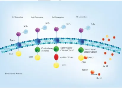
Figure 2. Four different CAR-T cell generations.
links to an intracellular signaling domain that
affords the downstream signaling, such as
phosphorylation of immunoreceptor tyrosine based
activation motifs (ITAMs) that are present in the
cytoplasmic domain, and lead to the activation of
CAR-T cells (27). According to the structure of
intracellular signaling domain, CARs have been
classified into four different generations.
Different generations of CARs
The first generation of CAR-T cells were
developed in 1989 by Gross et al. (28, 29). These kind of modified T cells contain a CD3ζ or FcεRIγ as an intracellular signal transmitter, which is
sufficient to induce T cell activation through the
phosphorylation of ITAMs (11, 12). This activation
triggers cytotoxic responses against tumor cells
besides secretion of cytokines, especially IL-2 and
IFN-γ (30). Some in vivo preclinical studies were
able to illustrate better survival for tumor-bearing
mice using the first generation of CAR-T cells (30,
31). However, this activation signal usually is
insufficient to completely activate CAR-T cells and
results in T cell anergy, poor cytotoxic activity, poor
T cell survival, weak proliferation, and poor
persistence of CAR-T cells (32-34).
Therefore, the second generation of CAR-T
cells was developed to improve the activation
signaling and also enhance the proliferation,
persistence and cytokine secretion of CAR-T cells.
These were achieved by adding a co-stimulatory
molecule (second signaling domain or signal 2) such
as CD28 into the intracellular domain of CARs
between the transmembrane domain and CD3-ζ
domain, which causes complete T cell activation,
and prevents apoptosis by promoting the expression
of anti-apoptotic proteins such as Bcl-xL (13, 14).
CD28 along with 4-1BB, as a part of the tumor
necrosis factor (TNF) receptor family are the most
broadly applied co-stimulatory molecules in CARs
structure (35). CD28-containing CARs have a great
potency to activate the T cells. Additionaly, CAR-T
cells with CD28 domain are generally able to secrete
high amounts of cytokines, such as IL-2, IL-10,
TNF-α, and IFN-γ in comparison with other CAR-T
cells (25, 36). Moreover, inducible co-stimulation
(ICOS) which is a member of CD28 super family
and OX40 (part of TNFR family) are two other
co-stimulatory molecules that are used as a second
signal domain in CARs construction (37). Currently,
the second generation of CAR-T cells is major
CARs used in clinical trial experiments (table 1).
Hitherto, most experiments have been
performed on different type of B-cell malignancy,
and CD19 (an extracellular glycoprotein) is the most
common targeted antigen on these cells. For
example, Kochenderfer et al. used second
generation anti-CD19-CAR-T cells containing
CD28 co-stimulator for treatment of advanced
follicular lymphoma. In this trial, infusion of
anti-CD19 CAR-T cells provided partial remission for up
to 32 weeks with no remarkable toxicity, and the
persistence of CAR-T cells was being monitored
using qPCR (38). In another phase I trial study with
four patients, Ritchie et al. used second generation
autologous anti-LeY CAR T-cells for treatment of
acute myeloid leukemia (AML) and tried to estimate
the persistence and safety of these adoptive T cells.
They delivered totally 1.3 × 10 9 T cells to the
patients, and observed a significant persistence of
infused CAR-T cells in the patients for up to 10
months (39). Recently, Kebriaei et al. expressed the
second-generation anti-CD19 CAR-T cells in a
non-viral system, through the use of Sleeping Beauty
(SB) transposon/transposase system, and applied
them as an adjuvant therapy in phase I trial for
treatment of 26 patients with advanced non-Hodgkin
lymphoma (NHL) and acute lymphoblastic
leukemia (ALL). The rate of CAR-T cell persistence
in these patients was in average 201 days for the
autologous recipient and 51 days for allogeneic
recipients. Furthermore, these results emphasized
safety of SB system for application in
immunotherapy of human diseases (40).
Enhancing the persistence of CAR-T cells after
infusion is the main issue to improve the CAR-T cell
function. Accordingly, Rossig et al. conducted a
multi-center phase I/II trial study and used
Epstein-Barr-virus (EBV)-specific cytotoxic T-cells (EBV
CTL) redirected with a CD19-CAR-T cells for
treatment of patients with relapsed pediatric ALL.
Additionally, they used EBV-directed vaccination
which was able to show a significant increase in the
persistence of infused CAR-T cells in comparison
with T-cells without vaccination (41).
Different co-stimulatory receptors provide
various functional properties for CAR-T cells. For
example, the second-generation CD28-specific
CARs immediately proliferate and are activated
against tumor, whereas 4-1BB-specific CARs have
the long-term persistence potency in tumor
environment (42). Therefore, discrete features of
co-stimulatory domains provide an opportunity to be
able to further develop CAR-based T cell therapies,
and improve their properties for a variety of cancers.
Following features can be provided by third
generation of CAR-T cells. This generation, in addition to CD3ζ, contain two co-stimulatory domains with different properties which are able to
enhance the activation status of CAR-T cells with
powerful cytokine production (for example:
CD3ζ-CD28-OX40 or CD3ζ-CD28-41BB) (15). Different
in vitro (43) and in vivo (44) preclinical studies have illustrated that third generation CAR-T cells
have a superior potential in activation, proliferation
and persistence but there is no sufficient clinical
study capable to demonstrate the safety and efficacy
of third generation CAR-T cells. Additionally,
enormous phenotypic heterogeneity of solid tumors
makes limitation on activity of these CAR-T cells.
Due to heterogeneity, there are significantly high
rate of antigen-negative tumor cells which are not
recognized by CAR-T cells which enhance the
probability of tumor relapse. This may be resolved
through the activation of native T-cells in
tumor environment mediated by infused CAR-T
cells.
Fourth generation of CAR-T cells with dual
anti-cancer cell activities through the inducible
IL-12 have been developed (45). Theae are second
generation of CAR-T cells additionally engineered
with an inducible expression cassette encoding
transgenic IL-12 and known as T cell redirected for
universal cytokine-mediated killing (TRUCKs)
(45). Thereby, inducible recombinant IL-12 (iIL-12)
(composed of the p40 and p35 chain) is under
control of the NFAT/IL-2 minimal promoter and
releasing of iIL-12 is promoted by CAR signaling
pathway (46). Releasing of IL-12 by TRUCKs
increases T cells activation in tumor environment,
and attracts innate immune system which causes the
eradication of antigen-negative tumor cells (45).
However, similar to third generation CAR-T cells, it
is needed to direct more and more experiments to be
able to illustrate the efficacy and usability of fourth
generation CAR-T cells.
Genome editing technologies enable the generation of allogenic universal CAR-T cells
Despite recent advances in the development of
autologous adoptive T-cell immunotherapy (T-cells
that are currently applied in the generation of
CAR-T cells are mainly achieved from the patients
themselves (autologous T cells)), some limitations
reduce the efficacy of CAR-T cells including the
insufficient quantity of autologous T cells that affect
the efficiency and quality of CAR-T cells, and also
the generation of autologous T cell production is not
cost-effective either in term of time or expense.
Therefore, a large number of cancer patients cannot
be treated through this approach.
Accordingly, the use of genetically engineered
allogenic T cells which are received from healthy
donors and known as universal T cells can be a
promising opportunity for the development of a
highly efficient approach in cancer therapy.
Moreover, universal CAR-T cells can be used for
treatment of various cancer patients without raising
graft-versus-host disease (GVHD). Additionally,
allogenic CAR-T cells can be improved regarding
their persistence and efficient elimination of the
tumor cells. GVHD disease occurs by the
recognition of recipient antigens via TCRs related to
infused T-cells. Conversely, recognition of foreign
HLAs of infused allogenic T-cell causes rapid
rejection. Thereby, it is necessary to be able to
silence both TCRs and HLAs in universal allogenic
CAR-T cells (47).
Currently, the development of genome editing
technologies provided an opportunity to precisely
manipulate the genome. Hence, it is possible to use
gene editing strategies for silencing of infused
T-cell-related HLAs and TCRs. These approaches may
provide an opportunity to generate reliable allogenic
universal CAR-T cells that can be used for the
treatment of different cancer patients without any
concern about GVHD or immune response against
foreign T-cells and early rejection. Up to now, three
different genome editing approaches have been
developed. Here we briefly describe the safety
concerns regarding their specificity in allogenic
CAR-T cells generation.
Zinc finger nuclease (ZFN)
Zinc finger nuclease (ZFNs) are artificial
hybrid proteins, and were primarily developed as
one of genome-editing tools for specific
modifyication of genome. ZFNs are mainly
composed of two different domains: a
sequence-specific repeated zinc finger protein that recognizes
and binds to a predetermined region of the genome,
and a non-specific FokI restriction endonuclease
domain (48). Each zinc finger protein can recognize
three or four base pairs. As a result of this, ZFNs are
typically comprised from three or four zinc finger
protein which are joined together in tandem repeat
to be able to target 9-18 base pairs of the genome
(49). Moreover, dimerization of FokI endonucleases
is necessary to create a site specific double strand
breakage in the genome. Thereby, two ZFN
molecules are needed to bind complementarily to
adjacent regions of the genome. This site-specific
binding and dimerization of two FokI endonucleases
provides a highly efficient cleavage of DNA.
Consequently, DNA repair systems which include
two different strategies: error-prone
non-homologues end joining repair (NHEJ) in the
absence of a template, and homology-directed repair
(HDR) in the presence of a DNA template, are
vactivated (50). NHEJ causes disruption in a
targeted gene but HDR system provides a
template-mediated repair of the targeted gene (51, 53).
Torikali et al. used ZFN for genome editing of
allogenic CD19-specific CAR-T cells, and
they could permanently eliminate the expression
of endogenous TCRs (53). However, the
difficulty of engineering ZFN monomers with high
affinity for a specific site of the DNA and low
efficacy with high off-target due to
heterodimerization of FokI enzymes, limited their
capability for further utilization in the genome
editing strategies (54).
Transcription activator-like effector nucleases
(TALEN)
In 2009, two independent experiments
described details of transcription activator-like
effector (TALE) DNA-binding proteins that are
secreted by plant-specific pathogens such as
Xanthomonas and Ralstonia sp (55, 56). DNA-binding domain of TALE proteins consist of tandem
repeats with highly conserved 34-35 amino acids.
Moreover, hypervariable amino acids, located at
positions 12 and 13 within each tandem repeats
referred to as the repeat variable di-residue (RVD)
show a high affinity to specific nucleotide targets.
Thereby, each of this tandem repeats are able to
recognize a single nucleotide target site. Thereby,
selective combination of these specific
DNA-binding tandem repeats allows to target a unique
sequence in the genome (55). Similar to ZFNs, FokI
endonuclease fuses to a reprogrammed TALE
domain, and provides a TALE nuclease (TALEN)
monomer. Binding of two engineered TALEN
monomers in an adjacent region enables FokI
enzyme dimerization, and creates a double
strand break in targeted sequence (57). Like
ZFNs, TALENs based cleavage induces NHEJ
or HDR system of targeted cells (58). Developing
of TALENs allows to target any specific site
of genome. Moreover, the ease of reprogram-
ming of TALE domains make TALENs more
interesting than ZFNs for targeted genome-
editing (54).
In a pre-clinical study TALEN have been used
for multiplex genome-editing of anti-CD19 CAR-T
cells (59). In this study, both TCR and CD52
(encodes a protein targeted by alemtuzumab) genes,
were knocked out. CD52 elimination, increases the
resistance of third-party T cells to lympho depleting/
immunosuppressive effects of alemtuzumab, and
significantly reduces their rejection chance by the
recipient patient. Ultimately, these results illustrated
the highly efficiency of TALEN system for the
generation of allogenic adoptive T-cells from
third-party healthy donors without raising GVHD (59).
Recently, Qasim et al. directed the first clinical trial
of TALEN-mediated genome-engineered universal
CD19-specific CAR-T cells, and have shown
remarkably remission of leukemia in an
11-month-old patient (60). However, TALENs based genome
editing is associated with some limitations. For
example, the large size of TALEN monomers that
requires 34-35 amino acids to specify a single base
pair, reduces the delivery performance of both
TALEN monomers in a single viral vector.
Moreover, unstable nature of TALE tandem repeats
makes difficult their packaging in viral vectors (54).
CRISPR/Cas9
An alternative strategy to protein-mediated
genome-editing technologies, the clustered
regularly interspaced short palindromic repeats
(CRISPR)/ CRISPR associated protein 9 (Cas9)
system has been developed as a novel RNA-guided
genome-editing tool. Because of the high efficiency
and ease of use, CRISPR/Cas9 system is the widely
applied strategy for precisely modifying of the
genome (61). Originally, CRISPR/Cas9 system
from Streptococcus pyogenes, was recognized as a
complex system that acts as a bacterial and archaeal
immune defense system against bacteriophage DNA
or other foreign DNAs like plasmids (62). Briefly,
when a foreign DNA was detected by type II
CRISPR immune system, some part of its sequence
was incorporated into the CRISPR repeat sequences
(63). The core components of engineered
CRISPR/Cas9 system are Cas9 nuclease and single
guide RNA (sgRNA). To date, more than 20 Cas9
homologs have been derived from different species
of bacteria and each of them have a specific
protospacer adjacent motif (PAM) (64).
Streptococcus pyogenes Cas9 (spCas9) is the most commonly utilized Cas9 for modifying the human
genome (65).
Commonly, Cas9 enzyme is composed
from two catalytically active domains consisting
of RuvC and HNH. As well as, chimeric
sgRNAs are generated by fusion of a programmable
CRISPR RNA(crRNA) and a trans-activating
crRNA (tracrRNA) (66) in which crRNA is
synthesized against a specific target and tracrRNA
acts as a scaffold for crRNA and facilitates the
interaction between crRNA and Cas9 (66).
Accordingly, complementary base paring of sgRNA
is necessary for precisely directing Cas9 to the
desired genomic region. Cas9-mediated cleavage
necessarily requires a PAM which should be
located precisely downstream of the
sgRNA-targeted sequence. PAM acts as a binding signal
for Cas9, and typically has 3-5 bp in length.
Binding of sgRNA to the targeted sequence
along with the presence of PAM provide an
opportunity to Cas9 to create a double strand
cleavage between the 3rd and 4th base upstream of
the PAM (67, 68).
Because of the use of sgRNA instead of protein
constructions, CRISPR/Cas9 system is much easier
in comparison with other genome editing
approaches. This system was developed as a great
therapeutic approach to the generation of allogenic
CAR-T cells. As well as, efficient multiplex
genome editing by CRISPR/Cas9 system
enables to simultaneously knock out different
types of gene such as programmed cell death
protein 1 (PD1), cytotoxic T-lymphocyte associated
protein 4 (CTLA4), TCR beta chain, and
beta-2-microglobulin (B2M), which reduce the expression
of HLA class I antigen on the CAR-T cells surface.
Up to now, several pre-clinical studies demonstrated
a significant efficacy of CRISPR/Cas9
system-based allogenic CAR-T cells (69). Most recently,
Ren et al. used one shot CRISPR/Cas9 protocol
which was enabled to integrate multiple sgRNAs
into a CAR-lentiviral vector (70). Consequently,
they could generate allogenic CAR-T cells that were
simultaneously disrupted in four gene loci
including TCR, HLAI, PD1, and CTLA-4 genes (71).
Thereby, double knocking out of TCR and HLAI
eliminates the risk of both GVHD and early
rejection. Moreover, disruption of PD1 and
CTLA4 which are considered as inhibitory
signals, provided inhibitory resistant universal
CAR-T cells (70). Currently, the main focus of
researches is the production of highly homogenous
population of allogenic CAR-T cells. Therefore, in
another recently performed pre-clinical study,
scientists developed a self-inactivating lentiviral
based vector in which a specific sgRNA was incorporated in 3’ long terminal repeat (LTR). In the mentioned investigation, they could generate
highly homogenous allogenic engineered-T
cells that were 96% CAR positive and 99% TCR
negative (72).
Even though variety of studies has been
directed to treat advanced refractory B cell
malignancy, there are significant challenges about
the use of this therapeutic strategy for treatment of
solid tumors (73). Zhang et al. demonstrated that
the disruption of lymphocyte activation gene 3
(LAG3), as a negatively regulating factor of T
cells, by CRISPR/Cas9 system, remarkably
increased the efficiency of CAR-T cells against
solid tumors (74). According to these and some
other successful pre-clinical studies, generation
of highly efficient allogenic CAR-T cells by
CRISPR/Cas9 system were carried out in
clinical trial stages, and now some different
clinical studies are ongoing in phase 1 and 2
(Table 1).
However, one of the crucial challenges
relatedto CRISPR/Cas9 system as a safe genome
editing approach, is undesirable cleavage of the
genome (off-targets) that should be reliably reduced
(75). Consequently, there are some suggestions
that could be performed to improve the efficacy of
this system. For example, designing proper
sgRNAs, using programmable systems such as
tet-on for controlling the expression of Cas9 or using Cas9 mRNA and protein instead of
plasmid containing Cas9 gene, because mRNA is
very sensitive and is rapidly degraded by
RNases after on-target cleavage, can be performed
(76). Moreover, the nickase form of Cas9, in
which Cas9 is able to cut just a single strand of
DNA, can be used. Therefore, creating a
double strand breakage in DNA requires two
sgRNAs and two Cas9 which significantly
reduces the off-target rate (67). As well as, it is
possible to use dead Cas9-FokI as a fused protein
to create an on-target double strand cleavage. In
this strategy, dCas9 is unable to cut DNA but will
direct the FokI enzyme to a specific region of the
genome that sgRNA have paired. Thereby, double
strand cleavage is needed to pair FokI domains
adequately close to create a dimer (two target
sites should not be more than 25 bp apart,
approximately 13-25 bp) (77).
Another utility of the CRISPR/Cas9 system is
its application as a gene knock-in
platform to insert a DNA sequence in a desired
region of genome. Viral delivery vectors such
as lentivirus and retrovirus are the traditional
approaches that are used to integrate CAR fragments
into the T-cells genome. Nevertheless, this
kind of uncontrolled integration has the great
risk of causing insertional mutagenesis (78).
CRISPR/Cas9 mediates effective gene
knock-in in various kinds of cells, but integration of
large fragments such as CARs into T-cells
genome through the knock-in approach is a
great challenge. However, most recently Sather
et al. showed that knock-in of CAR into T-cells is
possible through the megaTAL and an
adeno-associated virus HDR template (79).
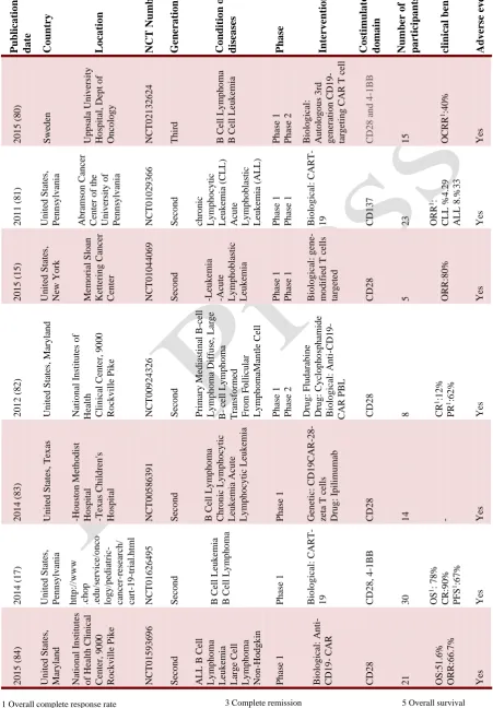
1 Overall complete response rate 2 Overall Response Rate
P ub lica tio n da te Co un try L o ca tio n NCT Nu mb er G ener a tio n Co nd it io n o r dis ea ses P ha se I nte rv ent io ns Co st imu la to ry do ma in Numb er o f pa rt icipa nts clinica l benef it s Adv er se ev ent s 2 0 1 5 ( 80 ) S we d en Up p sa la Un iv ersity Ho sp it al, De p t o f On co lo g y NCT0 2 1 3 2 6 2 4 Th ir d B Ce ll Ly m p h o m a B Ce ll Leu k em ia P h ase 1 P h ase 2 Bio lo g ica l: Au to lo g o u s 3 rd g en era ti o n CD1 9 - targ eti n g CAR T c ell CD2 8 a n d 4 -1 BB
15 OCRR 1:4 0 % Ye s 2 0 1 1 ( 81 ) Un it ed S tate s, P en n sy lv an ia Ab ra m so n Ca n ce r Ce n ter o f th e Un iv ersit y o f P en n sy lv an ia NCT0 1 0 2 9 3 6 6 S ec o n d c h ro n ic Ly m p h o cy tic Leu k em ia (CL L) Ac u te Ly m p h o b las ti c Leu k em ia (ALL ) P h ase 1 P h ase 1 Bio lo g ica l: CART -
19 CD1
3
7
23 ORR 1: CLL % 4 .2 9 ALL 8 .% 3 3 Ye s 2 0 1 5 ( 15 ) Un it ed S tate s, Ne w Yo rk M em o ria l S lo an Ke tt erin g Ca n ce r Ce n ter NCT0 1 0 4 4 0 6 9 S ec o n d -Leu k em ia -Ac u te Ly m p h o b las ti c Leu k em ia P h ase 1 P h ase 1 Bio lo g ica l: g en m o d ifi ed T c ell s targ eted CD2 8
5 ORR:8
0 % Ye s 2 0 1 2 ( 82 ) Un it ed S tate s, M ary la n d Na ti o n al In stit u tes o f He alt h Cli n ica l Ce n ter, 9 0 0 0 Ro ck v il le P ik e NCT0 0 9 2 4 3 2 6 S ec o n d P rima ry M ed ias ti n al B -c ell Ly m p h o m a Diffu se , Lar g e B - ce ll Ly m p h o m a Tran sfo rm ed F ro m F o ll icu lar Ly m p h o m aM an tl e Ce ll P h ase 1 P h ase 2 Dru g : F lu d ara b in e Dru g : Cy clo p h o sp h am id e Bio lo g ica l: A n ti -CD1 9 - CAR P BL CD2 8
8 CR
1:1 2 % PR 1:6 2 % Ye s 2 0 1 4 ( 83 ) Un it ed S tate s, Tex as -Ho u st o n M eth o d ist Ho sp it al -Tex as Ch il d re n 's Ho sp it al NCT0 0 5 8 6 3 9 1 S ec o n d B Ce ll Ly m p h o m a Ch ro n ic L y m p h o cy ti c Leu k em ia Ac u te Ly m p h o cy tic Leu k em ia P h ase 1 Ge n eti c: CD1 9 CAR -28 - z eta T c ell s Dru g : Ip il imu m ab CD2 8
14 - Ye
s 2 0 1 4 ( 17 ) Un it ed S tate s, P en n sy lv an ia h tt p :/ /www . ch o p . ed u /se rv ice /o n co lo g y /p ed iatric - c an ce r-re se ar ch / c art -19 -tri al. h tml NCT0 1 6 2 6 4 9 5 S ec o n d B Ce ll Leu k em ia B Ce ll Ly m p h o m a P h ase 1 Bio lo g ica l: CART -
19 CD2
8
,
4
-1
BB
30 OS 1: 7 8 % CR:9 0 % PFS 1:6 7 % Ye s 2 0 1 5 ( 84 ) Un it ed S tate s, M ary lan d Na ti o n al In stit u tes o f He alt h Cl in ica l Ce n ter, 9 0 0 0 Ro ck v il le P ik e NCT0 1 5 9 3 6 9 6 S ec o n d ALL B Ce ll Ly m p h o m a Leu k em ia Larg e Ce ll Ly m p h o m a No n -Ho d g k in Ly m p h o m a P h ase 1 Bio lo g ica l: A n ti - CD1 9 - CAR CD2 8
21 OS:
5 1 .6 % ORR:6 6 .7 % Ye s
Table 1. Overview of CAR-T cell clinical trials
5 Overall survival 6 Progression-free survival 3 Complete remission
4 Partial remission
Conclusion
Immunotherapy of cancer based on CAR-T
cells therapy has been found to be a novel and
hopeful strategy that is approved for commercial
application. For example, Kymriah is the first
CAR-T cell therapy approved in Canada which is used for
the treatment of adult patients with relapsed and
refractory (r/r) diffuse large B-cell lymphoma
(DLBCL). Because of highly number of clinical
trials which are registered in clinicaltrial.gov, it is
possible that other CAR-T cell-based therapies will
follow the next few years. However, in order to
produce CAR-T cells with minimum toxicity and
high number of complete responders, achieving
more knowledge about various elements impacting
the clinical outcome, as well as creating novel CAR
constructions with highly stability is required. In
addition, autologous CAR T-cell therapy is not
cost-effective, because of the high cost of bone marrow
transplantation. Thereby, emergence of allogenic
CAR-T cells as “off-the-shelf” approach is
considered as a solution to this challenge. Moreover,
genome editing by CRISPR/Cas9 provided a great
therapeutic opportunity to enhance the capability of
allogenic CAR-T-cell-based immunotherapy, but
there are some challenges that restrict the use of
CAR-T cell therapies based on genome editing
strategies. Some pre-clinical and early stage clinical
trials attempted to demonstrate the safety of
CRISPR/Cas9 based genome editing strategy
through minimizing off-target rate. However, more
clinical trials are required to achieve a high degree
of accuracy that confirm the efficiency and safety of
this technology.
Naturally occurring immune responses against
cancer require two signals that are provided by two
different types of receptors; the first signal which
is an antigen-specific and known as a recognition
signal, is provided by T-cell receptors (TCRs),
and the second signal is mediated by co-stimulatory
receptors (7, 8). Therefore, co-stimulatory receptors
are positive signals and their binding is required
for full T cells activation. In contrast, there
are alternative receptors on T cells with negative
effect and known as co-inhibitory receptors, because
they are able to act as modulator or through feedback
mechanisms and prevent T cells activation (7, 8).
For instance, programmed cell death protein 1 (PD1)
and cytotoxic T lymphocyte-associated antigen
(CTLA4) are the most important co-inhibitory
receptors and so many tumors upregulate related
ligands (PD-L1 and PD-L2 act as inhibitory ligands
for PD-1 receptors and CD80 and CD86 are
inhibitory ligands for CTLA-4) to inhibit T cells
function (9). Tumors may also downregulate major
histocompatibility complex (MHC) to avoid binding
of TCRs to their peptide-MHC ligands and evade an
efficient immune reaction (10).
Conflict of interest
The authors declared no conflict of interest.
References
1. Ruhlmann C H, Iversen T Z, Okera M, et al. Multinational study exploring patients' perceptions of side-effects induced by chemo-radiotherapy. Radiother Oncol. 2015;117:333-7. 2. Yousefi M, Bahrami T, Salmaninejad A, et al. Lung cancer-associated brain metastasis: Molecular mechanisms and therapeutic options. Cellular Oncol. 2017;40:419-41.
3. Yin Q, Shen J, Zhang Z, et al. Reversal of multidrug resistance by stimuli-responsive drug delivery systems for therapy of tumor. Adv Drug Deliv Rev. 2013;65:1699-715.
4. Li R, Pu X, Chang J Y, et al. MiRNA-Related Genetic Variations Associated with Radiotherapy-Induced Toxicities in Patients with Locally Advanced Non-Small Cell Lung Cancer. PLoS One. 2016;11:e0150467.
5. Crompton J G, Klemen N D, Kammula U S. Correction to: Metastasectomy for Tumor-Infiltrating Lymphocytes: An Emerging Operative Indication in Surgical Oncology. Ann Surg
Oncol. 2018..25-55:565;
6. Kunert A, Obenaus M, Lamers C H J, et al. T-cell Receptors for Clinical Therapy: In Vitro Assessment of Toxicity Risk. Clin Cancer Res. 2017;23:6012-20.
7. Hay K A and Turtle C J. Chimeric Antigen Receptor (CAR) T Cells: Lessons Learned from Targeting of CD19 in B-Cell Malignancies. Drugs. 2017;77:237-45.
8. Wu R, Forget M A, Chacon J, et al. Adoptive T-cell therapy
using autologous tumor-infiltrating lymphocytes for metastatic melanoma: current status and future outlook. Cancer J. 2012;18:160-75.
9. Feldman S A, Assadipour Y, Kriley I, et al. Adoptive Cell Therapy--Tumor-Infiltrating Lymphocytes, T-Cell Receptors, and Chimeric Antigen Receptors. Semin Oncol. 2015;42:626-39. 10. Billingham R E, Brent L, Medawar P B. Quantitative studies on tissue transplantation immunity. II. The origin, strength and duration of actively and adoptively acquired immunity. Proc R Soc Lond B. 1954;143:58-80.
11. Ruella M and Kalos M. Adoptive immunotherapy for cancer. Immunol Rev. 2014;257:14-38.
12. Rapoport A P, Stadtmauer E A, Binder-Scholl G K, et al. NY-ESO-1-specific TCR-engineered T cells mediate sustained antigen-specific antitumor effects in myeloma. Nat Med. 2015;21:914-21.
13. Ramos C A and Dotti G. Chimeric antigen receptor (CAR)-engineered lymphocytes for cancer therapy. Expert Opin Biol Ther. 2011;11:855-73.
14. Abken H, Chmielewski M, Hombach A A. Antigen-specific T-cell activation independently of the MHC: chimeric antigen receptor-redirected T cells. Front Immunol. 20l13;4:371. 15. Brentjens R J, Davila M L, Riviere I, et al. CD19-targeted T cells rapidly induce molecular remissions in adults with chemotherapy-refractory acute lymphoblastic leukemia. Sci Transl Med. 2013;5:177ra38.
16. Davila M L, Riviere I, Wang X, et al. Efficacy and toxicity management of 19-28z CAR T cell therapy in B cell acute lymphoblastic leukemia. Sci Transl Med. 2014;6:224ra25. 17. Maude S L, Frey N, Shaw P A, et al. Chimeric antigen receptor T cells for sustained remissions in leukemia. N Engl J Med. 2014;371:1507-17.
18. Dotti G, Gottschalk S, Savoldo B, et al. Design and development of therapies using chimeric antigen receptor-expressing T cells. Immunol Rev. 2014;257:107-26.
19. Hudecek M, Lupo-Stanghellini M-T, Kosasih P L, et al. Receptor affinity and extracellular domain modifications affect tumor recognition by ROR1-specific chimeric antigen receptor T cells. Clin Cancer Res. 2013;19:3153-64.
20. Chmielewski M, Hombach A, Heuser C, et al. T cell activation by antibody-like immunoreceptors: increase in affinity of the single-chain fragment domain above threshold does not increase T cell activation against antigen-positive target cells but
decreases selectivity. J Immunol. 2004;173:7647-53.
21. Lipowska-Bhalla G, Gilham D E, Hawkins R E, et al. Targeted immunotherapy of cancer with CAR T cells: achievements and challenges. Cancer Immunol Immunother. 2012;61:953-62.
22. Paubelle E, Rocher C, Julia E, et al. Chimeric Antigen Receptor-Engineered T Cell Therapy in Acute Myeloid Leukaemia. Eur Med J. 2018;3:11-9.
23. Guest R D, Hawkins R E, Kirillova N, et al. The role of extracellular spacer regions in the optimal design of chimeric immune receptors: evaluation of four different scFvs and antigens. J Immunother. 2005;28:203-11.
24. Romeo C, Amiot M, Seed B. Sequence requirements for induction of cytolysis by the T cell antigen/Fc receptor zeta chain. Cell. 1992;68:889-97.
25. Savoldo B, Ramos C A, Liu E, et al. CD28 costimulation improves expansion and persistence of chimeric antigen receptor–modified T cells in lymphoma patients. J Clin Invest. 2011;121:1822-6.
26. Zhang C, Liu J, Zhong J F, et al. Engineering CAR-T cells. Biomark Res. 2017;5:22.
27. Irving B A and Weiss A. The cytoplasmic domain of the T cell receptor ζ chain is sufficient to couple to receptor-associated signal transduction pathways. Cell. 1991;64:891-901.
28. Gross G, Gorochov G, Waks T, et al. Generation of effector T cells expressing chimeric T cell receptor with antibody type-specificity. Transplant Proc. 1989;21:127-30.
29. Rosenbaum L. Tragedy, Perseverance, and Chance - The Story of CAR-T Therapy. N Engl J Med. 2017;377:1313-5. 30. Chmielewski M, Rappl G, Hombach A A, et al. T cells redirected by a CD3zeta chimeric antigen receptor can establish self-antigen-specific tumour protection in the long term. Gene Ther. 2013;20:177-86.
31. Hwu P, Yang J C, Cowherd R, et al. In vivo antitumor activity of T cells redirected with chimeric antibody/T-cell receptor genes. Cancer Res. 1995;55:3369-73.
32. Lenschow D J, Walunas T L, Bluestone J A. CD28/B7 system of T cell costimulation. Annu Rev Immunol 1996;14:233-58. 33. Lamers C H, Sleijfer S, Vulto A G, et al. Treatment of metastatic renal cell carcinoma with autologous T-lymphocytes genetically retargeted against carbonic anhydrase IX: first clinical experience. J Clin Oncol. 2006;24:e20-e2.
34. Thistlethwaite F C, Gilham D E, Guest R D, et al. The clinical
efficacy of first-generation carcinoembryonic antigen (CEACAM5)-specific CAR T cells is limited by poor persistence and transient pre-conditioning-dependent respiratory toxicity. Cancer Immunol Immunother. 2017;66:1425-36.
35. Holohan D R, Lee J C, Bluestone J A. Shifting the evolving CAR T cell platform into higher gear. Cancer cell. 2015;28: 401-2.
36. Chmielewski M, Hombach A A, Abken H. CD28 cosignalling does not affect the activation threshold in a chimeric antigen receptor-redirected T-cell attack. Gene Ther. 2011;18:62-72. 37. Finney H M, Akbar A N, Lawson A D. Activation of resting human primary T cells with chimeric receptors: costimulation from CD28, inducible costimulator, CD134, and CD137 in series with signals from the TCRζ chain. J Immunol. 5004;125:104-13. 38. Kochenderfer J N, Wilson W H, Janik J E, et al. Eradication of B-lineage cells and regression of lymphoma in a patient treated with autologous T cells genetically engineered to recognize CD19. Blood. 2010;116:4099-102.
39. Ritchie D S, Neeson P J, Khot A, et al. Persistence and efficacy of second generation CAR T cell against the LeY antigen in acute myeloid leukemia. Mol Ther. 2013;21:2122-9. 40. Kebriaei P, Singh H, Huls M H, et al. Phase I trials using Sleeping Beauty to generate CD19-specific CAR T cells. J Clin Invest. 2016;126:3363-76.
41. Rossig C, Pule M, Altvater B, et al. Vaccination to improve the persistence of CD19CAR gene-modified T cells in relapsed
pediatric acute lymphoblastic leukemia. Leukemia.
2017;31:1087-95.
42. Van Der Stegen S J, Hamieh M, Sadelain M. The pharmacology of second-generation chimeric antigen receptors. Nat Rev Drug Discov. 2015;14:499-509.
43. Karlsson H, Svensson E, Gigg C, et al. Evaluation of
Intracellular Signaling Downstream Chimeric Antigen
Receptors. PLoS One. 2015;10:e0144787.
44. Kalaitsidou M, Kueberuwa G, Schutt A, et al. CAR T-cell therapy: toxicity and the relevance of preclinical models. Immunotherapy. 2015;7:487-97.
45. Chmielewski M and Abken H. TRUCKs: the fourth generation of CARs. Expert Opin Biol Ther . 2015;15:1145-54. 46. Chmielewski M, Kopecky C, Hombach A A, et al. IL-12 release by engineered T cells expressing chimeric antigen receptors can effectively Muster an antigen-independent macrophage response on tumor cells that have shut down tumor
antigen expression. Cancer Res. 2011;71:5697-706.
47. Okamoto S, Mineno J, Ikeda H, et al. Improved expression and reactivity of transduced tumor-specific TCRs in human lymphocytes by specific silencing of endogenous TCR. Cancer Res. 2009;69:9003-11.
48. Carroll D. Genome engineering with zinc-finger nucleases. Genetics. 2011;188:773-82.
49. Liu Q, Segal D J, Ghiara J B, et al. Design of polydactyl zinc-finger proteins for unique addressing within complex genomes. Proc Natl Acad Sci U S A. 1997;94:5525-30.
50. Zhang J H, Adikaram P, Pandey M, et al. Optimization of
genome editing through CRISPR-Cas9 engineering.
Bioengineered. 2016;7:166-74.
51. Liang F, Han M, Romanienko P J, et al. Homology-directed repair is a major double-strand break repair pathway in mammalian cells. Proc Natl Acad Sci U S A. 1998;95:5172-7. 52. Hefferin M L and Tomkinson A E. Mechanism of DNA double-strand break repair by non-homologous end joining. DNA repair. 2005;4:639-48.
53. Torikai H, Reik A, Liu P Q, et al. A foundation for universal T-cell based immunotherapy: T cells engineered to express a
CD19-specific chimeric-antigen-receptor and eliminate
expression of endogenous TCR. Blood. 2012;119:5697-705. 54. Gaj T, Gersbach C A, Barbas C F, 3rd. ZFN, TALEN, and CRISPR/Cas-based methods for genome engineering. Trends Biotechnol. 2013;31:397-405.
55. Moscou M J and Bogdanove A J. A simple cipher governs DNA recognition by TAL effectors. Science. 2009;326:1501. 56. Boch J, Scholze H, Schornack S, et al. Breaking the code of DNA binding specificity of TAL-type III effectors. Science. 2009;326:1509-12.
57. Christian M, Cermak T, Doyle E L, et al. Targeting DNA double-strand breaks with TAL effector nucleases. Genetics. 2010;186:757-61.
58. Miller J C, Tan S, Qiao G, et al. A TALE nuclease architecture for efficient genome editing. Nat Biotechnol. 2011;29:143-8. 59. Poirot L, Philip B, Schiffer-Mannioui C, et al. Multiplex Genome-Edited T-cell Manufacturing Platform for "Off-the-Shelf" Adoptive T-cell Immunotherapies. Cancer Res. 2015;75:3853-64.
60. Qasim W, Zhan H, Samarasinghe S, et al. Molecular remission of infant B-ALL after infusion of universal TALEN gene-edited CAR T cells. Sci Transl Med. 2017;9:eaaj2013.
61. Ran F A, Hsu P D, Wright J, et al. Genome engineering using the CRISPR-Cas9 system. Nat Protoc. 2013;8:2281.
62. Wiedenheft B, Sternberg S H, Doudna J A. RNA-guided genetic silencing systems in bacteria and archaea. Nature. 2012;482:331.
63. Horvath P and Barrangou R. CRISPR/Cas, the immune system of bacteria and archaea. Science. 2010;327:167-70. 64. Kleinstiver B P, Prew M S, Tsai S Q, et al. Engineered CRISPR-Cas9 nucleases with altered PAM specificities. Nature. 2015;523:481-5.
65. Wagner D L, Amini L, Wendering D J, et al. High prevalence of Streptococcus pyogenes Cas9-reactive T cells within the adult human population. Nat Med. 2019;25:242-8.
66. Esvelt K M, Mali P, Braff J L, et al. Orthogonal Cas9 proteins for RNA-guided gene regulation and editing. Nat Methods. 2013;10:1116-21.
67. Jinek M, Chylinski K, Fonfara I, et al. A programmable dual-RNA–guided DNA endonuclease in adaptive bacterial immunity. Science. 2012;337:816-21.
68. Salmaninejad A, Valilou S F, Bayat H, et al. Duchenne muscular dystrophy: an updated review of common available therapies. Int J Neurosci. 2018;128:854-64.
69. Salmaninejad A, Khoramshahi V, Azani A, et al. PD-1 and
cancer: molecular mechanisms and polymorphisms.
Immunogenetics. 2018;70:73-86.
70. Ren J, Zhang X, Liu X, et al. A versatile system for rapid multiplex genome-edited CAR T cell generation. Oncotarget. 2017;8:17002.
71. Zamani M R, Aslani S, Salmaninejad A, et al. PD-1/PD-L and
autoimmunity: a growing relationship. Cell Immunol.
2016;310:27-41.
72. Georgiadis C, Preece R, Nickolay L, et al. Long Terminal Repeat CRISPR-CAR-Coupled "Universal" T Cells Mediate Potent Anti-leukemic Effects. Mol Ther. 2018;26:1215-27. 73. Mondino A, Vella G, Icardi L. Targeting the tumor and its associated stroma: One and one can make three in adoptive T cell therapy of solid tumors. Cytokine Growth Factor Rev.
2017;36:57-65.
74. Zhang Y P, Zhang X Y, Cheng C, et al. CRISPR-Cas9 mediated LAG-3 disruption in CAR-T cells. Front Med. 2017;11:554-62.
75. Tsai S Q, Zheng Z, Nguyen N T, et al. GUIDE-seq enables genome-wide profiling of off-target cleavage by CRISPR-Cas nucleases. Nat Biotechnol. 2015;33:187-97.
76. Hsu P D, Scott D A, Weinstein J A, et al. DNA targeting specificity of RNA-guided Cas9 nucleases. Nat Biotechnol. 2013;31:827-32.
77. Guilinger J P, Thompson D B, Liu D R. Fusion of catalytically inactive Cas9 to FokI nuclease improves the specificity of genome modification. Nat Biotechnol. 2014;32:577-82. 78. Rossi A and Salvetti A. (Integration of AAV vectors and insertional mutagenesis). Med Sci (Paris). 2016;32:167-74. 79. Sather B D, Ibarra G S R, Sommer K, et al. Efficient modification of CCR5 in primary human hematopoietic cells using a megaTAL nuclease and AAV donor template. Sci Transl Med. 2015;7:307ra156.
80. Enblad G, Karlsson H, Wikstrom K, et al., A Phase I/IIa Trial Using CD19-Targeted Third-Generation CAR T Cells for Lymphoma and Leukemia. Clin Cancer Res.2018;24:6185-94. 81. Porter D L, Levine B L, Kalos M, et al. Chimeric antigen receptor–modified T cells in chronic lymphoid leukemia. N Eng J Med. 2011;365:725-33.
82. Kochenderfer J N, Dudley M E, Feldman S A, et al. B-cell depletion and remissions of malignancy along with cytokine-associated toxicity in a clinical trial of anti-CD19 chimeric-antigen-receptor–transduced T cells. Blood. 2012;119:2709-20. 83. Yang X, Zhang M, Ramos C, et al. Closely-related T-memory stem cells correlate with in-vivo expansion of CAR. CD19-T cells in patients and are preserved by IL-7 and IL-15. Blood. 2014;123:3750-9.
84. Lee D W, Kochenderfer J N, Stetler-Stevenson M, et al. T cells expressing CD19 chimeric antigen receptors for acute lymphoblastic leukaemia in children and young adults: a phase 1 dose-escalation trial. The Lancet. 2015;385:517-28.