T E C H N I C A L A D V A N C E
Open Access
Computer-aided transrectal ultrasound:
does prostate HistoScanning
™
improve
detection performance of prostate cancer
in repeat biopsies?
Moritz Franz Hamann
*, C. Hamann, A. Trettel, K P Jünemann and C M Naumann
Abstract
Background:An imaging tool providing reliable prostate cancer (PCa) detection and localization is necessary to improve common diagnostic pathway with ultrasound targeted biopsies. To determine the performance of transrectal ultrasound (TRUS) augmented by prostate HistoScanningTManalysis (PHS) we investigated the detection of prostate cancer (PCa) foci in repeat prostate biopsies (Bx).
Methods:97 men with a mean age of 66.2 (44–82) years underwent PHS augmented TRUS analysis prior to a repeat Bx. Three PHS positive foci were defined in accordance with 6 bilateral prostatic sectors. Targeted Bx (tBx) limited to PHS positive foci and a systematic 14-core backup Bx (sBx) were taken. Results were correlated to biopsy outcome. Sensitivity, specificity, predictive accuracy, negative predictive value (NPV) and positive predictive value (PPV) were calculated.
Results:PCa was found in 31 of 97 (32 %) patients. Detection rate in tBx was significantly higher (p < .001). Detection rate in tBx and sBx did not differ on patient level(p≥0.7). PHS sensitivity, specificity, predictive accuracy, PPV and NPV were 45 %, 83 %, 80 %, 19 % and 95 %, respectively.
Conclusions:PHS augmented TRUS identifies abnormal prostatic tissue. Although sensitivity and PPV for PCa are low, PHS information facilitates Bx targeting to vulnerable foci and results in a higher cancer detection rate. PHS targeted Bx should be considered in patients at persistent risk of PCa.
Keywords:Prostate, Prostate cancer, Transrectal ultrasonography, HistoScanning, Prostate biopsy
Background
Transrectal ultrasound (TRUS) imaging and systematic TRUS-guided biopsies (Bx) are gold standard procedures for prostate diagnostics and detection of prostate cancer (PCa) [1, 2]. However, the power to identify - and in par-ticular to exclude - cancer reliably is limited due to low PCa specificity of grey scale ultrasound patterns [3]. Up to one-third of men with an initial negative systematic Bx are found to have prostate cancer in subsequent Bx [3, 4]. The question whether to pursue further repeat TRUS guided Bx in patients with a rising prostate-specific antigen (PSA) level subsequent to an initially
negative Bx is a common clinical dilemma and remains a diagnostic challenge in urology. On the other hand, multiparametric magnetic resonance imaging (mp MRI) of the prostate is rapidly gaining significance due to its capability to detect PCa. Targeted biopsies of suspicious lesions, using MRI fused with real-time ultrasound, show encouraging rates of detection of clinically significant PCa [5]. In contrast to widely-used ultrasound, MRI hard-ware is expensive and time-consuming diagnostic proto-cols limit its availability. Further developments in ultrasound techniques, like contrast enhancement, elasto-graphy or prostate HistoScanning™(PHS), improve TRUS capability to detect pathology confined to the prostatic gland and increase the validity of TRUS-guided Bx [6]. Initial data on PHS computer-aided ultrasonography have * Correspondence:[email protected]
Department of Urology and Pediatric Urology, University Hospital Schleswig Holstein, Campus Kiel, Arnold Heller Str. 3, 24105 Kiel, Germany
shown favorable results [7]. But evidence from existing Bx studies is scarce and controversial [8–11].
To generate a greater diagnostic yield than systematic needle Bx, we integrated the results of HS into our re-peat prostate biopsy program. We report the cancer de-tection rate and characteristics of this approach in a prospective series of 97 consecutive patients with previ-ous negative prostate Bx.
Methods
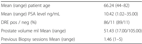
At one center, data was collected from 97 consecutive men with a mean age of 66.2 (44 –82) years [Table 1]. All of them were at a persistent risk of prostate cancer and had at least one previous set of TRUS-guided pros-tate Bx, yielding a non-cancerous diagnosis. All of them had suspicious findings at the digital rectal examination (DRE), or serum prostate-specific antigen (PSA) level >10 ng/mL, or both. Elevated serum PSA levels >4 ng/ mL a PSA-velocity of >0.75 ng/mL p.a. and free-to-total PSA ratio < 15 % were seen as the indication for prostate biopsies. Rescanning of the prostate was scheduled at least three months after the previous manipulation of the gland in order to minimize impairment of data qual-ity through earlier diagnostic procedures. All patients were informed of the mode of the extended prostate Bx scheme and its potential complications. All patients pro-vided written informed consent for the procedures. Pa-tients were advised that information collected from their Bx would be used for internal analysis and medical re-search as approved by the local Ethics Committee of the medical faculty of the University of Schleswig-Holstein, and was subsequently analyzed retrospectively.
HistoScanning technique and prostate biopsy technique
After indication diagnostics and before starting the Bx procedure, all patients underwent an automatic stan-dardized 3-dimensional (3D) transrectal ultrasound (TRUS) using a BK ProFocus UV ultrasound system with 8818 tri-plane probe (Analogic Corp, Peabody MA, USA). To facilitate appropriate data acquisition and a standardized scanning process, an external motor sweeps the ultrasound probe's sagittal array. Beside the signals from macroscopic tissue boundaries, which are used to create anatomical images, ultrasound produces a continuous stream of echoes emanating from the
tis-sue`s underlying microscopic features, known as
ultrasound backscatter. PHS analyses these backscatter signals by using numerical descriptors of multipara-metric measures from the individual 3D raw data scan, which vary in its properties due to suspicious (malig-nant) or normal prostatic tissue characteristics. A statis-tical classifier categorizes corresponding prostatic regions as normal or suspicious. Displaying these infor-mations as a colored (red) overlay in the ultrasound im-ages complements the conventional ultrasound grey-scale diagnostics.
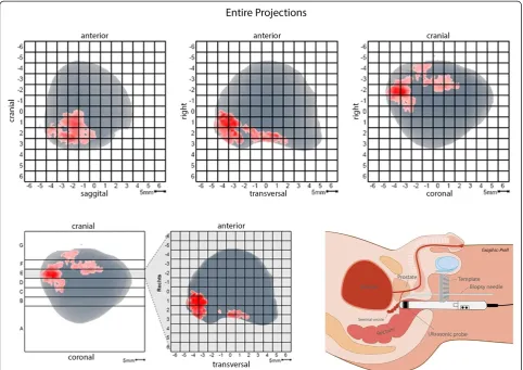
Computer-aided analysis of the data was performed on a PHS workstation with software version 2.3 (Advanced Medical Diagnostics, Waterloo BE). Based on the PHS image, the physician defined the most prominent (lar-gest) target regions, up to a maximum of three. In turn, a structured scaffold was created from PHS projection reports of the prostate, which was used at a later point for guidance of targeted Bx procedures (Fig. 1). The lo-cation of the suspicious/target regions was defined ac-cording to 12 peripheral sectors and two central sectors: a bilateral transition zone, apex, center, and base (each of the latter three medially and laterally).
The biopsy procedures were performed consecutively during a single visit with the patient in a dorsal lithot-omy position under general anesthesia. Three targeted cores were taken transperineally from each suspicious region based on PHS analysis (maximum of 3). A brachytherapy template grid fixed to a cradle was placed next to the perineum and used as a guide. Using the in-formation from the PHS projection reports, the biopsy needle was directed through the brachytherapy template to obtain the Bx under direct TRUS guidance using a BK 8848 ultrasound probe (Analogic Corp, Peabody MA, USA). Sampling a target lesion, neither the needle position (grid perforation) nor the depth of the biopsies was changed (Fig. 1). Thereafter, three targeted cores were taken by a transrectal approach from each suspi-cious region using the tri-plane 8818 probe. Using the information from PHS analysis, Bx was directed cogni-tively to the PHS positive volumes. Finally, a standard-ized 14-core transrectal-guided Bx was performed by sampling the corresponding seven sectors bilaterally, as mentioned above. Biopsies were performed randomly by four senior surgeons with at least five years of biopsy experience.
The local pathologists of the University Hospital Schleswig Holstein, Campus Kiel, performed histopath-ology analysis of Bx cores.
Statistical analysis
All data were registered in a Microsoft Access database (2010) and tabulated subsequently in Microsoft Excel (2010), with statistical analysis performed using PC SAS (Version 9.3 or higher) and R (version 3.0.1). For Table 1Patient characteristics
Mean (range) patient age 66.24 (44–82)
Mean (range) PSA level ng/mL 10.42 (1.02–35.00)
DRE pos / neg (%) 86/11 (89/11)
Prostate volume ml Mean (range) 51.43 (17.00/105.00)
calculation on paired data the McNemar test was used. A two-sample Chi-squared test of proportions was used for binomial data. Differences in total PHS suspicious volume were compared using the Wilcoxon signed rank test. A p-value of 0.05 was considered to indicate statis-tical significance.
The cancer detection rate and procedure characteris-tics for all cases were evaluated. Table 1 summarizes the patient characteristics.
Results
PHS analysis detected abnormalities in all prostate scans of the 97 patients included. 224 sectors contained PHS positive ultrasound patterns. In 31 of 97 (32 %) patients prostate adenocarcinoma was diagnosed, and 40 of 248 (16 %) sectors harbored cancer with respect to biopsy re-sults. Additionally, histopathological examination de-tected atypical small acinar proliferation in seven patients (7 %), high-grade prostatic intraepithelial neo-plasia in 20 (21 %) and chronic active inflammation in 72 (74 %) respectively. Based on the combined outcome,
rate for each PHS positive region was significantly higher in both targeted transperineal and targeted transrectal Bx compared to systematic Bx(p < .001). Table 4 shows an analysis of the detection rates of different procedures. Twenty two cancers were detected by systematic biop-sies, while 20 and 21 cancers were detected in PHS tar-geted perineal and transrectal biopsies, respectively. The detection rate of significant cancer (GS≥7) in PHS-targeted biopsies was superior in comparison to system-atic biopsies. Perineal PHS targeted biopsies missed 1 significant cancer, PHS targeted transrectal biopsies missed 3 significant cancers, respectively. Most of the cancers (18/23, 78 %) detected by systematic biopsy were insignificant.
Discussion
Cancer specific accuracy in standard prostate Bx tends to be determined by PSA, prostate texture recorded by digital rectal examination as well as the number of biopsy cores in relation to prostate size, rather than by transrectal grey scale ultrasound displaying malignant foci [12]. Thus, a substantial number of patients undergo repeat biopsy, indicated by weak prognostic factors, still facing the same limited cancer-specific imaging characteristics of TRUS. With respect to initial data, PHS seems to overcome these limitations by analyzing radiofrequency backscattered raw data of prostatic ultrasound [13]. Our data is unable to confirm a comparable high prognostic validity for prostate cancer. In the present study, PHS revealed at least 2 to 3
aberrations in 94 % and 59 % of the ultrasound scans, but histopathological analysis confirms malignant tissue in only 32 % of the patients. Although, these detection re-sults are within the range of previous studies on repeat bi-opsies [4], calculations of PHS predictive characteristics show weak results with respect to PPV (17 %) and sensi-tivity (44 %). These findings are in line with recent studies which also failed to show significant accuracy for PHS to predict positive biopsy results: According to sextant ana-lysis, PHS had a sensitivity, specificity, PPV and NPV of 32.0-100 %, 5.9-32.7 %, 19.6-22.7 % and 86.4-84 %, re-spectively [9, 10]. Further analysis found no correlation between pathology and the whole gland tumor volumes estimated by PHS, nor between the PHS volumes and prostate cancer GS [10, 14]. Table 3 shows comparable re-sults. It has to be taken into account, that PHS algorithms allow exclusively two opposed classifications (suspicious vs. non-suspicious) for ultrasound properties in virtual subvolumes. Based on a repeated but uniform calculation, the number of decisions as displayed by the PHS volume does not display varying characteristics or allow risk strati-fication concerning Gleason grading. Additionally, it must be assumed that acoustic similarity in the ultrasound backscatter properties of malignant and nonmalignant structures might account for further limitations in the analysis. With respect to specificity and NPV of 83 % and 95 %, respectively, PHS results need careful interpretation before the results can make a contribution to decision-making like biopsy indication or planning.
Table 2Histopathological results in prostate biopsies
Systematic HS-template targeted transperineal HS- targeted transrectal All biopsies
Bx+ 23 20 21 31
GS sum in Bx+ Mean (range) 6.5 (5–9) 6.5 (5–9) 6.38 (5–9) 6.35 (5–9)
GS sum≥7 in Bx+ N (%) 5 (5) 10(10) 8(8) 11 (11)
GS I = 2/3 in Bx+ N (%) 19(19) 14(14) 16(16) 24 (25)
GS I = 4/5 in Bx+ N (%) 4(4) 6(6) 5(5) 7 (7)
ASAP N (%) 3(3) 2(2) 2(2) 7 (7)
HG-PIN positive N (%) 12(12) 9(9) 12(12) 20 (21)
Prostatitis N (%) 68(70) 59(61) 59(61) 72 (74)
Bx+, prostate cancer positive biopsy; GS, Gleason Score; ASAP, atypical small acinar proliferations; HG PIN, high-grade intra epithelial proliferations
Table 3Relation between Prostate HistoScanning™suspicious volume and Bx outcome
Bx + Bx- p-value
Total PHS suspicious volume in ml N 31 66
median(range) 1.58 (0.20-9.32) 1.26 (0.22-6.59) n.s.
Gleason sum <7 Gleason sum > =7
Total PHS suspicious volume in ml N 20 11
median (range) 1.55 (0.20-9.32) 1.86 (0.22-4.35) n.s.
On the other hand, the detection rate in our cohort improved significantly by PHS-targeted biopsies. On pa-tient level, the PHS template-targeted biopsies account for a 27 % increase in the detection rate, transrectal-targeted biopsies for 23 %, and the combined transrectal-targeted biopsies for an increase by 41 % compared to systematic biopsy alone. PHS perineal targeted biopsies detect twice as much significant cancer foci than systematic ultrasound-guided biopsies. De Coninck et al. reported comparable findings in 41 men who underwent prostate biopsies supplemented with cognitive-targeted biopsies in suspicious regions based on prostate PHS. Targeted biopsies were 4.5-fold more likely to be positive than the random biopsies [11]. In our series, the rate of non-significant cancers detected by PHS-targeted perineal vs. systematic Bx was lower (Table 2). This rebuts po-tential concerns of an increased detection of clinically insignificant cancers caused by more substantial sam-pling. PHS-targeted biopsies detected 6 significant pros-tate cancers that were overseen by systematic biopsy. Additionally, the absolute numbers of atypical histo-pathological findings like ASAP, HG PIN and prostatitis in PHS-positive sectors do not differ significantly from systematic Bx despite the limited sampling.
Our data might suggest a reduction of biopsies in non-PHS positive sectors and to focus on more vulner-able or suspect regions in the prostate. The correspond-ing PB scheme would theoretically achieve appropriate sampling by targeting certain locations rather than by increasing the core numbers. In an analysis of 164 aut-opsies, which had not previously undergone clinical bi-opsy, step-section analysis revealed that the common 12-core biopsy technique detects the majority of clinic-ally significant cancers with a sensitivity of 80 % [15]. The authors conclude that the chance of detecting can-cer was correlated more closely to the location of the sampling (peripheral, lateral and apical cores) than to the number of biopsy cores taken.
Prostate cancer detection rates from perineal targeted biopsies differed from the transrectal targeted biopsy re-gime. This difference occurred independently from pre-vious tissue analysis, for the reason that both targeted approaches are aligned to the same scanning process. In comparison to the transrectal approach, the perineal
biopsy technique might reduce variables that can influ-ence the needle placement. Furthermore, longitudinal bi-opsy punches following the axis of the prostate seem to allow more accurate sampling of the anterior part. The-oretically, because previous studies reported inhomogen-eous results comparing transrectal and transperineal prostate biopsies [8, 16].
There are several limitations in this study. Firstly, the analysis was performed on a regional basis. Dependen-cies between neighboring regions, as well as correlation of the different biopsy procedures with the HS results are difficult to account for and may incur misleading re-sults. Secondly, there is no minimum threshold defining when a PHS signal is to be called positive; this may also affect the results. For example, the targeted regions could stretch across different sections meaning that even a small amount in one sector would lead to it being seen as a positive PHS sector, but it might still yield negative biopsies because the few cores would not necessarily pick up the cancer. Moreover, the knowledge of“ abnor-mal” regions might influence the surgeon’s decisions during the subsequent systematic biopsy as a result of the non-blinded, single-surgeon biopsy procedure. Nevertheless, we did not find any significant inter-operator variability with respect to the detection rate or the biopsy approach.
Generally, it is difficult to compare the diagnostic ac-curacy of Bx techniques in different patient series. Eth-ical concerns will always prevent verification of negative Bx results by radical prostatectomy, and positive biopsies are not always and necessarily followed by prostatec-tomy. Therefore, the clinical setting will always account for a potential verification bias, not knowing what the exact location of the cancer is and how many cancers were missed by biopsy.
Finally, recent results from multi-parametric MRI pro-cedures pull into question all ultrasound-based techniques including color Doppler imaging, real-time elastography, C-TRUS / ANNA and Prostate HS. The application of in-tegrated interpretation strategies for mp MRI variables, such as the PIRADS score, improves the risk stratification for prostate cancer and allows for effective fusion guid-ance of targeted prostate biopsy [17, 18]. Likewise, all ultrasound modalities have been devised to increase the Table 4Analysis of detection rate per prostate region of different procedures
N = 97 Systematic Bx PHS-template targeted transperineal Bx PHS- targeted transrectal Bx
patients with Bx+ 23 20 n.s. 21 n.s.
regions biopsied 1358 248 248
regions with Bx+ 73 32 28
detection rate per region 5 % 13 % <0.001 11 % <0.001
diagnostic yield of prostate biopsy, using ultrasound data to visualize and locate tissues suspected of harboring pros-tate cancer [19–21]. Despite their promising characteris-tics, none of them are established in clinical practice because of missing evidence, lack of standardization and significant user-dependent performance. Similarity in the prostate tissue features such as the degree of elasti-city/firmness, irregular patterns of blood vessels and ultrasound backscatter properties of malignant and other non-malignant microstructures might account for these limitations. So far, each technique in itselfis lim-ited in its predictive impact on prostate cancer diagnos-tics, but might be combined into an effective diagnostic tool. Accordingly, a multiparametric approach similar to MRI algorithms might improve future performance and increase the diagnostic yield by specifically visualizing and locating prostate cancer tissue.
Conclusions
HS-augmented ultrasound analysis improves the inter-pretation of transrectal prostate imaging by identifica-tion of abnormal prostatic tissue. Although prostate cancer specificity remains low, the additional informa-tion facilitates improved biopsy targeting and results in a higher cancer detection rate in selective prostate sectors. The high NPV might help to reduce biopsies in PHS-negative prostate sectors and to focus on more vulner-able regions in the prostate. With the help of this strategy, increased but unsought detection of clinically insignificant cancers through too substantial sampling can be avoided. The PHS technique should be consid-ered at leastwith respect to patients who had previously negative prostate biopsy results despite elevated PSA levels. Further multicenter analysis has to confirm the advantages of PHS in primary Bx-settings.
Abbreviations
TRUS:Transrectal ultrasound; PHS: Prostate HistoScanning™; PCa: Prostate cancer; Bx: Prostate biopsies; tBx: Targeted prostate biopsies; sBx: Systematic prostate biopsies; NPV: Negative predictive value; PPV: Positive predictive value; PSA: Prostate specific antigen; mp MRI: Multiparametric magnet resonance imaging.
Competing interests
The authors declare that they have no competing interests.
Authors´ contributions
All of the authors have made substantial contributions to the study. MH has developed the concept and design of the study, performed the statistical analysis and interpretation of data and drafted the manuscript. CH has made substantial contributions to concept of the study, data acquisition and was involved in drafting the manuscript. AT was involved in acquisition, analysis and interpretation of data. KJ has made substantial contributions to concept of the study and was involved in revising the manuscript critically and adding important content. MN has participated in the design of the study, the interpretation of data and was involved in drafting the manuscript and revising it critically and substantially. All authors have read and approved the final manuscript.
Acknowledgements
We thank Almut Kalz who has provided medical writing services and linguistic amendments. Further, we thank Felix Prell who set up the figures. They both belong to our institutional study center, a part of the department of Urology and pediatric Urology, University Hospital Schleswig Holstein, Campus Kiel.
Received: 6 February 2015 Accepted: 20 July 2015
References
1. Heidenreich A, Bastian PJ, Bellmunt J, Bolla M, Joniau S, van der Kwast T, et al. EAU guidelines on prostate cancer. part 1: screening, diagnosis, and local treatment with curative intent-update 2013. Eur Urol.
2014;65(1):124–37.
2. Carter HB, Albertsen PC, Barry MJ, Etzioni R, Freedland SJ, Greene KL, et al. Early detection of prostate cancer: AUA Guideline. J Urol.
2013;190(2):419–26.
3. Ukimura O, Coleman JA, de la Taille A, Emberton M, Epstein JI, Freedland SJ, et al. Contemporary role of systematic prostate biopsies: indications, techniques, and implications for patient care. Eur Urol. 2013;63(2):214–30. 4. Roehl KA, Antenor JA, Catalona WJ. Serial biopsy results in prostate cancer
screening study. J Urol. 2002;167:2435–9.
5. Kasivisvanathan V, Dufour R, Moore CM, Ahmed HU, Abd-Alazeez M, Charman SC, et al. Transperineal magnetic resonance image targeted prostate Biopsy versus transperineal template prostate Biopsy in the detection of clinically significant prostate cancer. J Urol. 2013;189:860–6. 6. Aigner F, Pallwein L, Schocke M, Lebovici A, Junker D, Schäfer G, et al.
Comparison of Real-time Sonoelastography With T2-weighted endorectal magnetic resonance imaging for prostate cancer detection. JUM. 2011;30:643–9.
7. Simmons LA, Autier P, Zát'ura F, Braeckman J, Peltier A, Romic I, et al. Detection, localisation and characterisation of prostate cancer by Prostate HistoScanningTM. BJU Int. 2011;110:28–35.
8. Hamann MF, Hamann C, Schenk E, Al-Najar A, Naumann CM, Jünemann KP. Computer-aided (HistoScanningTM) Biopsies versus conventional transrectal ultrasound-guided prostate biopsies: Do targeted biopsy schemes improve the cancer detection rate? Urology. 2013;81:370–5.
9. Schiffmann J, Tennstedt P, Fischer J, Tian Z, Beyer B, Boehm K, et al. Does HistoScanning™predict positive results in prostate biopsy? A retrospective analysis of 1,188 sextants of the prostate. World J Urol. 2014;32(4):925–30. 10. Javed S, Chadwick E, Edwards AA, Beveridge S, Laing R, Bott S, et al. Does HistoScanningTM play a role in detecting prostate cancer in routine clinical practice? Results from three independent studies. BJU Int. 2014;114(4):541–8. 11. De Coninck V, Braeckman J, Michielsen D. Prostate HistoScanning: a
screening tool for prostate cancer? Int J Urol. 2013;20:1184–90. 12. Chun FK, Epstein JI, Ficarra V, Freedland SJ, Montironi R, Montorsi F, et al.
Optimizing performance and interpretation of prostate biopsy: a critical analysis of the literature. Eur Urol. 2010;58:851–64.
13. Braeckman J, Autier P, Soviany C, Nir R, Nir D, Michielsen D, et al. The accuracy of transrectal ultrasonography supplemented with computer-aided ultrasonography for detecting small prostate cancers. BJU Int.
2008;102:1560–5.
14. Schiffmann J, Fischer J, Tennstedt P, Beyer B, Böhm K, Michl U, et al. Comparison of prostate cancer volume measured by HistoScanning and final histopathological results. World J Urol. 2014;32(4):939–44. 15. Haas GP, Delongchamps NB, Jones RF, Chandan V, Serio AM, Vickers AJ,
et al. Needle biopsies on autopsy prostates: sensitivity of cancer detection based on true prevalence. J Natl Cancer Inst. 2007;99:1484–9.
16. Abdollah F, Novara G, Briganti A, Scattoni V, Raber M, Roscigno M, et al. Trans-rectal versus trans-perineal saturation rebiopsy of the prostate: is there a difference in cancer detection rate? Urology. 2011;77:921–5.
17. Somford DM, Hoeks CM, de Kaa CA H-v, Hambrock T, Fütterer JJ, Witjes JA, et al. Evaluation of diffusion-weighted MR imaging at inclusion in an active surveillance protocol for low-risk prostate cancer. Invest Radiol.
2013;48:152–7.
18. Barentsz JO, Richenberg J, Clements R, Choyke P, Verma S, Villeirs G, et al. ESUR prostate MR guidelines 2012. Eur Radiol. 2012;22:746–57.
20. Salomon G, Drews N, Autier P, Beckmann A, Heinzer H, Hansen J, et al. Incremental detection rate of prostate cancer by real-time elastography targeted biopsies in combination with a conventional 10-core biopsy in 1024 consecutive patients. BJU Int. 2014;113(4):548–53.
21. Grabski B, Baeurle L, Loch A, Wefer B, Paul U, Loch T. Computerized transrectal ultrasound of the prostate in a multicenter setup (C-TRUS-MS): detection of cancer after multiple negative systematic random and in primary biopsies. World J Urol. 2011;29(5):573–9.
Submit your next manuscript to BioMed Central and take full advantage of:
• Convenient online submission
• Thorough peer review
• No space constraints or color figure charges
• Immediate publication on acceptance
• Inclusion in PubMed, CAS, Scopus and Google Scholar
• Research which is freely available for redistribution