OncoTargets and Therapy 2017:10 4821–4828
OncoTargets and Therapy
Dove
press
submit your manuscript | www.dovepress.com
4821
R e v i e w
open access to scientific and medical research
Open Access Full Text Article
Theranostics in nuclear medicine practice
Anna Yordanova
1elisabeth eppard
2Stefan Kürpig
2Ralph A Bundschuh
3Stefan Schönberger
4Maria Gonzalez-Carmona
5Georg Feldmann
6Hojjat Ahmadzadehfar
1Markus essler
11Department of Nuclear Medicine
(Clinical Nuclear Medicine),
2Department of Nuclear Medicine
(Radiochemistry), 3Department
of Nuclear Medicine (Medical Physics and PeT), 4Department
of Paediatric Haematology and Oncology, 5Department of Medicine 1, 6Department of Medicine 3, University
Hospital Bonn, Bonn, Germany
Abstract: The importance of personalized medicine has been growing, mainly due to a more urgent need to avoid unnecessary and expensive treatments. In nuclear medicine, the theranostic approach is an established tool for specific molecular targeting, both for diagnostics and therapy. The visualization of potential targets can help predict if a patient will benefit from a particular treatment. Thanks to the quick development of radiopharmaceuticals and diagnostic techniques, the use of theranostic agents has been continually increasing. In this article, important milestones of nuclear therapies and diagnostics in the context of theranostics are highlighted. It begins with a well-known radioiodine therapy in patients with thyroid cancer and then progresses through various approaches for the treatment of advanced cancer with targeted therapies. The aim of this review was to provide a summary of background knowledge and current applications, and to identify the advantages of targeted therapies and imaging in nuclear medicine practices.
Keywords: theranostics, nuclear medicine, personalized medicine, PET/CT, therapy, diagnostics
Introduction: goals of theranostics in nuclear
medicine
Challenges in modern oncology include the fact that patients are getting older and are
typically unfit for conventional chemotherapy regimens because of comorbidities or
poor performance status.
1Furthermore, the occurrence of side effects may aggravate
treatment compliance in both young and elderly patients.
2To manage these problems,
it is important to improve patient selection, reduce side effects, and enhance therapeutic
efficacy. Taking these factors into consideration, the combination of targeted cancer
imaging and therapy is a considerable achievement for personalized medicine.
The theranostic approach in nuclear medicine couples diagnostic imaging and
therapy using the same molecule or at least very similar molecules (Figure 1), which are
either radiolabeled differently or given in different dosages. For example, iodine-131
and lutetium-177 are gamma and beta emitters; thus, these agents can be used for
both imaging and therapy. Furthermore, different isotopes of the same element, for
example, iodine-123 (gamma emitter) and iodine-131 (gamma and beta emitters), can
also be used for theranostic purposes.
3,4Newer examples are yttrium-86/yttrium-90 or
terbium isotopes (Tb):
152Tb (beta plus emitter),
155Tb (gamma emitter),
149Tb (alpha
emitter), and
161Tb (beta minus particle).
5,6The detection of potential targets can help predict whether a patient will benefit from
a particular treatment. Theranostics can be useful for estimating the potential response
and eventual toxicity. During the treatment, theranostics can be applied in monitoring
the therapy course. However, one cause of concern is the safety of high cumulative
doses of radioactive agents after multiple repeated cycles. For instance, reports
con-cerning irreversible high-grade toxicity followed soon after the first treatments with
radioiodine (iodine-131).
7However, there have been remarkable advances in nuclear
Correspondence: Anna Yordanova Department of Nuclear Medicine, University Hospital Bonn, Sigmund-Freud-Str 25, Bonn 53127, Germany
Tel +49 228 287 16814 Fax +49 228 287 90 19107 email [email protected]
Journal name: OncoTargets and Therapy Article Designation: Review
Year: 2017 Volume: 10
Running head verso: Yordanova et al
Running head recto: Theranostics in nuclear medicine DOI: http://dx.doi.org/10.2147/OTT.S140671
OncoTargets and Therapy downloaded from https://www.dovepress.com/ by 118.70.13.36 on 25-Aug-2020
For personal use only.
Number of times this article has been viewed
This article was published in the following Dove Press journal: OncoTargets and Therapy
Dovepress Yordanova et al
medicine, especially in the field of targeted therapies. After a
proper preselection of candidates, targeted nuclear therapies
have proven to be effective in the majority of cases with a
favorable safety profile.
8–11Recent studies have shown no
evidence of grade 3/4 toxicity in patients with neuroendocrine
tumors after repeated (
$
8 cycles) radiopeptide therapy with
a cumulative dose of up to 97 GBq.
12Good tolerability has
also been observed in patients with prostate cancer (PC)
after a cumulative dose of 36 GBq and up to six cycles of
radioligand therapy.
13,14Nuclear imaging utilizes gamma and positron emitters
(
β
+). Gamma emitters, such as technetium-99m (
99mTc) or
iodine-123 (
123I), can be located using gamma cameras
(pla-nar imaging) or SPECT (single photon emission computed
tomography).
15However, better resolution can be achieved
via PET (positron emission tomography) using positron
emit-ters, such as gallium-68 (
68Ga) and fluor-18 (
18F).
16Most therapeutic radiopharmaceuticals are labeled with
beta-emitting isotopes (
β
−). The tissue penetration of these
particles is proportional to the energy of the radioisotopes.
17Beta particles have a potential cytocidal effect, but they also
spare the surrounding healthy tissue due to having a tissue
penetration of only a few millimeters.
8Commonly used
beta emitters in routine nuclear oncology practices include
lutetium-177 (
177Lu, tissue penetration: 0.5–0.6 mm,
maxi-mum: 2 mm, 497 keV, half-life: 6.7 days) and yttrium-90
(
90Y, tissue penetration: mean 2.5 mm, maximum: 11 mm,
935 keV, half-life: 64 hours).
8,10,13,18–20The first theranostic radiopharmaceutical in nuclear
medicine history was radioiodine, which was used for therapy
and imaging in thyroid diseases.
21Since then, the use of
theranostic agents has been consistently increasing. Nuclear
targeted therapies play an essential role, especially in patients
with advanced neuroendocrine tumors, such as
gastroentero-pancreatic (GEP) tumors, bronchopulmonary neuroendocrine
tumors, pheochromocytoma, and neuroblastoma.
10,11,20,22–27Furthermore, there are positive results with radioligand
thera-pies in metastatic PC and metastatic melanoma.
8,13,19,28,29The aim of this review was to discuss the most
impor-tant milestones of nuclear theranostics in current practice
(Table 1), and to provide a summarized background and
overview of current applications and advantages of targeted
therapies and imaging.
Radioiodine therapy: “the gold standard”
in thyroid diseases
Iodine (stable isotope: iodine-127) is taken up by the
thy-roid gland for the production of thythy-roid hormones, namely,
thyroxine (T4) and triiodothyronine (T3).
30Thyroid hormones
are vital for the embryonic and neonatal development of the
brain, normal growth, and metabolic balance.
9,31–33In 1936,
Dr Saul Hertz, director of the Thyroid Clinic in Massachusetts
(1931–1943), developed the idea of administering radioactive
iodine in patients with thyroid diseases. Iodine and external
beam radiation were well-known tools in thyroid disease
therapy, but the combination of these was a considerably
innovative approach. This idea followed a few years of
preclinical studies in collaboration with the Massachusetts
Institute of Technology (MIT), where the first cyclotron for
medical use had been built. The MIT Cyclotron produced
90% iodine-130 (
130I, half-life: 12 hours) and 10% iodine-131
(
131I, half time: 8 days). Subsequently, on March 31, 1941,
Dr Hertz treated the first patient with radioiodine (
130I).
21,34,35The first radioiodine therapy (RAI) with
131I in patients
with thyroid cancer (TC) was undertaken by Seidlin et al in
1946.
36This group studied the use of RAI in patients with
metastatic thyroid carcinomas.
36,37Seidlin et al also reported
one of the first cases of acute myeloid leukemia after repeated
RAI treatments.
7To date,
131I is still the gold standard for the therapy and
diagnosis of differentiated TC.
38It is a low-cost nuclear
reactor product from the neutron bombardment of
tellurium-131.
131I combines the characteristics of a beta (
β
−,
approxi-mately 90% of the radiation, mean: 192 keV, mean tissue
penetration: 0.4 mm) and gamma (approximately 10% of the
radiation, mean: 383 keV) emitter. In this way, it irradiates
Figure 1 The theranostic principle in nuclear medicine involves combining diagnosticimaging and therapy with the same molecule, which is radiolabeled differently, or administered in other dosages. in case of radioiodine therapy (RAi), the radioisotope (131i or 123i) can be directly mediated by the sodium-iodide symporter in the thyroid
cells. In other cases, it can be more complex. The image shows a simplified model of a radiopharmaceutical, which consists of a binding molecule that binds the target, and a linking molecule, which binds the radioisotope. examples of such theranostic molecules are DOTA-TOC, DOTA-TATe, and PSMA-617.
5DGLRQXFOLGH
/LQNLQJPROHFXOH
&HOO
%LQGLQJPROHFXOH
7DUJHW
)RUWKHUDS\
β±RUDOSKD
HPLWWHUV
)RUGLDJQRVWLFV
βRUJDPPD
HPLWWHUV
OncoTargets and Therapy downloaded from https://www.dovepress.com/ by 118.70.13.36 on 25-Aug-2020
Dovepress Theranostics in nuclear medicine
the TC and the thyroid remnant from the inside and, at the
same time, targeted lesions can be visualized using a gamma
camera or SPECT.
9,32,33Another radioisotope is
123I, which is a
pure gamma emitter and is used for pre- and post-therapeutic
diagnostics. The advantages of imaging with
123I include a
higher quality of whole-body scans, which improves the
sensitivity in detecting thyroid remnants.
3,4Figure 2A shows the initial
131I-planar images of a
74-year-old female with metastatic TC (lung, bone, and
intracranial soft-tissue metastases). The level of tumor
marker thyroglobulin (Tg) before RAI was 572 ng/mL. After
two administrations of RAI (cumulative activity: 14.3 GBq),
the patient was in complete remission with a Tg level of
0.2 ng/mL (Figure 2B).
Diagnostics and therapy with
metaiodobenzylguanidine
Metaiodobenzylguanidine (mIBG), or iobenguane, is a
molecule similar to noradrenaline and enters
neuroendo-crine cells from the sympathetic nervous system, either
by endocytosis or passive diffusion before being stored in
neurosecretory granules.
39Among the used radiolabeled
molecules, [
123I]I-mIBG has a lower gamma energy than
[
131I]I-mIBG (159 keV vs 360 keV), which makes it more
suitable for planar imaging/SPECT. Furthermore, the pure
gamma emitter [
123I]I-mIBG consists of a shorter half-time
of 13 hours compared to 8 days for the combined beta and
gamma emitter [
131I]I-mIBG, leading to a smaller
radia-tion burden. Thus, higher quantities of [
123I]I-mIBG can be
injected.
40Both [
131I]I-mIBG and [
123I]I-mIBG are used in
mIBG scintigraphy for the detection of neuroendocrine
tumors, such as neuroblastomas, pheochromocytomas,
paragangliomas, medullary thyroid carcinomas, and other
neuroendocrine neoplasias.
41In patients with inoperable or
advanced tumors with distant metastases, mIBG imaging
plays an essential role in response assessment after therapy
and in the evaluation of potential [
131I]I-mIBG therapy.
42,43In patients with neuroblastoma and pheochromocytoma,
[
123I]I-mIBG has high sensitivity (97% and 94%) and
speci-ficity (up to 96% and 92%), respectively.
44–46If available,
[
124I]I-mIBG-PET can be equally used for the planning of
mIBG targeted therapy.
47,48Targeted therapy with [
131I]I-mIBG presents an
encour-aging efficacy with tolerable toxicity in relapsed or
refrac-tory neuroblastomas with response rates between 20% and
40% being used alone or in combination with high-dose
Table 1 Overview of theranostic agents
Theranostic molecule
Iodine mIBG SSA PSMA-ligands Benzamide/
arylcarboxamide Target Thyroid cancer cells Neurosecretory granules SSTR, especially the
subtype SSTR2
PSMA Melanin
Planar imaging/ SPeCT or PeT
131i and 123i [131i]i-miBG, [123i]i-miBG SSA labeled with
indium-111
[123i]i-MiP-1072 [123i]i-BA52
124i [124i]i-miBG [68Ga]Ga-DOTA-TATe
[68Ga]Ga-DOTA-TOC
[68Ga]Ga-DOTA-NOC
[68Ga]Ga-PSMA-11
[68Ga]Ga-PSMA-617
[18F]F-iCF15002
Therapeutic agent
131i [131i]i-miBG [177Lu]Lu-DOTA-TATe
[177Lu]Lu-DOTA-TOC
[90Y]Y-DOTA-TOC
[90Y]Y-DOTA-TATe
[177Lu]Lu-J591
[90Y]Y-J591
[131i]i-MiP-1095
[177Lu]Lu-PSMA-617
[131i]i-BA52
[131i]i-iCF15002
indication Thyroid cancer Neuroblastomas, pheochromocytomas, paragangliomas, medullary thyroid carcinomas, and other NeN
NeN, especially GeP-NeN
Metastatic prostate cancer
Metastatic melanoma
Abbreviations: miBG, metaiodobenzylguanidine; SSA, somatostatin analogs; SSTR, somatostatin receptors; NeN, neuroendocrine neoplasia; GeP, gastroenteropancreatic system; SPeCT, single photon emission computed tomography; PeT, positron emission tomography.
7JQJP/
$3 3$
5$,FXPXODWLYH DFWLYLW\*%T
7JQJP/ $3
3$
$
%
Figure 2 (A) initial 131i-planar images of a 74-year-old female with metastatic
thyroid cancer (lung, bone, and intracranial soft-tissue metastases, marked with arrows). The tumor marker thyroglobulin (Tg) before radioiodine therapy (RAi) was 572 ng/mL. (B) After two administrations of RAi (cumulative activity: 14.3 GBq), the patient was in complete remission with the Tg at 0.2 ng/mL. The planar images show only a physiological uptake of the radiotracer in the gastrointestinal tract and pharyngeal mucosa (marked with an asterisk).
OncoTargets and Therapy downloaded from https://www.dovepress.com/ by 118.70.13.36 on 25-Aug-2020
Dovepress Yordanova et al
chemotherapy followed by autologous stem cell
transplantation.
23–25,49Recently, the NB2004 Trial for Risk
Adapted Treatment of Children with Neuroblastoma closed,
and further analysis of the usage of mIBG therapy as a
first-line therapy is outstanding.
23,25Palliation of bone metastases
Radiolabeled phosphonates have a high bone affinity and can
be used for imaging and palliation of painful bone
metas-tases. Depending on the degree of osseous metabolism, the
tracer accumulates via adhesion to bones and, preferably, to
osteoblastic bone metastases.
8Therapy planning requires a bone scintigraphy with
technetium-99m-hydroxyethylidene diphosphonate (HEDP)
to estimate the metabolism and the extent of the metastases
involvement. Bisphosphonate HEDP can be labeled for
therapy either with rhenium-186 (beta-emitter, half-life:
89 hours, 1.1 MeV maximal energy, maximal range: 4.6 mm)
or rhenium-188 (beta-emitter [to 85%, 2.1 MeV] and
gamma-emitter [to 15%,155 keV], half-life: 16.8 hours, maximal
range in soft tissue: 10 mm).
50Both agents induce pain relief in
$
90% of patients.
51However, rhenium-188 is of particular interest because
it can be cost-effectively produced using a tungsten-188/
rhenium-188 generator. The gamma component of the agent
allows post-therapeutic imaging of good quality.
8Further-more, patients with prostatic cancer have shown, after two
[
188Re]Re-HEDP injections, a significantly higher response
rate (92%), a prostate-specific antigen (PSA) decline
.
50%
(38%), and a longer PFS (7 months) compared to patients
who have had a single injection.
52,53Biersack et al showed
an improved OS of 15.7 months in patients after repeated
injections (
$
3 injections) compared to patients with a
single therapy.
54Besides transient bone marrow toxicity
(thrombocytopenia grade III), the reported toxicity is low
to moderate.
51New promising radiopharmaceuticals for bone palliation
therapy include radiolabeled complexes of zoledronic acid.
Zoledronic acid belongs to a new, most potent generation of
bisphosphonates with cyclic side chains. The bone affinity of
zoledronic acid labeled with scandium-46 or lutetium-177 has
shown excellent absorption (98% for [
177Lu]Lu-zoledronate
and 82% for [
46Sc]Sc-zoledronate), which is much higher
than of bisphosphonates labeled with samarium-153
(maximum: 67%).
55Radiolabeled somatostatin analogs
Neuroendocrine neoplasia (NEN) of the GEP system
origi-nates most frequently from the pancreas, jejunum, ileum,
cecum, rectum, appendix, and colon. The common
character-istic of all GEP-NEN is the compound features of endocrine
and nerve cells.
56–58Well-differentiated NEN overexpresses
somatostatin receptors (SSTRs), especially the SSTR-2
subtype. Thus, SSTRs are theranostic targets in NEN that
have been known for almost three decades and have become
well established.
59–61SSTR imaging is necessary for staging, therapy planning,
and follow-up. In the PET diagnostics, there are three routine
somatostatin analog (SSA) tracers labelled with gallium-68:
DOTA-TATE, DOTA-TOC, and DOTA-NOC. All three
tracers bind specifically with SSTRs.
62,63Gallium-68-labeled
SSTRs have high sensitivity (82%–97%) and specificity
(80%–92%) in the detection of small primary tumors or
metastases of GEP-NEN.
64–66Alternatively, a conventional
whole-body scintigraphy (planat plus SPECT) or SPECT
alone can be performed using indium-111-labeled SSTR,
which is available at many institutions. Even if the
111In
imaging does not have as good a quality as PET, this method
has been shown to be superior to chromogranin A
measure-ments alone for the management of GEP-NET patients and
is better than computed tomography or ultrasound.
67,68Peptide receptor radionuclide therapy (PRRT) is a
sys-temic therapy in patients with advanced metastatic NEN.
PRRT requires a good tumor uptake in the SSTR imaging.
For therapeutic purposes, the peptides DOTA-TATE and
DOTA-TOC can be labeled with either
90Y or
177Lu. Because
of the renal excretion and the proximal tubular
reabsorp-tion of these tracers, the kidneys are one of the primary
limiting organs for this therapy.
69Due to the smaller range
(2 mm) and lower energy,
177Lu is less nephrotoxic than
90Y (range maximum: 11 mm, pure beta emitter, higher
energy).
177Lu is also less hematotoxic.
70This allows for the
performance of several PRRT cycles without any relevant
toxicity. Furthermore, the overall survival is not significantly
different between the therapies. For that reason, [
177Lu]
Lu-DOTA-TATE and DOTA-TOC are, in many centers, the
preferred agents for NEN therapy.
10,11,71,72PRRT has been increasingly gaining attention. The first
randomized controlled phase III study, NETTER-1 (started
2012), compared the standard therapy of Sandostatin
®LAR
(somatostatin analog) with PRRT in patients with midgut
NEN. The study showed a significant clinical benefit from
the therapy, achieving a prolonged progression-free survival
(median not reached, approximately 40 months,
p
,
0.001),
an overall response of 18%, and a presumably longer overall
survival (median not reached,
p
=
0.004).
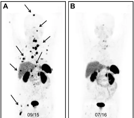
11Figure 3 represents the image of a male patient with a
neuroendocrine tumor (unknown origin), with a recurrence of
OncoTargets and Therapy downloaded from https://www.dovepress.com/ by 118.70.13.36 on 25-Aug-2020
Dovepress Theranostics in nuclear medicine
the disease nearly 2 years after the baseline PRRT. After a
sal-vage therapy of PRRT, the patient had a partial response.
Radiolabeled PSMA-ligands
PC is the most common cancer in men in Western countries.
73Hormone- and chemotherapy-refractory patients with
metastatic PC have a poor prognosis.
8,74,75The main cause
of death in these patients is progression to the
androgen-independent stage.
8PC cells overexpress prostate-specific membrane antigen
(PSMA) on the cell surface.
76–79There are several available
radiopharmaceuticals that target PSMA including [
68Ga]Ga-
PSMA-HBED-CC (also known as [
68Ga]Ga-PSMA-11
[PET]), a monoclonal antibody (mAb) [
177Lu]Lu/[
90Y]Y-J591
(therapy), [
123I]I-MIP-1072 (planar/SPECT), [
131I]I-MIP-1095
(therapy), and the theranostic agents PSMA-I&T and
DKFZ-PSMA-617 (DKFZ-PSMA-617), which are labeled with
68Ga for
PET or with
177Lu for therapy.
The specificity of the two commercially available
PET-tracers, [
68Ga]Ga-PSMA-617 and [
68Ga]Ga-PSMA-11, is
similar – 99% and 100%, respectively.
80–82However, due to
slower kinetics, PSMA-617 was suggested for labeling with
the long-half-life Lutetium-177 for therapy and, therefore,
there is sparse systematic evaluation of [
68Ga]Ga-PSMA-617
for diagnostics.
83The high kidney uptake of PSMA-11 in the
kidneys makes it unsuitable for therapy and does not fit the
definition “theranostics.”
84Still, it is at issue if molecules with
affinity to identical target structures might already present a
“theranostic surrogate” even if more than the isotope differs
between the diagnostics and the therapeutics.
The latest studies have shown that treatment with [
177Lu]
Lu-PSMA-617 is effective and well tolerated. In fact, nearly
70% of patients have benefited from this therapy.
13,28,85–90Dosimetry studies have shown that the most critical organs
are the kidneys, with a maximum kidney radiation dose of
0.88 Gy/GBq for [
177Lu]Lu-PSMA-617 and 0.93 Gy/GBq for
[
177Lu]Lu-PSMA-I&T.
91,92Okamoto et al stated that a
cumu-lative activity of 40 GBq [
177Lu]Lu-PSMA-I&T could be
safely applied in patients.
91Furthermore, [
177Lu]Lu-PSMA-617 has been shown to produce no relevant increase in renal
toxicity in the salvage setting or in patients with kidney
radiation doses
.
19 Gy.
14Figure 4A shows the pre-therapeutic [
68Ga]Ga-PSMA-11
images of a patient with multiple lymph node, peritoneal,
and bone metastases (arrows), and a history of chemotherapy
(first and second line), enzalutamide, and abiraterone. After
three cycles of [
177Lu]Lu-PSMA-617, the follow-up images
show a very good response with a substantial PSA decline
(Figure 4B).
Melanin targeting in patients with
metastatic melanoma
A promising approach in patients with metastatic melanoma
is the specific targeting of melanin. The newly developed
theranostic agents include [
123I]I-/[
131I]I-BA52 and [
18F]F-/
[
131I]F-ICF15002, which may play a considerable role in
the future.
$
%
Figure 3 (A) [68Ga]Ga-DOTA-TOC-image* of a male patient with a neuroendocrine
tumor (unknown origin) 23 months after the baseline PRRT (three cycles, cumulative activity: 19.6 GBq). The patient had a recurrence of the disease with multiple metastases in the bone, lung, liver, and lymph nodes (marked with arrows). (B) After another course of PRRT (three cycles, cumulative: 43.4 GBq), the PeT images showed a partial response.
Note: *Maximum-intensity projection (MiP) PeT image is a visualization technique that provides an initial overview of the case.
Abbreviations: PeT, positron emission tomography; PRRT, peptide receptor radionuclide therapy.
36$QJP/
F\FOHVRI>/X@/X360$
36$QJP/
$
%
Figure 4 (A) Pre-therapeutic [68Ga]Ga-PSMA-11 images* of a patient with multiple
lymph node, peritoneal, and bone metastases (arrows), and history of chemotherapy (first and second line) with enzalutamide and abiraterone. (B) After three cycles of [177Lu]Lu-PSMA-617, the follow-up images showed a very good response with a
substantial PSA decline.
Note: *Maximum-intensity projection (MiP) PeT image is a visualization technique that provides an initial overview of the case.
Abbreviations: PET, positron emission tomography; PSA, prostate-specific antigen.
OncoTargets and Therapy downloaded from https://www.dovepress.com/ by 118.70.13.36 on 25-Aug-2020
Dovepress Yordanova et al
BA52 is a melanin-binding benzamide. Labeled with
123
I, it shows a specific binding of the pigmented metastases
in planar imaging/SPECT and can help select patients who
would probably benefit from the therapy. In a pilot study,
[
131I]I-BA52 was effective in three of five patients who were
treated with more than 4.3 GBq.
29ICF15002 is an arylcarboxamide derivative and, as
a small molecule, can passively enter the cell and bind
to melanin. The PET tracer is radiolabeled with
18F and
[
131I]I-ICF15002 is designed for the therapy. However, both
tracers are still in the preclinical phase of their studies. One
major problem may be the absorbed dose in melanin-rich
tissues, such as skin, dark eyes, and the brain. For instance,
in a murine model, there was a 30% decrease of the retinal
thickness after two cycles of [
131I]I-ICF15002.
93Conclusion
In nuclear medicine, theranostics combine diagnostic
imaging and therapy with the same, but differently
radio-labeled, molecule, or the same agent in different dosages.
The visualization of potential targets can help predict if a
patient would benefit from a particular treatment. In properly
preselected patients, targeted nuclear therapies have proven
to be effective with a favorable safety profile. To conclude,
the combination of targeted cancer imaging and therapy is a
considerable contribution to personalized medicine and may
play an increasingly important role in the future.
Disclosure
The authors report no conflicts of interest in this work.
References
1. Gridelli C. Does chemotherapy have a role as palliative therapy for unfit or elderly patients with non-small-cell lung cancer? Lung Cancer. 2002;38(Suppl 2):S45–S50.
2. Richardson JL, Marks G, Levine A. The influence of symptoms of disease and side effects of treatment on compliance with cancer therapy. J Clin Oncol. 1988;6(11):1746–1752.
3. Gerard SK, Cavalieri RR. I-123 diagnostic thyroid tumor whole-body scanning with imaging at 6, 24, and 48 hours. Clin Nucl Med. 2002; 27(1):1–8.
4. Alzahrani AS, AlShaikh O, Tuli M, Al-Sugair A, Alamawi R, Al-Rasheed MM. Diagnostic value of recombinant human thyrotropin-stimulated 123I
whole-body scintigraphy in the follow-up of patients with differentiated thyroid cancer. Clin Nucl Med. 2012;37(3):229–234.
5. Walrand S, Hesse M, Renaud L, Jamar F. The impact of image recon-struction bias on PET/CT 90Y dosimetry after radioembolization. J Nucl
Med. 2015;56(3):494–495.
6. Müller C, Zhernosekov K, Köster U, et al. A unique matched quadru-plet of terbium radioisotopes for PET and SPECT and for alpha- and beta-radionuclide therapy: an in vivo proof-of-concept study with a new receptor-targeted folate derivative. J Nucl Med. 2012;53(12):1951–1959. 7. Seidlin SM, Siegel E, Yalow AA, Melamed S. Acute myeloid leukemia
following prolonged iodine-131 therapy for metastatic thyroid carcinoma.
Science. 1956;123(3201):800–801.
8. Ahmadzadehfar H. Targeted therapy for metastatic prostate cancer with radionuclides. In: Mohan R, editor. Prostate Cancer – Leading-edge Diagnostic Procedures and Treatments. InTech; 2016. DOI: 10.5772/64016. Available from: https://www.intechopen.com/books/ prostate-cancer-leading-edge-diagnostic-procedures-and-treatments/ targeted-therapy-for-metastatic-prostate-cancer-with-radionuclides. Accessed August 23, 2017.
9. Baum RP, Kulkarni HR. THERANOSTICS: from molecular imaging using Ga-68 labeled tracers and PET/CT to personalized radionu-clide therapy – the Bad Berka experience. Theranostics. 2012;2(5): 437–447.
10. Kwekkeboom DJ, de Herder WW, Kam BL, et al. Treatment with the radiolabeled somatostatin analog [177 Lu-DOTA 0, Tyr3] octreotate: toxicity, efficacy, and survival. J Clin Oncol. 2008;26(13): 2124–2130.
11. Strosberg J, El-Haddad G, Wolin E, et al; NETTER-1 Trial Investiga-tors. Phase 3 trial of 177Lu-dotatate for midgut neuroendocrine tumors.
N Engl J Med. 2017;376(2):125–135.
12. Yordanova A, Mayer K, Brossart P, et al. Safety of multiple repeated cycles of 177Lu-octreotate in patients with recurrent neuroendocrine tumour. Eur J Nucl Med Mol Imaging. 2017;44(7):1207–1214. 13. Ahmadzadehfar H, Eppard E, Kurpig S, et al. Therapeutic response
and side effects of repeated radioligand therapy with 177Lu-PSMA-DKFZ-617 of castrate-resistant metastatic prostate cancer. Oncotarget. 2016;7(11):12477–12488.
14. Yordanova A, Becker A, Eppard E, et al. The impact of repeated cycles of radioligand therapy using [177Lu]Lu-PSMA-617 on renal function in patients with hormone refractory metastatic prostate cancer. Eur J Nucl Med Mol Imaging. 2017;44(9):1473–1479.
15. Holman BL, Tumeh SS. Single-photon emission computed tomog-raphy (SPECT): applications and potential. JAMA. 1990;263(4): 561–564.
16. Eckelman WC, Gibson RE. The design of site-directed radiophar-maceuticals for use in drug discovery. In: Nuclear Imaging In Drug Discovery, Development, and Approval: Springer; 1993:113–134. 17. Kramer-Marek G, Capala J. The role of nuclear medicine in modern
therapy of cancer. Tumour Biol. 2012;33(3):629–640.
18. Pillai MR, Chakraborty S, Das T, Venkatesh M, Ramamoorthy N. Production logistics of 177Lu for radionuclide therapy. Appl Radiat Isot. 2003;59(2–3):109–118.
19. Ahmadzadehfar H, Biersack HJ, Ezziddin S. Radioembolization of liver tumors with yttrium-90 microspheres. Semin Nucl Med. 2010; 40(2):105–121.
20. Teunissen JJM, Kwekkeboom DJ, de Jong M, Esser JP, Valkema R, Krenning EP. Endocrine tumours of the gastrointestinal tract. Peptide receptor radionuclide therapy. Best Pract Res Clin Gastroenterol. 2005;19(4):595–616.
21. Hertz B. Dr. Saul Hertz (1905–1950) discovers the medical uses of radio-active iodine: the first targeted cancer therapy. In: Ahmadzadehfar H, editor. Thyroid Cancer-Advances in Diagnosis and Therapy. Intechopen; 2016:1.
22. Klingebiel T, Bader P, Bares R, et al. Treatment of neuroblastoma stage 4 with 131I-meta-iodo-benzylguanidine, high-dose chemo-therapy and immunochemo-therapy. A pilot study. Eur J Cancer. 1998;34(9): 1398–1402.
23. French S, DuBois SG, Horn B, et al. 131I-MIBG followed by con-solidation with busulfan, melphalan and autologous stem cell trans-plantation for refractory neuroblastoma. Pediatr Blood Cancer. 2013; 60(5):879–884.
24. Matthay KK, Yanik G, Messina J, et al. Phase II study on the effect of disease sites, age, and prior therapy on response to iodine-131-metaiodobenzylguanidine therapy in refractory neuroblastoma. J Clin Oncol. 2007;25(9):1054–1060.
25. Yanik GA, Villablanca JG, Maris JM, et al. 131I-metaiodobenzyl-guanidine with intensive chemotherapy and autologous stem cell transplantation for high-risk neuroblastoma. A new approaches to neuroblastoma therapy (NANT) phase II study. Biol Blood Marrow Transplant. 2015;21(4):673–681.
OncoTargets and Therapy downloaded from https://www.dovepress.com/ by 118.70.13.36 on 25-Aug-2020
Dovepress Theranostics in nuclear medicine
26. Stoelben E, Yordanova A, Gossmann A, et al. [Carcinoids of the lungs].
Best Practice Onkologie. 2016;11(5):22–28. German [with English abstract].
27. Yordanova A, Ahmadzadehfar H, Gonzalez-Carmona M, et al. A step-by-step clinical approach for the management of neuroendocrine tumours. Horm Metab Res. 2017;49(2):77–85.
28. Ferdinandus J, Eppard E, Gartner F, et al. Predictors of response to radioligand therapy of metastatic castrate-resistant prostate cancer with 177Lu-PSMA-617. J Nucl Med. 2017;58(2):312–319.
29. Mier W, Kratochwil C, Hassel JC, et al. Radiopharmaceutical therapy of patients with metastasized melanoma with the melanin-binding benzamide 131I-BA52. J Nucl Med. 2014;55(1):9–14.
30. Zhang J, Lazar MA. The mechanism of action of thyroid hormones.
Annu Rev Physiol. 2000;62(1):439–466.
31. Porterfield SP, Hendrich CE. The role of thyroid hormones in prenatal and neonatal neurological development – current perspectives. Endocr Rev. 1993;14(1):94–106.
32. Bauer FK, Barrett TF, Cassen BC. Scintigrams of the thyroid gland; the diagnosis of morphologic abnormalities with I131. Calif Med. 1952;77(6):380–382.
33. Hänscheid H, Lassmann M. Dosimetrie bei der Radioiodtherapie benigner und maligner Schilddrüsenerkrankungen. Nuklearmediziner. 2012;35(01):30–36.
34. Hertz B, Schuller K. Saul Hertz, MD (1905–1950): a pioneer in the use of radioactive iodine. Endocrine Pract. 2010;16(4):713–715. 35. Hertz S, Roberts A. Radioactive iodine as an indicator in thyroid
physiology: V. The use of radioactive iodine in the differential diagnosis of two types of Graves’ disease. J Clin Invest. 1942;21(1):31–32. 36. Seidlin SM, Rossman I, Oshry E, Siegel E. Radioiodine therapy of
metastases from carcinoma of the thyroid; a 6-year progress report.
J Clin Endocrinol Metab. 1949;9(11):1122–1137.
37. Seidlin SM. Radioiodine in the treatment of metastatic thyroid carci-noma. Med Clin North Am. 1952;36(3):663–680.
38. Luster M, Clarke SE, Dietlein M, et al; European Association of Nuclear Medicine (EANM). Guidelines for radioiodine therapy of dif-ferentiated thyroid cancer. Eur J Nucl Med Mol Imaging. 2008;35(10): 1941–1959.
39. Nakajo M, Shapiro B, Copp J, et al. The normal and abnormal distribu-tion of the adrenomedullary imaging agent m-[I-131]iodobenzylguani-dine [I-131 MIBG] in man: evaluation by scintigraphy. J Nucl Med. 1983;24(8):672–682.
40. Shapiro B, Gross MD. Radiochemistry, biochemistry, and kinetics of 131I-metaiodobenzylguanidine (MIBG) and 123I-MIBG: clinical implications of the use of 123I-MIBG. Med Pediatr Oncol. 1987;15(4): 170–177.
41. Bombardieri E, Giammarile F, Aktolun C, et al; European Association for Nuclear Medicine. 131I/123I-metaiodobenzylguanidine (mIBG) scintigraphy: procedure guidelines for tumour imaging. Eur J Nucl Med Mol Imaging. 2010;37(12):2436–2446.
42. Decarolis B, Schneider C, Hero B, et al. Iodine-123 metaiodoben-zylguanidine scintigraphy scoring allows prediction of outcome in patients with stage 4 neuroblastoma: results of the Cologne interscore comparison study. J Clin Oncol. 2013;31(7):944–951.
43. Schmidt M, Simon T, Hero B, Schicha H, Berthold F. The prognostic impact of functional imaging with (123)I-mIBG in patients with stage 4 neuroblastoma .1 year of age on a high-risk treatment protocol: results of the German Neuroblastoma Trial NB97. Eur J Cancer. 2008;44(11):1552–1558.
44. Jacobson AF, Deng H, Lombard J, Lessig HJ, Black RR. 123 I-meta-iodobenzylguanidine scintigraphy for the detection of neuroblastoma and pheochromocytoma: results of a meta-analysis. J Clin Endocrinol Metab. 2010;95(6):2596–2606.
45. Rufini V, Fisher GA, Shulkin BL, Sisson JC, Shapiro B. Iodine-123-MIBG imaging of neuroblastoma: utility of SPECT and delayed imaging. J Nucl Med. 1996;37(9):1464–1468.
46. Leung A, Shapiro B, Hattner R, et al. Specificity of radioiodinated MIBG for neural crest tumors in childhood. J Nucl Med. 1997;38(9): 1352–1357.
47. Ott RJ, Tait D, Flower MA, Babich JW, Lambrecht RM. Treatment planning for 131I-mIBG radiotherapy of neural crest tumours using 124I-mIBG positron emission tomography. Br J Radiol. 1992;65(777): 787–791.
48. Huang SY, Bolch WE, Lee C, et al. Patient-specific dosimetry using pretherapy [124I]m-iodobenzylguanidine ([124I]mIBG) dynamic PET/
CT imaging before [131I]mIBG targeted radionuclide therapy for
neu-roblastoma. Mol Imaging Biol. 2015;17(2):284–294.
49. Zhou MJ, Doral MY, DuBois SG, Villablanca JG, Yanik GA, Matthay KK. Different outcomes for relapsed versus refractory neuroblastoma after therapy with (131)I-metaiodobenzylguanidine ((131)I-MIBG). Eur J Cancer. 2015;51(16):2465–2472.
50. Palmedo H. Radionuclide therapy of bone metastases. In: Biersack HJ, Freeman LM, editors. Clinical Nuclear Medicine. Berlin, Heidelberg: Springer Berlin Heidelberg; 2007:433–442.
51. Liepe K, Kotzerke J. A comparative study of 188Re-HEDP, 186Re-HEDP, 153Sm-EDTMP and 89Sr in the treatment of painful skeletal metastases. Nucl Med Commun. 2007;28(8):623–630.
52. Palmedo H, Manka-Waluch A, Albers P, et al. Repeated bone-targeted therapy for hormone-refractory prostate carcinoma: tandom-ized phase II trial with the new, high-energy radiopharmaceutical rhenium-188 hydroxyethylidenediphosphonate. J Clin Oncol. 2003; 21(15):2869–2875.
53. Palmedo H, Guhlke S, Bender H, et al. Dose escalation study with rhenium-188 hydroxyethylidene diphosphonate in prostate cancer patients with osseous metastases. Eur J Nucl Med. 2000;27(2):123–130. 54. Biersack HJ, Palmedo H, Andris A, et al. Palliation and survival after
repeated (188)Re-HEDP therapy of hormone-refractory bone metas-tases of prostate cancer: a retrospective analysis. J Nucl Med. 2011; 52(11):1721–1726.
55. Majkowska A, Neves M, Antunes I, Bilewicz A. Complexes of low energy beta emitters 47Sc and 177Lu with zoledronic acid for bone pain therapy. Appl Radiat Isot. 2009;67(1):11–13.
56. Kloppel G. Classification and pathology of gastroenteropancreatic neuroendocrine neoplasms. Endocr Relat Cancer. 2011;18(Suppl 1): S1–S16.
57. Kloppel G, Rindi G, Anlauf M, Perren A, Komminoth P. Site-specific biology and pathology of gastroenteropancreatic neuroendocrine tumors. Virchows Arch. 2007;451(Suppl 1):S9–S27.
58. Rindi G, Kloppel G, Couvelard A, et al. TNM staging of midgut and hindgut (neuro) endocrine tumors: a consensus proposal including a grading system. Virchows Arch. 2007;451(4):757–762.
59. Lamberts SW, Bakker WH, Reubi JC, Krenning EP. Treatment with Sandostatin and in vivo localization of tumors with radiolabeled soma-tostatin analogs. Metabolism. 1990;39(9 Suppl 2):152–155. 60. Lamberts SW, Reubi JC, Bakker WH, Krenning EP. Somatostatin
receptor imaging with 123I-Tyr3-Octreotide. Z Gastroenterol. 1990; 28(Suppl 2):20–21.
61. Krenning EP, Bakker WH, Breeman WA, et al. Localisation of endocrine-related tumours with radioiodinated analogue of somatosta-tin. Lancet. 1989;1(8632):242–244.
62. Poeppel TD, Binse I, Petersenn S, et al. 68Ga-DOTATOC versus 68Ga-DOTATATE PET/CT in functional imaging of neuroendocrine tumors. J Nucl Med. 2011;52(12):1864–1870.
63. Wild D, Schmitt JS, Ginj M, et al. DOTA-NOC, a high-affinity ligand of somatostatin receptor subtypes 2, 3 and 5 for labelling with various radiometals. Eur J Nucl Med Mol Imaging. 2003;30(10):1338–1347. 64. Gabriel M, Decristoforo C, Kendler D, et al.
68Ga-DOTA-Tyr3-octreotide PET in neuroendocrine tumors: comparison with somatosta-tin receptor scintigraphy and CT. J Nucl Med. 2007;48(4):508–518. 65. Haug A, Auernhammer CJ, Wangler B, et al. Intraindividual
com-parison of 68Ga-DOTA-TATE and 18F-DOPA PET in patients with well-differentiated metastatic neuroendocrine tumours. Eur J Nucl Med Mol Imaging. 2009;36(5):765–770.
66. Kayani I, Bomanji JB, Groves A, et al. Functional imaging of neu-roendocrine tumors with combined PET/CT using 68Ga-DOTATATE (DOTA-DPhe1, Tyr3-octreotate) and 18F-FDG. Cancer. 2008;112(11): 2447–2455.
OncoTargets and Therapy downloaded from https://www.dovepress.com/ by 118.70.13.36 on 25-Aug-2020
OncoTargets and Therapy
Publish your work in this journal
Submit your manuscript here: http://www.dovepress.com/oncotargets-and-therapy-journal OncoTargets and Therapy is an international, peer-reviewed, open access journal focusing on the pathological basis of all cancers, potential targets for therapy and treatment protocols employed to improve the management of cancer patients. The journal also focuses on the impact of management programs and new therapeutic agents and protocols on
patient perspectives such as quality of life, adherence and satisfaction. The manuscript management system is completely online and includes a very quick and fair peer-review system, which is all easy to use. Visit http://www.dovepress.com/testimonials.php to read real quotes from published authors.
Dovepress
Dove
press
Yordanova et al
67. Chiti A, Fanti S, Savelli G, et al. Comparison of somatostatin receptor imaging, computed tomography and ultrasound in the clinical manage-ment of neuroendocrine gastro-entero-pancreatic tumours. Eur J Nucl Med. 1998;25(10):1396–1403.
68. Cimitan M, Buonadonna A, Cannizzaro R, et al. Somatostatin recep-tor scintigraphy versus chromogranin A assay in the management of patients with neuroendocrine tumors of different types: clinical role.
Ann Oncol. 2003;14(7):1135–1141.
69. Sabet A, Ezziddin K, Pape UF, et al. Accurate assessment of long-term nephrotoxicity after peptide receptor radionuclide therapy with (177) Lu-octreotate. Eur J Nucl Med Mol Imaging. 2014;41(3):505–510. 70. Romer A, Seiler D, Marincek N, et al. Somatostatin-based
radiopep-tide therapy with [177Lu-DOTA]-TOC versus [90Y-DOTA]-TOC in neuroendocrine tumours. Eur J Nucl Med Mol Imaging. 2014; 41(2):214–222.
71. Wong FC, Kim EE. Therapeutic applications of radiopharmaceuticals. In: Kim EE, editor. Handbook of Nuclear Medicine and Molecular Imaging: Principles and Clinical Applications. New Jersey: World Scientific; 2012:401.
72. Cwikla JB, Sankowski A, Seklecka N, et al. Efficacy of radionuclide treatment DOTATATE Y-90 in patients with progressive metastatic gastroenteropancreatic neuroendocrine carcinomas (GEP-NETs): a phase II study. Ann Oncol. 2010;21(4):787–794.
73. Ferlay J, Soerjomataram I, Dikshit R, et al. Cancer incidence and mor-tality worldwide: sources, methods and major patterns in GLOBOCAN 2012. Int J Cancer. 2015;136(5):E359–E386.
74. Sonpavde G, Periman PO, Bernold D, et al. Sunitinib malate for meta-static castration-resistant prostate cancer following docetaxel-based chemotherapy. Ann Oncol. 2010;21(2):319–324.
75. Tolcher AW, Quinn DI, Ferrari A, et al. A phase II study of YM155, a novel small-molecule suppressor of survivin, in castration-resistant taxane-pretreated prostate cancer. Ann Oncol. 2012;23(4):968–973. 76. Ghosh A, Heston WDW. Tumor target prostate specific membrane
antigen (PSMA) and its regulation in prostate cancer. J Cell Biochem. 2004;91(3):528–539.
77. Mhawech-Fauceglia P, Zhang S, Terracciano L, et al. Prostate-specific membrane antigen (PSMA) protein expression in normal and neoplastic tissues and its sensitivity and specificity in prostate adenocarcinoma: an immunohistochemical study using mutiple tumour tissue microarray technique. Histopathology. 2007;50(4):472–483.
78. Santoni M, Scarpelli M, Mazzucchelli R, et al. Targeting prostate-specific membrane antigen for personalized therapies in prostate cancer: morphologic and molecular backgrounds and future promises. J Biol Regul Homeost Agents. 2014;28(4):555–563.
79. Silver DA, Pellicer I, Fair WR, Heston WD, Cordon-Cardo C. Prostate-specific membrane antigen expression in normal and malignant human tissues. Clin Cancer Res. 1997;3(1):81–85.
80. Afshar-Oromieh A, Avtzi E, Giesel FL, et al. The diagnostic value of PET/CT imaging with the (68)Ga-labelled PSMA ligand HBED-CC in the diagnosis of recurrent prostate cancer. Eur J Nucl Med Mol Imaging. 2015;42(2):197–209.
81. Hijazi S, Meller B, Leitsmann C, et al. Pelvic lymph node dissection for nodal oligometastatic prostate cancer detected by 68Ga-PSMA-positron emission tomography/computerized tomography. Prostate. 2015; 75(16):1934–1940.
82. Perera M, Papa N, Christidis D, et al. Sensitivity, specificity, and pre-dictors of positive 68Ga-prostate-specific membrane antigen positron emission tomography in advanced prostate cancer: a systematic review and meta-analysis. Eur Urol. 2016;70(6):926–937.
83. Afshar-Oromieh A, Hetzheim H, Kratochwil C, et al. The theranostic PSMA ligand PSMA-617 in the diagnosis of prostate cancer by PET/ CT: biodistribution in humans, radiation dosimetry, and first evaluation of tumor lesions. J Nucl Med. 2015;56(11):1697–1705.
84. Afshar-Oromieh A, Hetzheim H, Kubler W, et al. Radiation dosim-etry of (68)Ga-PSMA-11 (HBED-CC) and preliminary evaluation of optimal imaging timing. Eur J Nucl Med Mol Imaging. 2016;43(9): 1611–1620.
85. Rahbar K, Ahmadzadehfar H, Kratochwil C, et al. German multicenter study investigating 177Lu-PSMA-617 radioligand therapy in advanced
prostate cancer patients. J Nucl Med. 2017;58:85–90.
86. Rahbar K, Bode A, Weckesser M, et al. Radioligand therapy with 177Lu-PSMA-617 as a novel therapeutic option in patients with meta-static castration resistant prostate cancer. Clin Nucl Med. 2016;41(7): 522–528.
87. Rahbar K, Schmidt M, Heinzel A, et al. Response and tolerability of a single dose of 177Lu-PSMA-617 in patients with metastatic castration-resistant prostate cancer: a multicenter retrospective analysis. J Nucl Med. 2016;57(9):1334–1338.
88. Ahmadzadehfar H, Rahbar K, Kurpig S, et al. Early side effects and first results of radioligand therapy with (177)Lu-DKFZ-617 PSMA of castrate-resistant metastatic prostate cancer: a two-centre study.
EJNMMI Res. 2015;5(1):114.
89. Becker A, Eppard E, Kuerpig S, et al. Nephro- and hepatotoxicity after radioligand therapy of metastatic castrate-resistant prostate cancer with 177Lu-PSMA-617. J Nucl Med. 2016;57(Suppl 2):1430.
90. Zimbelmann S, Eppard E, Hauser S, et al. Hematotoxicity after radioligand therapy of metastatic castrate-resistant prostate cancer with 177Lu-PSMA-617. J Nucl Med. 2016;57(Suppl 2):1429.
91. Okamoto S, Thieme A, Allmann J, et al. Radiation dosimetry for 177Lu-PSMA I&T in metastatic castration-resistant prostate cancer: absorbed dose in normal organs and tumor lesions. J Nucl Med. 2017;58(3): 445–450.
92. Kabasakal L, AbuQbeitah M, Aygun A, et al. Pre-therapeutic dosimetry of normal organs and tissues of (177)Lu-PSMA-617 prostate-specific membrane antigen (PSMA) inhibitor in patients with castration-resistant prostate cancer. Eur J Nucl Med Mol Imaging. 2015;42(13):1976–1983.
93. Rbah-Vidal L, Vidal A, Billaud EMF, et al. Theranostic approach for metastatic pigmented melanoma using ICF15002, a multimodal radiotracer for both PET imaging and targeted radionuclide therapy.
Neoplasia. 2017;19(1):17–27.
OncoTargets and Therapy downloaded from https://www.dovepress.com/ by 118.70.13.36 on 25-Aug-2020