Complications of Central Venous Access
Devices: A Systematic Review
Amanda J. Ullman, RN, MAppScia,b,c, Nicole Marsh, RN, MAdvPracb,c,d, Gabor Mihala, MEng, GCert(Biostat)c,e,f, Marie Cooke, RN, PhDa,b,c, Claire M. Rickard, RN, PhDa,b,c
abstract
CONTEXT:The failure and complications of central venous access devices (CVADs) result ininterrupted medical treatment, morbidity, and mortality for the patient. The resulting insertion of a new CVAD further contributes to risk and consumes extra resources.
OBJECTIVE:To systematically review existing evidence of the incidence of CVAD failure and
complications across CVAD types within pediatrics.
DATA SOURCES:Central Register of Controlled Trials, PubMed, and Cumulative Index to Nursing
and Allied Health databases were systematically searched up to January 2015.
STUDY SELECTION:Included studies were of cohort design and examined the incidence of CVAD failure and complications across CVAD type in pediatrics within the last 10 years. CVAD failure was defined as CVAD loss of function before the completion of necessary treatment, and complications were defined as CVAD-associated bloodstream infection, CVAD local infection, dislodgement, occlusion, thrombosis, and breakage.
DATA EXTRACTION:Data were independently extracted and critiqued for quality by 2 authors.
RESULTS:Seventy-four cohort studies met the inclusion criteria, with mixed quality of reporting and
methods. Overall, 25% of CVADs failed before completion of therapy (95% confidence interval [CI] 20.9%–29.2%) at a rate of 1.97 per 1000 catheter days (95% CI 1.71–2.23). The failure per CVAD device was highest proportionally in hemodialysis catheters (46.4% [95% CI 29.6%–63.6%]) and per 1000 catheter days in umbilical catheters (28.6 per 1000 catheter days [95% CI 17.4–39.8]). Totally implanted devices had the lowest rate of failure per 1000 catheter days (0.15 [95% CI 0.09–0.20]).
LIMITATIONS:The inclusion of nonrandomized and noncomparator studies may have affected the
robustness of the research.
CONCLUSIONS:CVAD failure and complications in pediatrics are a significant burden on the health
care system internationally.
aSchool of Nursing and Midwifery, andeSchool of Medicine, Griffith University, Queensland, Australia;bNational Health and Medical Research Council, Centre of Research Excellence in Nursing,
and Centre for Health Practice Innovation,cAlliance for Vascular Access Teaching and Research Group, andfCentre for Applied Health Economics, Menzies Health Institute, Queensland,
Australia; anddCentre for Clinical Nursing, Royal Brisbane and Women’s Hospital, Queensland, Australia
Ms Ullman conceptualized and designed the study, carried out the initial analysis, and drafted the initial manuscript; Ms Marsh assisted with the acquisition of data and critically reviewed the manuscript; Mr Mihala carried out the subsequent analysis; Drs Cooke and Rickard assisted with the conception and design of the study; Mr Mihala and Drs Cooke and Rickard assisted with the interpretation of the data and reviewed and revised the manuscript; and all authors approved thefinal manuscript as submitted and agree to be accountable for all aspects of the work.
www.pediatrics.org/cgi/doi/10.1542/peds.2015-1507
DOI:10.1542/peds.2015-1507 Accepted for publication Aug 5, 2015
Address correspondence to Amanda Ullman, School of Nursing and Midwifery, Griffith University, N48 Kessels Road, Nathan, Queensland, 4111, Australia. E-mail: a.ullman@griffith.edu.au
PEDIATRICS (ISSN Numbers: Print, 0031-4005; Online, 1098-4275).
Worldwide, millions of central venous access devices (CVADs) are used in health care facilities to provide supportive and interventional therapies during acute and chronic illness. Within pediatrics, the therapies that CVADs facilitate are diverse, varying from lifelong administration of nutrition to the aggressive treatment of oncological conditions.1Children with CVADs are already vulnerable to complications and disability because of their underlying health condition. This vulnerability to complications is worsened by the risk of adverse events associated with the insertion and management of CVADs.2,3
A range of CVADs are available. Health care professionals choose a CVAD on the basis of the predicted duration of clinical necessity (short, medium, or long term), risk of adverse outcomes, treatment requirements (eg, hemodialysis), frequency of use, and vein availability. Traditionally, nontunneled and umbilical CVADs have been recommended for short-term use (7 to 10 days),4–6peripherally inserted central catheters (PICCs) for short- to medium-term use (4 weeks to 6 months),4,6and tunneled CVADs and totally implantable catheters for long-term use (months to years).6,7The goal for all CVADs is to provide safe and reliable vascular access to facilitate necessary treatment, without complications related to insertion, maintenance, or removal.
CVADs provide a vital contribution to each child’s treatment, and their failure can result in significant harm. Each failure also places a significant burden on the health care system. The immediate interruption to necessary treatment results in an inability to receive prescribed chemotherapy,fluids, nutrition, antibiotics, or other necessary medicines.8,9CVAD reinsertions are costly, requiring highly skilled staff, large amounts of sterile and disposable equipment, theater time,
monitoring devices, and radiologic confirmation of placement.10Their insertion can result in complications including pneumothorax, arterial puncture, hemorrhage, and cardiac rhythm dysfunction,11with overall CVAD insertion–related complications reported in 7% to 18% of CVAD insertions.12,13 The more CVADs a child has had, the more complex the procedure becomes, as CVAD failures can result in venous damage and insufficiency.12Even after successful CVAD insertion, many mechanisms may result in CVAD failure or complications, many of which are considered preventable.14,15
CVADs place patients at risk for local and systemic infectious complications, including local site infection (eg, exit site) and
bloodstream infection (BSI).5,16The multifocal path of microbial
transmission of bacteria or fungi can be as a result of skin organisms at the insertion site, contamination of the internal device hub, hematogenous seeding, or infusate contamination.5,17,18Microbial colonization of the entry or exit site of CVADs can result in local infection. This infection is commonly caused by resident skinflora and results in inflammation of the skin (dermatitis), subcutaneous tissue (cellulitis), or vein (phlebitis). CVAD failure related to local infection is normally due to poor response to topical therapy, tunnel infection, and purulent drainage.19
CVAD-associated BSIs are prevalent worldwide, with an estimated 41 000 occurring in US hospitals each year.20 CVAD-associated BSI is associated with a prolonged hospital stay (∼10 days) and an increase in the relative risk of death by 1.06 (absolute 1% attributable increase).21 CVAD-associated BSIs have an attributable cost of between US$5821 and $60 536 per event22–24and frequently result in device failure.
Because CVADs remain partially exogenous to the body, CVAD failure may also occur as a result of
dislodgement and breakage. Breakage of a CVAD is most commonly due to the use of excessive force, causing a split in the structure of the device, as a result of drag from multiple heavy infusion tubes, catching on environmental structures (eg, clothing, bedrails), intentional or accidental removal by patients, or the use of inappropriately small syringe size for the injection of infusates.3
CVAD occlusion may also result in device failure and is caused by the presence of afibrin sheath,
medication precipitate, or catheter tip thrombus or the catheter tip being positioned against a vessel or chamber wall.8,25CVAD-associated thrombosis may be as a result of fibrin deposited inside the CVADs (intraluminal thrombosis), adhering to the vein wall (mural thrombosis), or around the intravascular portion of the CVADs (fibrin sheath).9,25 Fibrin sheaths cause malfunction only when the sheath extends around or over the tip of the CVADs, and in many cases CVAD-associated thromboses are asymptomatic and the device continues to function.25
Individual studies have examined the rate and incidence rate of CVAD failure and complications in pediatrics, but an overall estimation per CVAD type throughout this population has not been established. This systematic review aims to examine the proportion and rate of CVAD failure and complications in pediatrics across CVAD types.
METHODS
The study used standard methods for systematic review and is reported in accordance with Meta-analysis of Observational Studies in
Epidemiology26where applicable.
Eligibility Criteria
A systematic search for cohort studies examining failure and complications of CVADs in pediatrics was
inclusion criteria1: (1) cohort design (prospective or retrospective)2; (2) study participants aged 0 to 18 years3; and (3) failure and/or complications of CVADs included as outcome measures4and reported as outcomes per PICCs, umbilical catheters, nontunneled percutaneous CVADs, hemodialysis (HD) catheters, tunneled CVADs, or totally
implantable CVADs. The review was limited to observational studies to describe the failure and complications statistics across CVADs in pediatrics, without confounding the description with the comparative effectiveness of various interventions. There were no restrictions placed in terms of the patient’s underlying condition. We excluded studies if they were not written in English and were.10 years old, to reflect and maximize relevance to current practices.
Outcome Measures
The primary outcome of the review was defined a priori, in accordance with landmark intravascular research, as CVAD failure before the completion of necessary treatment.27–29The secondary outcomes were CVAD complications after successful CVAD insertion. These were as follows: (1) CVAD-associated BSI: minimum definition of a laboratory-confirmed BSI that is not secondary to an infection at another body site, with a CVAD in place for.2 days20; (2) CVAD-associated thrombosis: development of thrombosed vessel (partial or complete) at the CVAD site diagnosed via ultrasound30; (3) occlusion or blockage: as defined by study investigators, including partial and full blockage of the CVAD lumen or lumens, irrespective of occlusion treatment30; (4) dislodgement or migration: as defined by study investigators, including partial, complete, and accidental removal resulting in the CVAD tip no longer being placed in the inferior or superior vena cava5; (5) breakage or rupture: as defined by study investigators, including a visible split in CVAD
material diagnosed by leakage or radiographic evidence of extravasation from a portion of the CVAD into tissue13; and (6) local infection and phlebitis: as defined by the study investigators, including exit, entrance, and tunnel infections and phlebitis.5
Search Strategy and Study Selection
The Cochrane Central Register of Controlled Trials (the Cochrane Library), US National Library of Medicine National Institutes of Health (PubMed), and Cumulative Index to Nursing and Allied Health databases were systematically and independently searched on January 27, 2015. Medical subject headings were developed by a health care librarian and were
“vascular access devices,” “central venous catheters,”and“pediatrics.” Additional studies were identified through searches of bibliographies.
Data Extraction and Missing Data
All data were extracted by 2 independent investigators (AJU, NM) using a standardized data extraction form. Study data were extracted regarding the number of patients, catheters, patient population, CVAD type, study method, frequency of CVAD failures and complications, catheter days, and country of origin. For studies with missing data (eg, CVAD catheter days), the study authors were contacted via e-mail if possible.
Statistical Methods
Because only cohort studies were included, descriptive statistics have been used to provide summative information of the study population and results. Score confidence intervals (CIs) with Freeman–Tukey double arcsine transformations were
calculated for individual studies where the outcome was dichotomous (failure/no failure; binomial data),31 and Poisson confidence intervals and standard errors were calculated for incidence rate (IR) outcomes. Pooled estimates were generated with random-effects meta-analysis, with results summarized per device type
using proportion (%) and 95% CI. IR outcomes (continuous data) were pooled by using inverse variance, with the DerSimonian and Laird method, per 1000 catheter days and 95% CI. Heterogeneity (between studies) was assessed by using theI2measure, categorized as low (,25%), moderate (25% to 75%), or high (.75%). Subgroup analysis was completed with random-effects meta-regression. Subgroup analysis and tests for overall effect (null hypothesis: no treatment effect) were assessed with thePvalue, categorized as significant at,.05. Extreme or obviously incorrect data were rechecked for accuracy. Stata32 was used for all analyses.
Subgroup Analysis
Given the predicted heterogeneity of the study populations, subgroup analyses were planned to compare CVAD failure rates by CVAD types in populations involving neonates and pediatrics; oncology/hematology and all others; and outpatient and inpatient managed devices. Results of the subgroup analyses are described using CVAD failure proportion, IR per 1000 catheter days, and 95% CI, where possible.
Risk of Bias Assessment and Sensitivity Analyses
In accordance with the Meta-analysis of Observational Studies in
RESULTS
Systematic Search Results
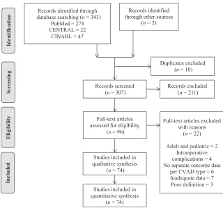
Figure 1 describes theflow of inclusion and exclusion for the study selection, in accordance with the referred Reporting Items for Systematic Reviews and Meta-Analyses (PRISMA) guidelines.34 After removal of duplicates, 307 records were identified, with 96 requiring full text for review. From the full-text articles, 22 were excluded because they included both adult and pediatric participants,35,36 reported intraoperative CVAD complications,11,37–39did not provide separate outcome data per CVAD type,7,40–44provided inadequate information to facilitate data extraction,45–51or had outcome definitions not in accordance with the review.52–54Seventy-four studies were assessed as meeting the inclusion criteria.
Thirty-two study authors were contacted to provide additional information regarding the research results, most commonly for the total CVAD catheter days per CVAD type. Eleven authors were able to provide the additional information,55–65 3 were unable to provide the requested data,66–68and 17 did not respond.69–85
Characteristics of Included Studies
The review includes 24 prospective and 50 retrospective cohort studies. These studies were undertaken in Europe,2,3,30,57–60,66,68,71,73,75,77,79–81,86–99 North America,62–65,67,72,76,83–85,100–114 Asia,55,61,69,70,74,82,115–125and South America.56,78Included subjects were ages 0 to 17 years and required treatment of oncologic or hematologic conditions, support during
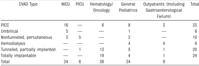
intensive care admission, or central access for hemodialysis, postsurgical, or general infusion therapy or parenteral nutrition. Table 1 describes the populations and CVAD types described in the included studies.
Study Qualities
The quality of the studies was mixed, with incomplete reporting of denominators of outcomes and poor outcome definition consistency to benchmarked standards evident. Twelve
studies59,61,66,67,72,76,82,83,89,97,105,121 described the outcome of CVAD-associated BSI without the clarity and rigor of benchmarked standards, which meant that their CVAD-associated BSI data were not included in the review. Two studies30,116provided combined “all-type”infection or mechanical failure outcomes, instead of providing separated local and systemic infection, occlusion, and dislodgement data. These data also were not included in the review. We were unable to ascertain the number of catheter days in 23 studies,66–82,84,85,92,97,99,122 which excluded their data from contributing to the meta-analysis reporting failure and complications per 1000 catheter days.
Synthesis of Results
CVAD Failure
Table 2 reports the overall pooled proportion and IR of CVAD failure and complications across CVAD types. Supplemental Figures 2 and 3 report the individual and pooled proportions and IRs of CVAD failure across CVAD types. Overall, 25% (95% CI
20.9%–29.2%) of pediatric CVADs failed before completion of therapy, with an IR of 1.97 per 1000 catheter days (95% CI 1.71–2.23; 60 studies; 16 859 CVADs; 1 282 332 catheter days). Overall heterogeneity of studies reporting proportions of failure was high (I2= 97.1%) and moderate to high (I2= 75.0% to 97.3%) when examined per device group. Umbilical catheters had the lowest pooled proportion of CVAD failure (11% [95% CI 0.7%–30.2%]) and the highest pooled IR per 1000 catheter days (28.6 [95% CI 17.4–39.8]), with 2 studies and 1 study, respectively (426 CVADs; FIGURE 1
979 catheter days) contributing to the analysis. HD catheters had the highest pooled proportion of CVAD failure (46.4% [95% CI 29.6%–63.6%]; 323 CVAD). PICCs had the second-highest pooled IR per 1000 catheter days (12.4 [95% CI 10.0–14.9]; 241 019 catheter days). Totally implanted devices had the lowest pooled IR of failure per 1000 catheter days (0.15 [95% CI 0.09–0.20]; 819 022 catheter days). Because of a lack of data, the rate of CVAD failure per 1000 catheter days for nontunneled, percutaneous CVADs could not be estimated.
CVAD Complications
Table 2 and Supplemental Figures 4 and 5 report the individual and pooled proportions and IRs per 1000 catheter days of CVAD-associated BSI, stratified by CVAD type. Overall, 10.3% (95% CI 8.9%–11.6%;I2= 95.0%; 75 studies; 31 933 CVADs) of pediatric CVADs developed a CVAD-associated BSI, with an IR of 1.63 per 1000 catheter days (95% CI
1.40–1.86;I2= 97.3%; 50 studies; 1 338 756 catheter days). Tunneled CVADs had the highest pooled proportion of CVAD-associated BSIs (19.9% [95% CI 12.6%–27.2%]; 1992 CVADs), whereas umbilical catheters had the highest pooled IR per 1000 catheter days (IR 33.7 [95% CI 21.6–45.8]; 979 catheter days).
Tunneled CVADs had the highest pooled proportion of occlusion or blockage (12.1% [95% CI 0.4%–23.5%]; 1485 CVADs), but PICCs had the highest pooled IR per 1000 catheter days (2.2 [95% CI
1.7–2.8]; 269 774 catheter days). Tunneled CVADs had the highest proportion of local infection or phlebitis (4.8% [95% CI 1.4%–9.6%]; 1827 catheter days), with
umbilical catheters having the highest IR per 1000 catheter days (9.2 [95% CI 2.6–15.8]; 979 catheter days). Totally implanted devices had the lowest proportion and rate of dislodgement per 1000 catheter days (2.0% [95% CI 0.1%–5.1%], 1902 CVADs; 0.02 [95% CI 0.00–0.04]; 256 962 catheter days), and the lowest proportion of
breakage/rupture (0.0% [95% CI 0.0%–0.0%]; 2179 CVADs).
Subgroup Analyses
The results of the subgroup analyses describing the pooled proportions and IRs of CVAD failure per 1000 catheter days across study populations are shown in Table 3. Due to availability of data, subgroup analyses were carried out only on PICCs (neonates and pediatrics; oncology/hematology and all others; outpatients and inpatients), tunneled CVADs (oncology/hematology and all others; outpatients and inpatients), and totally implantable CVADs (oncology/hematology and all others). The rate of PICC failure per 1000 catheter days was significantly (P,.001) higher for neonates (IR 25.9 [95% CI 21.2–30.5]) compared with pediatric patients (IR 5.6 [95% CI 3.2–8.1]). PICCs managed in outpatient facilities had significantly (P= .007) lower proportions of failure (24.5% [95% CI
16.9%–32.8%]) in comparison with
inpatient facilities (35.1% [95% CI 27.3%–43.2%]). Comparatively, tunneled CVADs that were managed in outpatient facilities had
significantly (P= .016) higher proportions of failure (37.8% [95% CI 17.0%–61.3%]) than those managed in inpatient facilities (12.6% [95% CI 8.5%–17.3%]).
Sensitivity Analyses
Table 4 describes the results of sensitivity analyses comparing pooled proportions and IRs of CVAD failure across study methods. The majority of studies reporting CVAD failure used retrospective methods (73.6% of studies; 64.8% of CVADs; 78.9% of catheter days). Overall IRs of failure were not different between prospective and retrospective studies. There was a significant difference between study types in reported proportions of failure of nontunneled devices (prospective, 22.4% [95% CI 20.0%–25.0%]; retrospective, 8.8% [95% CI 1.2%–21.2%];P= .046). Sensitivity analyses could not be undertaken for HD catheters, as all included studies used retrospective study methods.
DISCUSSION
This study has, for thefirst time, carefully found, critiqued, and synthesized CVAD failure rates across CVAD types and pediatric
populations. The results clearly show that failure of CVADs throughout pediatrics is a substantial and significant problem, with 1 in 4 failing. This is especially prevalent within the lifespan of umbilical catheters and PICCs. These devices have been traditionally recommended for short- to medium-term use,4,6but the PICCs and umbilical catheters described within the included studies failed before the completion of therapy in 11% to 30% of cases, with a pooled incidence failure rate of 12 to 29 per 1000 catheter days. This high rate of pediatric umbilical catheter and PICC failure is also evident in reported rates of catheter-associated
TABLE 1 Studies Included, With Patient Population and CVAD Type CVAD Type NICU PICU Hematology/
Oncology
General Pediatrics
Outpatients (Including Gastroenterological
Failure)
Total
PICC 16 — 6 8 3 33
Umbilical 5 — — 1 — 6
Nontunneled, percutaneous 3 5 — 2 — 10
Hemodialysis — — — 4 4 8
Tunneled, partially implanted — 1 13 5 1 20
Totally implantable — — 19 4 1 24
Total 24 6 38 24 9
TABLE 2 Proportions and Incidence Rates of CVAD Complications Across Device Type in Included Studies
Event and CVAD Type Proportion of Complications Incidence Rates of Complications per 1000 Catheter Days
Studies CVADs Outcomes Pooled % 95% CI Studies Catheter Days Outcomes Pooled IR 95% CI
Failure
All 60 16 859 4121 25.0d,e,g 20.9–29.2 37 1 282 332 2370 1.97d,e,g 1.71–2.23
PICC 23 10 163 2771 30.1d,e 24.4–36.1 17 241 019 2006 12.43d,e 9.98–14.89
Umbilical 2 426 41 11.0d,e 0.7–30.2 1 979 28 28.60e 17.44–39.77
Nontunneled 2 1126 248 16.7c,e 6.1–30.9 0 — — — —
HD 4 323 124 46.4d,e 29.6–63.6 2 53 828 86 1.57b,e 1.16–1.99
Tunneled 10 1501 424 29.2d,e 15.9–44.6 6 167 484 123 0.86d,e 0.41–1.32
Totally implanted 19 3320 493 15.8d,e 9.4–23.5 10 819 022 127 0.15c,e 0.09–0.20
CVAD-associated BSI
All 75 31 933 2899 10.3d,e,h 8.9–11.6 50 1 338 756 2164 1.63d,e,g 1.40–1.86
PICC 27 16 428 1081 8.6d,e 7.0–10.2 22 363 208 861 3.06d,e 2.39–3.72
Umbilical 6 498 60 8.7d,e 1.5–15.9 1 979 33 33.7e 21.64–45.78
Nontunneled 10 11 020 1028 8.7d,e 3.6–13.8 7 144 885 887 5.86d,e 3.38–8.34
HD 4 323 31 10.4b,e 3.7–17.0 2 53 828 23 0.41a,e 0.22–0.60
Tunneled 13 1992 413 19.9d,e 12.6–27.2 8 188 807 189 1.13d,e 0.65–1.61
Totally implanted 15 1672 286 15.9d,e 10.2–21.7 10 587 049 171 0.28d,e 0.14–0.42
Thrombosis
All 53 15 979 471 1.7d,e,h 0.8–2.8 30 1 168 248 153 0.08c,e,h 0.04–0.11
PICC 16 8482 317 2.1d,e 0.5–4.7 12 226 931 61 0.17c,e 0.06–0.29
Umbilical 3 402 12 3.7d,f 0.0–12.2 0 — — — —
Nontunneled 4 1370 23 3.7d,e 0.0–11.1 2 7689 13 9.06d,f 0.00–28.4
HD 3 264 7 2.9d,f 0.0–12.8 1 30 936 2 0.07f 0.00–0.18
Tunneled 12 3019 42 0.6b,e 0.2–1.2 7 335 689 20 0.04b,e 0.01–0.07
Totally implanted 15 2442 70 1.9d,e 0.1–4.9 8 567 003 57 0.06d, 0.01–0.12
Occlusion/blockage
All 53 15 344 1321 7.4d,e,h 5.5–9.6 32 698 836 823 1.06d,e,g 0.85–1.27
PICC 23 9786 837 8.2d,e 5.9–10.9 18 269 774 543 2.21d,e 1.66–2.77
Umbilical 3 472 2 0.2a,f 0.0–1.2 1 979 0 0.00f 0.00–1.88
Nontunneled 2 1126 118 8.0c,e 2.7–15.5 0 — — — —
HD 2 233 17 11.1d,e 0.0–34.8 1 30 936 8 0.26d,e 0.06–0.46
Tunneled 10 1485 227 12.1d,e 0.4–23.5 7 280 516 216 0.85d,e 0.48–1.23
Totally implanted 13 2242 120 5.0d,e 1.5–9.9 5 116 631 56 0.30d,e 0.04–0.57
Dislodgement/migration
All 39 9784 686 4.7d,e,h 3.2–6.4 23 645 611 437 0.43d,e,g 0.30–0.56
PICC 14 5389 389 5.4d,e 3.3–8.0 11 203 619 383 1.42d,e 0.70–2.14
Umbilical 1 140 4 2.9e 0.6–6.4 1 979 4 4.09e 0.0–8.76
Nontunneled 2 1126 91 3.5d,f 0.0–15.2 0 — — — —
HD 3 264 14 8.8d,e 0.1–26.0 1 30 936 3 0.10f 0.00–0.23
Tunneled 8 963 89 7.0d,e 1.7–15.0 5 154 725 41 0.24d,e 0.03–0.46
Totally implanted 11 1902 99 2.0d,e 0.1–5.2 5 256 962 6 0.02a,f 0.00–0.04
Breakage/rupture
All 45 12 092 313 1.6d,e,g 0.9–2.5 29 841 359 240 0.14d,e,g 0.08–0.19
PICC 19 8154 279 3.9d,e 2.5–5.5 15 226 990 218 0.88d,e 0.51–1.26
Umbilical 1 140 0 0.0f 0.0–1.2 1 979 0 0.00f 0.00–1.88
Nontunneled 1 34 3 8.8e 1.2–21.2 0 — — — —
HD 3 264 2 0.5c,f 0.0–5.3 1 30 936 0 0.00f 0.00–0.06
Tunneled 8 963 21 1.1c,e 0.0–3.1 5 154 725 18 0.08c,f 0.00–0.17
Totally implanted 13 2179 8 0.0a,f 0.0–0.0 7 427 729 4 0.01a,f 0.00–0.02
Local infection/phlebitis
All 39 8217 404 3.1d,e,g 2.0–4.4 28 900 192 304 0.19d,e,g 0.12–0.26
PICC 15 4191 220 4.5c,e 3.3–5.8 13 176 590 219 1.32d,e 0.85–1.79
Umbilical 1 140 9 6.4e 2.9–11.2 1 979 9 9.19e 2.57–15.82
Nontunneled 2 259 0 0.0b,f 0.0–0.4 1 7303 0 0.00f 0.00–0.25
HD 2 233 3 0.9a,f 0.0–2.9 1 30 936 3 0.10f 0.00–0.23
Tunneled 9 1827 113 4.8d,e 1.4–9.6 5 154 725 64 0.38d,e 0.10–0.65
Totally implanted 10 1567 59 1.5d,e 0.0–4.5 7 529 659 9 0.01a,f 0.00–0.02
Heterogeneity of studiesanegligible,blow,cmoderate, ordhigh.
Effect-size testesignificant orfnonsignificant.
BSI, occlusion, dislodgement, and local infection/phlebitis. There is no previous umbilical catheter meta-analysis to benchmark these results, and only small studies are included within this review. However, our results in PICCs and recent studies by Chopra and colleagues in adults126,127have demonstrated that PICCs are substantially more problematic than originally thought. The outcomes of PICCs used in clinical practice need to be cautiously and systematically monitored. Clinicians should be made aware of the high rates of failure associated with their use and should question whether PICCs are the suitable intravascular device for their patient group. Research needs to be undertaken to discover and evaluate innovative strategies to reduce PICC and umbilical catheter failures, through examining insertion procedures, securement devices, and patency practices.
Totally implanted devices were frequently associated with the lowest pooled incidence rate of failure and complications. These devices have previously been credited with
improved ease of medication administration, decreased infectious risks, and improved patient quality of life.76Although the insertion of totally implanted and other tunneled CVADs requires the skills and resources of an experienced surgeon and an operating theater, fatal complications from cardiac tamponade and major vessel injury are rare.10It may be that, because of lower rates of failure and complications, the cost-effectiveness of totally implanted devices is superior to that of other CVAD types for some population groups. Randomized controlled trials (RCTs) studying comparative clinical and cost-effectiveness of totally implanted devices, compared with other intravascular device types in suitable populations (eg, cystic fibrosis), are urgently needed.
Given that many cases of CVAD failure and complications are thought to be avoidable,6the overall rate of CVAD failure and complication for children across all CVAD types appears variable, but remains unacceptably high. There are no current
benchmarked targets for clinicians to
compare their current rates of CVAD failure and complications, with the exception of CVAD-associated BSI in the ICU.128,129Quality improvement studies have previously
demonstrated a marked reduction in complication rates associated with CVAD in pediatrics and neonates, indicating that complication rates depend on the care provided by multidisciplinary clinicians.130–132 Previous international focus on the prevention of CVAD complications from organizations such as the World Health Organization and the Centers for Disease Control and Prevention has been solely on CVAD-associated BSI, and generally in the ICU setting. However, our data demonstrate that there is also a high rate of failure due to occlusion, thrombosis, breakage, and dislodgement. The prevalence of thrombosis is likely to be higher than described, as some included studies relied on clinical suspicion of thrombosis, rather than routine imaging, significantly
underestimating the true proportion/ rate of CVAD-related thrombosis. These mechanical complications also
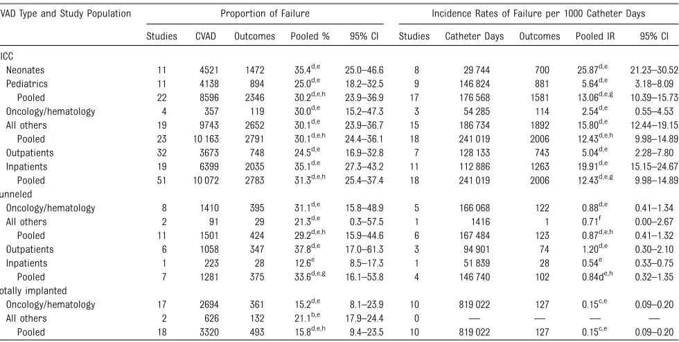
TABLE 3 Subgroup Analyses: Proportions and Incidence Rates of CVAD Failure per Device Across Study Populations
CVAD Type and Study Population Proportion of Failure Incidence Rates of Failure per 1000 Catheter Days
Studies CVAD Outcomes Pooled % 95% CI Studies Catheter Days Outcomes Pooled IR 95% CI
PICC
Neonates 11 4521 1472 35.4d,e 25.0–46.6 8 29 744 700 25.87d,e 21.23–30.52
Pediatrics 11 4138 894 25.0d,e 18.2–32.5 9 146 824 881 5.64d,e 3.18–8.09
Pooled 22 8596 2346 30.2d,e,h 23.9–36.9 17 176 568 1581 13.06d,e,g 10.39–15.73
Oncology/hematology 4 357 119 30.0d,e 15.2–47.3 3 54 285 114 2.54d,e 0.55–4.53
All others 19 9743 2652 30.1d,e 23.9–36.7 15 186 734 1892 15.80d,e 12.44–19.15
Pooled 23 10 163 2791 30.1d,e,h 24.4–36.1 18 241 019 2006 12.43d,e,h 9.98–14.89
Outpatients 32 3673 748 24.5d,e 16.9–32.8 7 128 133 743 5.04d,e 2.28–7.80
Inpatients 19 6399 2035 35.1d,e 27.3–43.2 11 112 886 1263 19.91d,e 15.15–24.67
Pooled 51 10 072 2783 31.3d,e,h 25.4–37.4 18 241 019 2006 12.43d,e,g 9.98–14.89
Tunneled
Oncology/hematology 8 1410 395 31.1d,e 15.8–48.9 5 166 068 122 0.88d,e 0.41–1.34
All others 2 91 29 21.3d,e 0.3–57.5 1 1416 1 0.71f 0.00–2.67
Pooled 11 1501 424 29.2d,e,h 15.9–44.6 6 167 484 123 0.87d,e,h 0.41–1.32
Outpatients 6 1058 347 37.8d,e 17.0–61.3 3 94 901 74 1.20d,e 0.30–2.10
Inpatients 1 223 28 12.6e 8.5–17.3 1 51 839 28 0.54e 0.33–0.75
Pooled 7 1281 375 33.6d,e,g 16.1–53.8 4 146 740 102 0.84de,h 0.32–1.35
Totally implanted
Oncology/hematology 17 2694 361 15.2d,e 8.1–23.9 10 819 022 127 0.15c,e 0.09–0.20
All others 2 626 132 21.1b,e 17.9–24.4 0 — — — —
Pooled 18 3320 493 15.8d,e,h 9.4–23.5 10 819 022 127 0.15c,e 0.09–0.20
Heterogeneity of studiesanegligible,blow,cmoderate, ordhigh.
Effect-size testesignificant orfnonsignificant.
result in an interruption to necessary treatment and the insertion of new CVADs and should be the focus of the next generation of multidisciplinary international CVAD campaigns for improvement.
The subgroup analyses demonstrated the variation in CVAD failure based on patient age. The variation was most evident in comparisons involving PICCs, where neonates had a
significantly higher rate of failure (P,
.001) than the remaining pediatric population, with a failure rate of 25.5 per 1000 catheter days. PICCs are extensively used to provide hyperosmolar solutions, inotropic medicines, and parenteral nutrition within the neonatal period.124The neonates requiring PICCs are often very low birth weight (,1500 g) or extremely low birth weight (,1000 g) and are at greatest risk for failure and sequelae.60,117The increasing use of PICCs within the neonatal population requires caution and careful
surveillance and should be the focus of significant innovation for
improvement.
Our study has demonstrated current gaps in the breadth and quality of research into pediatric CVAD failure and complications. Further prospective
cohort studies estimating the rates of failure and complications of CVADs in pediatrics are necessary to provide benchmarking targets and inform practice innovations. Meta-synthesis of the 2 studies reporting nontunneled CVAD failure showed a failure proportion of 16.7%; however, no studies reported an estimation of catheter days. In accordance with previous international focus, the majority of nontunneled CVAD studies that reported CVAD complications reported only CVAD-associated BSI. The failure and complications associated with HD and umbilical catheters were also inadequately reported, with only 6 studies of 749 CVADs available. Considering the prevalence and importance of umbilical, HD, and nontunneled CVADs within pediatric health care management, reliable measurement of their failure and complications is essential. Additionally, although multiple cohort studies described the failure of PICCs and totally implanted devices, the majority used retrospective methods, less reliable means of data collection. Future descriptions of CVAD failure need to be planned prospectively, use validated definitions for outcome measures, and report denominator information including catheter days.
Our study results should be interpreted in the context of some limitations. Not all study authors were able to provide the total number of catheter days, which limited their data being included in the meta-analysis per 1000 catheter days. Such time-based analysis is the more valid way to compare CVAD complication incidence, since the different dwell times typical of the CVAD types already expose the patient to more or less risk of complications. Second, the unavoidable heterogeneity of the populations in the included studies may have affected the
generalizability of the results; subgroup analyses were used to reduce this problem. The levels of statistical heterogeneity of thefinal analyses are indicative of the heterogeneous group of pediatric patients who require CVADs. This heterogeneity needs to be recognized before applying the results to local individual health care
institutions. Third, although our review was limited to include studies
#10 years old, many quality improvement activities have been instituted in pediatric facilities to prevent complications and failures associated with CVAD within that period. It is therefore possible that the pooled data may overestimate the
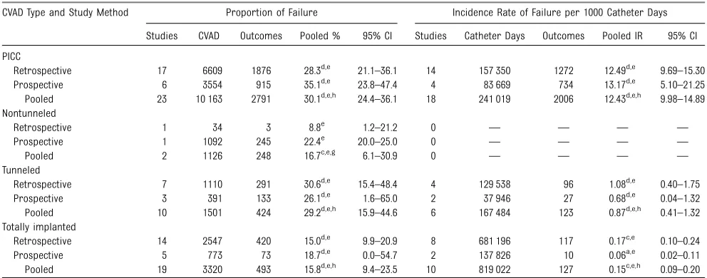
TABLE 4 Sensitivity Analyses: Proportions and Incidence Rates of CVAD Failure per Device Reported in Prospective and Retrospective Studies CVAD Type and Study Method Proportion of Failure Incidence Rate of Failure per 1000 Catheter Days
Studies CVAD Outcomes Pooled % 95% CI Studies Catheter Days Outcomes Pooled IR 95% CI
PICC
Retrospective 17 6609 1876 28.3d,e 21.1–36.1 14 157 350 1272 12.49d,e 9.69–15.30
Prospective 6 3554 915 35.1d,e 23.8–47.4 4 83 669 734 13.17d,e 5.10–21.25
Pooled 23 10 163 2791 30.1d,e,h 24.4–36.1 18 241 019 2006 12.43d,e,h 9.98–14.89
Nontunneled
Retrospective 1 34 3 8.8e 1.2–21.2 0 — — — —
Prospective 1 1092 245 22.4e 20.0–25.0 0 — — — —
Pooled 2 1126 248 16.7c,e,g 6.1–30.9 0 — — — —
Tunneled
Retrospective 7 1110 291 30.6d,e 15.4–48.4 4 129 538 96 1.08d,e 0.40–1.75
Prospective 3 391 133 26.1d,e 1.6–65.0 2 37 946 27 0.68d,e 0.04–1.32
Pooled 10 1501 424 29.2d,e,h 15.9–44.6 6 167 484 123 0.87d,e,h 0.41–1.32
Totally implanted
Retrospective 14 2547 420 15.0d,e 9.9–20.9 8 681 196 117 0.17c,e 0.10–0.24
Prospective 5 773 73 18.7d,e 0.0–54.7 2 137 826 10 0.06a,e 0.02–0.11
Pooled 19 3320 493 15.8d,e,h 9.4–23.5 10 819 022 127 0.15c,e,h 0.09–0.20
Heterogeneity of studiesanegligible,blow,cmoderate, ordhigh.
Effect-size testesignificant orfnonsignificant.
burden of device failure in 2015. Finally, whereas our study presents the association between CVAD types and failure and complications, these results do not reflect causation. Without RCTs comparing the various CVAD types, it is impossible to assert that one CVAD type reduces complications and failure in comparison with another.
Future updates of this review may also consider inclusion of the“standard care”arms of RCTs that have evaluated various interventions.
Comparison With Other Studies
No previous systematic review in pediatrics has examined the failure and complications associated with different CVADs. Landmark work by McGee and Gould133described the prevention, treatment, and incidence of CVAD failure in the adult population. Although primarily focused on describing strategies to prevent and treat CVAD complications, their systematic review reported an overall incidence of CVAD failure of
.15%, with mechanical
complications reported in 5% to 19% of patients and infectious
complications in 5% to 26%. Our study describes a higher rate of mechanical and infectious
complications, which may be due to differing synthesis methodology and
the underlying vulnerability and other clinical characteristics of the population studied.
CONCLUSIONS AND FUTURE RESEARCH
International health care institutions have highlighted the significance of CVAD failure associated with BSI. This systematic review has described the broader, multifocal rate of CVAD failure and complications across CVAD types in pediatrics within the international health care community. As context, from the 82 US hospitals reporting to the National Healthcare Safety Network in 2013,.2.7 million CVAD catheter days in the pediatric and neonatal population were
registered.134Applying the rate of failure described in our study, 5457 pediatric and neonatal CVADs in US hospitals failed before completion of treatment in 1 year alone. These failures place a massive economic and physical burden on the US health care system, patients, and families.
Strategies have been developed to prevent CVAD failure by focusing on different aspects of infectious and mechanical complications and their pathogenesis. The current rate of CVAD failure in pediatrics
demonstrates that further evidence-based improvements to their insertion and maintenance are necessary. This includes insertion and maintenance practices surrounding CVAD dressing and securement, needleless access devices,flushing procedures, and CVAD materials. Research is required urgently to develop and apply innovative and effective solutions to prevent CVAD failure in this vulnerable pediatric group.
ACKNOWLEDGMENTS
Thank you to all the study authors who were able to contribute additional data.
ABBREVIATIONS
BSI: bloodstream infection CI: confidence interval CVAD: central venous access
device HD: hemodialysis IR: incidence rate
PICC: peripherally inserted central catheter
PRISMA: Preferred Reporting Items for Systematic Reviews and Meta-Analyses
RCT: randomized controlled trial
FINANCIAL DISCLOSURE:The authors have indicated they have nofinancial relationships relevant to this article to disclose.
FUNDING:This research has been undertaken as part of Ms Ullman’s PhD program. She has received PhD scholarship funding from the Menzies Health Institute Queensland, National Health and Medical Research Council Centre of Research Excellence in Nursing, and Centurion Medical Products.
POTENTIAL CONFLICT OF INTEREST:Ms Ullman, Ms Marsh, and Dr Rickard have received funding through Griffith University for their research from central venous access device dressing manufacturers (3M, Carefusion, Centurion Medical Products), but these medical products were not included within the scope of this review.
Mr Mihala and Dr Cooke have no conflicts of interest relevant to this article to disclose.
REFERENCES
1. Carraro F, Cicalese MP, Cesaro S, et al. Guidelines for the use of long-term central venous catheter in children with hemato-oncological disorders. On
behalf of supportive therapy working group of Italian Association of Pediatric Hematology and Oncology
(AIEOP).Ann Hematol. 2013;92(10): 1405–1412
2. Perdikaris P, Petsios K, Vasilatou-Kosmidis H, Matziou V. Complications of Hickman-Broviac catheters in children with malignancies.Pediatr Hematol Oncol. 2008;25(5):375–384
3. Cesaro S, Corrò R, Pelosin A, et al. A prospective survey on incidence and outcome of Broviac/Hickman catheter-related complications in pediatric
4. Pittiruti M, Hamilton H, BiffiR, MacFie J, Pertkiewicz M; ESPEN. ESPEN Guidelines on Parenteral Nutrition: central venous catheters (access, care, diagnosis and therapy of complications).Clin Nutr. 2009;28(4):365–377
5. O’Grady NP, Alexander M, Burns LA, et al; Healthcare Infection Control Practices Advisory Committee (HICPAC). Guidelines for the prevention of intravascular catheter-related infections.Clin Infect Dis. 2011;52(9): e162–e193
6. Loveday HP, Wilson JA, Pratt RJ, et al; UK Department of Health. epic3: national evidence-based guidelines for preventing healthcare-associated infections in NHS hospitals in England.
J Hosp Infect. 2014;86(Suppl 1):S1–S70 7. Ruebner R, Keren R, Coffin S, Chu J,
Horn D, Zaoutis TE. Complications of central venous catheters used for the treatment of acute hematogenous osteomyelitis.Pediatrics. 2006;117(4). Available at: www.pediatrics.org/cgi/ content/full/117/4/e1210
8. Peng C, Monagle P, Newall F. Clinical outcomes of management of CVAD occlusions.Arch Dis Child. 2011;96(9): 885–887
9. van Miert C, Hill R, Jones L.
Interventions for restoring patency of occluded central venous catheter lumens.Cochrane Database Syst Rev. 2012;4:CD007119
10. Askegard-Giesmann JR, Caniano DA, Kenney BD. Rare but serious
complications of central line insertion.
Semin Pediatr Surg. 2009;18(2):73–83 11. Rey C, Alvarez F, De La Rua V, et al.
Mechanical complications during central venous cannulations in pediatric patients.Intensive Care Med. 2009;35(8):1438–1443
12. Gibson F, Bodenham A. Misplaced central venous catheters: applied anatomy and practical management.
Br J Anaesth. 2013;110(3):333–346 13. Alexandrou E, Spencer TR, Frost SA,
Mifflin N, Davidson PM, Hillman KM. Central venous catheter placement by advanced practice nurses
demonstrates low procedural complication and infection rates– a report from 13 years of service.Crit Care Med. 2014;42(3):536–543
14. Ullman AJ, Long DA, Rickard CM. Prevention of central venous catheter infections: a survey of paediatric ICU nurses’knowledge and practice.Nurse Educ Today. 2014;34(2):202–207 15. Miller MR, Niedner MF, Huskins WC,
et al; National Association of Children’s Hospitals and Related Institutions Pediatric Intensive Care Unit Central Line-Associated Bloodstream Infection Quality Transformation Teams. Reducing PICU central line-associated bloodstream infections: 3-year results.
Pediatrics. 2011;128(5). Available at: www.pediatrics.org/cgi/content/full/ 128/5/e1077
16. Ullman AJ, Mitchell M, Lin F, et al. Dressings and securement devices for central venous catheters (CVC).
Cochrane Database Syst Rev. 2015. Available at: http://dx.doi.org/10.1002/ 14651858.CD010367. Accessed August 2015
17. Maki DG, Weise CE, Sarafin HW. A semiquantitative culture method for identifying intravenous-catheter-related infection.N Engl J Med. 1977;296(23): 1305–1309
18. Mermel LA. What is the predominant source of intravascular catheter infections?Clin Infect Dis. 2011;52(2): 211–212
19. Mermel LA, Allon M, Bouza E, et al. Clinical practice guidelines for the diagnosis and management of intravascular catheter-related infection: 2009 Update by the Infectious Diseases Society of America.Clin Infect Dis. 2009; 49(1):1–45
20. Centers for Disease Control and Prevention. National Healthcare Safety Network Device Associated Module: CLABSI. Atlanta: CDC; 2014:1–9
21. Halton KA, Cook D, Paterson DL, Safdar N, Graves N. Cost-effectiveness of a central venous catheter care bundle.
PLoS One. 2010;5(9):e12815
22. Cooper K, Frampton G, Harris P, et al. Are educational interventions to prevent catheter-related bloodstream infections in intensive care unit cost-effective?J Hosp Infect. 2014;86(1): 47–52
23. Kim JS, Holtom P, Vigen C. Reduction of catheter-related bloodstream infections through the use of a central venous line bundle: epidemiologic and economic
consequences.Am J Infect Control. 2011;39(8):640–646
24. Schwebel C, Lucet JC, Vesin A, et al. Economic evaluation of chlorhexidine-impregnated sponges for preventing catheter-related infections in critically ill adults in the Dressing Study.Crit Care Med. 2012;40(1):11–17 25. Barnacle A, Arthurs OJ, Roebuck D,
Hiorns MP. Malfunctioning central venous catheters in children: a diagnostic approach.Pediatr Radiol. 2008;38(4):363–378, quiz 486–487
26. Stroup DF, Berlin JA, Morton SC, et al. Meta-analysis of observational studies in epidemiology: a proposal for reporting. Meta-analysis Of
Observational Studies in Epidemiology (MOOSE) group.JAMA. 2000;283(15): 2008–2012
27. Rickard CM, Webster J, Wallis MC, et al. Routine versus clinically indicated replacement of peripheral intravenous catheters: a randomised controlled equivalence trial.Lancet. 2012; 380(9847):1066–1074
28. Prieto-Merino D, Smeeth L, Staa TP, Roberts I. Dangers of non-specific composite outcome measures in clinical trials.BMJ. 2013;347:f6782 29. Schulz KF, Altman DG, Moher D;
CONSORT Group. CONSORT 2010 statement: updated guidelines for reporting parallel group randomised trials.Int J Surg. 2011; 9(8):672–677
30. Fratino G, Molinari AC, Parodi S, et al. Central venous catheter-related complications in children with oncological/hematological diseases: an observational study of 418 devices.Ann Oncol. 2005;16(4):648–654
31. Nyaga VN, Arbyn M, Aerts M. Metaprop: a Stata command to perform meta-analysis of binomial data.Arch Public Health. 2014;72(1):39
32. StataCorp, Stata Statistical Software: Release 12.1.201, StataCorp LP: College Station, TX
33. Vandenbroucke JP, von Elm E, Altman DG, et al; STROBE Initiative.
Strengthening the Reporting of Observational Studies in Epidemiology (STROBE): explanation and elaboration.
34. Moher D, Liberati A, Tetzlaff J, Altman DG; PRISMA Group. Preferred reporting items for systematic reviews and meta-analyses: the PRISMA statement.Int J Surg. 2010;8(5):336–341
35. Dal Molin A, Di Massimo DS, Braggion C, et al. Totally implantable central venous access ports in patients with cystic
fibrosis: a multicenter prospective cohort study.J Vasc Access. 2012;13(3): 290–295
36. Zaritsky JJ, Salusky IB, Gales B, et al. Vascular access complications in long-term pediatric hemodialysis patients.
Pediatr Nephrol. 2008;23(11): 2061–2065
37. Dheer G, Chaudhry GK, Singh T. Immediate complications of percutaneous central venous
cannulation in children.J Indian Assoc Pediatr Surg. 2011;16(4):145–147 38. Dzierzega M, Ossowska M, Chmiel D,
Wieczorek A, Balwierz W. The
malposition of central venous catheters in children.Pol J Radiol. 2014;79: 275–278
39. Malbezin S, Gauss T, Smith I, et al. A review of 5434 percutaneous pediatric central venous catheters inserted by anesthesiologists.Paediatr Anaesth. 2013;23(11):974–979
40. Wiegering V, Schmid S, Andres O, et al. Thrombosis as a complication of central venous access in pediatric patients with malignancies: a 5-year single-center experience.BMC Hematol. 2014;14(1):18
41. Fallon SC, Kim ME, Fernandes CJ, Vasudevan SA, Nuchtern JG, Kim ES. Identifying and reducing early complications of surgical central lines in infants and toddlers.J Surg Res. 2014;190(1):246–250
42. Haddad H, Lee KS, Higgins A, McMillan D, Price V, El-Naggar W. Routine surveillance ultrasound for the management of central venous catheters in neonates.J Pediatr. 2014; 164(1):118–122
43. Robinson JL, Casey LM, Huynh HQ, et al. Prospective cohort study of the outcome of and risk factors for intravascular catheter-related bloodstream infections in children with intestinal failure.J Parenter Enteral Nutr. 2013;38(5):625–630
44. Journeycake JM, Buchanan GR. Catheter-related deep venous thrombosis and other catheter complications in children with cancer.
J Clin Oncol. 2006;24(28):4575–4580 45. Allen RC, Holdsworth MT, Johnson CA,
et al. Risk determinants for catheter-associated blood stream infections in children and young adults with cancer.
Pediatr Blood Cancer. 2008;51(1):53–58 46. Blanchard AC, Fortin E, Rocher I, et al.
Central line-associated bloodstream infection in neonatal intensive care units.Infect Control Hosp Epidemiol. 2013;34(11):1167–1173
47. McLean TW, Fisher CJ, Snively BM, Chauvenet AR. Central venous lines in children with lesser risk acute lymphoblastic leukemia: optimal type and timing of placement.J Clin Oncol. 2005;23(13):3024–3029
48. Yang RY, Moineddin R, Filipescu D, et al. Increased complexity and
complications associated with multiple peripherally inserted central catheter insertions in children: the tip of the iceberg.J Vasc Interv Radiol. 2012; 23(3):351–357
49. Elihu A, Gollin G. Complications of implanted central venous catheters in neutropenic children.Am Surg. 2007; 73(10):1079–1082
50. Colacchio K, Deng Y, Northrup V, Bizzarro MJ. Complications associated with central and non-central venous catheters in a neonatal intensive care unit.J Perinatol. 2012;32(12):941–946 51. Fadel FI, Abdel Mooty HN, Bazaraa HM,
Sabry SM. Central venous catheters as a vascular access modality for pediatric hemodialysis.Int Urol Nephrol. 2008;40(2):489–496
52. Hussain S, Gomez MM, Wludyka P, Chiu T, Rathore MH. Survival times and complications of catheters used for outpatient parenteral antibiotic therapy in children.Clin Pediatr (Phila). 2007; 46(3):247–251
53. Jain A, Deshpande P, Shah P. Peripherally inserted central catheter tip position and risk of associated complications in neonates.J Perinatol. 2013;33(4):307–312
54. Xia B, Tang J, Xiong Y, Li XH, Mu DZ. Peripherally inserted central catheters and the incidence of candidal sepsis in
VLBW and ELBW infants: is sepsis increased?World J Pediatr. 2010;6(2): 154–157
55. Revel-Vilk S, Yacobovich J, Tamary H, et al. Risk factors for central venous catheter thrombotic complications in children and adolescents with cancer.
Cancer. 2010;116(17):4197–4205 56. Costa P, Bueno M, Alves AM, Kimura AF.
Incidence of nonelective removal of percutaneously inserted central catheters according to tip position in neonates.J Obstet Gynecol Neonatal Nurs. 2013;42(3):348–356
57. Yumani DF, van den Dungen FA, van Weissenbruch MM. Incidence and risk factors for catheter-associated bloodstream infections in neonatal intensive care.Acta Paediatr. 2013; 102(7):e293–e298
58. Wagner M, Bonhoeffer J, Erb TO, et al. Prospective study on central venous line associated bloodstream infections.
Arch Dis Child. 2011;96(9):827–831 59. Uygun I, Okur MH, Otcu S, Ozturk H.
Peripherally inserted central catheters in the neonatal period.Acta Cir Bras. 2011;26(5):404–411
60. Ozkiraz S, Gokmen Z, Anuk Ince D, et al. Peripherally inserted central venous catheters in critically ill premature neonates.J Vasc Access. 2013;14(4): 320–324
61. Nam SH, Kim DY, Kim SC, Kim IK. Complications and risk factors of infection in pediatric hemato-oncology patients with totally implantable access ports (TIAPs).Pediatr Blood Cancer. 2010;54(4):546–551
62. Casner M, Hoesli SJ, Slaughter JC, Hill M, Weitkamp JH. Incidence of catheter-related bloodstream infections in neonates following removal of peripherally inserted central venous catheters.Pediatr Crit Care Med. 2014; 15(1):42–48
63. Barrier A, Williams DJ, Connelly M, Creech CB. Frequency of peripherally inserted central catheter complications in children.Pediatr Infect Dis J. 2012; 31(5):519–521
routes.Pediatr Crit Care Med. 2012; 13(5):549–553
65. Chelliah A, Heydon KH, Zaoutis TE, et al. Observational trial of antibiotic-coated central venous catheters in critically ill pediatric patients.Pediatr Infect Dis J. 2007;26(9):816–820
66. White AD, Othman D, Dawrant MJ, Sohrabi S, Young AL, Squire R. Implantable versus cuffed external central venous catheters for the management of children and adolescents with acute lymphoblastic leukaemia.Pediatr Surg Int. 2012; 28(12):1195–1199
67. Mangum DS, Verma A, Weng C, et al. A comparison of central lines in pediatric oncology patients: Early removal and patient centered outcomes.Pediatr Blood Cancer. 2013; 60(11):1890–1895
68. Tercier S, Gapany C, Diezi M, Clément C, Lemay K, Joseph JM. Incidents and complications of totally implanted vascular access devices in children: a prospective study.Patient Saf Surg. 2008;2(1):30
69. Adler A, Yaniv I, Steinberg R, et al. Infectious complications of implantable ports and Hickman catheters in paediatric haematology-oncology patients.J Hosp Infect. 2006;62(3): 358–365
70. Almuneef MA, Memish ZA, Balkhy HH, Hijazi O, Cunningham G, Francis C. Rate, risk factors and outcomes of catheter-related bloodstream infection in a paediatric intensive care unit in Saudi Arabia.J Hosp Infect. 2006;62(2): 207–213
71. Bezzio S, Scolfaro C, Broglia R, et al. Prospective incidence study of bloodstream infection in infants and children with central venous catheters after cardiac surgery in Italy.Infect Control Hosp Epidemiol. 2009;30(7): 698–701
72. Bratton J, Johnstone PA, McMullen KP. Outpatient management of vascular access devices in children receiving radiotherapy: complications and morbidity.Pediatr Blood Cancer. 2014; 61(3):499–501
73. Can E, Salihoglu O, Oztürk A, Güngör A, Güler E, Hatipoglu S. Complication profiles of central and non-central 1 Fr PICCs in neonates weighing,1500 g.
J Matern Fetal Neonatal Med. 2014; 27(15):1522–1525
74. Cheng HY, Lu CY, Huang LM, Lee PI, Chen JM, Chang LY. Increased frequency of peripheral venipunctures raises the risk of central-line associated bloodstream infection in neonates with peripherally inserted central venous catheters [published online ahead of print July 24, 2014.J Microbiol Immunol Infect. doi:10.1016/j.jmii.2014.06.001 75. Delarbre B, Dabadie A, Stremler-Lebel
N, et al. Introduction of the use of a pediatric PICC line in a French University Hospital: review of thefirst 91 procedures.Diagn Interv Imaging. 2014;95(3):277–281
76. Fallon SC, Larimer EL, Gwilliam NR, et al. Increased complication rates
associated with Port-a-Cath placement in pediatric patients: location matters.
J Pediatr Surg. 2013;48(6):1263–1268 77. Gapany C, Tercier S, Diezi M, Clement C,
Lemay K, Joseph JM. Frequent accesses to totally implanted vascular ports in pediatric oncology patients are associated with higher infection rates.
J Vasc Access. 2011;12(3):207–210 78. Lopez PJ, Troncoso B, Grandy J, et al.
Outcome of tunnelled central venous catheters used for haemodialysis in children weighing less than 15 kg.
J Pediatr Surg. 2014;49(8):1300–1303 79. Njere I, Islam S, Parish D, Kuna J,
Keshtgar AS. Outcome of peripherally inserted central venous catheters in surgical and medical neonates.
J Pediatr Surg. 2011;46(5):946–950 80. Peynircioglu B, Ozkan F, Canyigit M, et al. Radiologically placed tunneled internal jugular catheters in the management of chronic hemodialysis and long-term infusion therapies in the pediatric population.J Vasc Interv Radiol. 2007;18(7):875–881 81. Pinon M, Bezzio S, Tovo PA, et al. A
prospective 7-year survey on central venous catheter-related complications at a single pediatric hospital.Eur J Pediatr. 2009;168(12):1505–1512 82. Rouzrokh M, Shamsian BS,
KhaleghNejad Tabari A, et al. Totally implantable subpectoral vs.
subcutaneous port systems in children with malignant diseases.Arch Iran Med. 2009;12(4):389–394
83. Sharp NE, Knott EM, Thomas P, Rivard DC, St Peter SD. Burden of
complications from needle penetration of plastic ports in children.J Pediatr Surg. 2014;49(5):763–765
84. Smitherman AB, Weston BW. Catheter-associated thrombosis in children: single-institution experience and review of pediatric venous thromboembolic disease.J Infus Nurs. 2014;37(2): 103–107
85. Thornburg CD, Smith PB, Smithwick ML, Cotten CM, Benjamin DK Jr. Association between thrombosis and bloodstream infection in neonates with peripherally inserted catheters.Thromb Res. 2008; 122(6):782–785
86. Albisetti M, Kellenberger CJ, Bergsträsser E, et al. Port-a-cath-related thrombosis and postthrombotic syndrome in pediatric oncology patients.J Pediatr. 2013;163(5): 1340–1346
87. Bartram JL, O’Driscoll S, Kulasekararaj AG, et al. Portacaths are safe for long-term regular blood transfusion in children with sickle cell anaemia.Arch Dis Child. 2011;96(11):1082–1084 88. Bucki B, Tomaszewska R, Karpe J,
Stoksik P, Sonta-Jakimczyk D, Szczepanski T. Central venous access ports in children treated for hematopoietic malignancies.Pediatr Hematol Oncol. 2008;25(8):751–755 89. Castagnola E, Fratino G, Valera M,
Giacchino M, Haupt R, Molinari AC. Correlation between“malfunctioning events”and catheter-related infections in pediatric cancer patients bearing tunneled indwelling central venous catheter: results of a prospective observational study.Support Care Cancer. 2005;13(9):757–759 90. Cil BE. Radiological placement of
chest ports in pediatric oncology patients.Eur Radiol. 2004;14(11): 2015–2019
91. Fratino G, Avanzini S, Molinari AC, Buffa P, Castagnola E, Haupt R. Incidence of indwelling central venous catheter-related complications using the Sri Paran technique for devicefixation in children with cancer.Pediatr Surg Int. 2009;25(7):591–594
Group. Infectious complications of percutaneous central venous catheterization in pediatric patients: a Spanish multicenter study.Intensive Care Med. 2007;33(3):466–476 93. Handrup MM, Møller JK, Frydenberg M,
Schrøder H. Placing of tunneled central venous catheters prior to induction chemotherapy in children with acute lymphoblastic leukemia.Pediatr Blood Cancer. 2010;55(2):309–313
94. Hengartner H, Berger C, Nadal D, Niggli FK, Grotzer MA. Port-A-Cath infections in children with cancer.Eur J Cancer. 2004;40(16):2452–2458
95. Krishnaiah A, Soothill J, Wade A, Mok QQ, Ramnarayan P. Central venous catheter-associated bloodstream infections in a pediatric intensive care unit: effect of the location of catheter insertion.Pediatr Crit Care Med. 2012; 13(3):e176–e180
96. Ruggiero A, Barone G, Margani G, Nanni L, Pittiruti M, Riccardi R. Groshong catheter-related complications in children with cancer.Pediatr Blood Cancer. 2010;54(7):947–951 97. Haumont D, de Beauregard VG, Van
Herreweghe I, Delanghe G, Ciardelli R, Haelterman E. A new technique for transumbilical insertion of central venous silicone catheters in newborn infants.Acta Paediatr. 2008;97(7): 988–990
98. Arnts IJ, Bullens LM, Groenewoud JM, Liem KD. Comparison of complication rates between umbilical and peripherally inserted central venous catheters in newborns.J Obstet Gynecol Neonatal Nurs. 2014;43(2): 205–215
99. Unal S, Ekici F, Cetin II, Bilgin L. Heparin infusion to prevent umbilical venous catheter related thrombosis in neonates.Thromb Res. 2012;130(5): 725–728
100. Adeb M, Baskin KM, Keller MS, et al. Radiologically placed tunneled hemodialysis catheters: a single pediatric institutional experience of 120 patients.J Vasc Interv Radiol. 2012; 23(5):604–612
101. Advani S, Reich NG, Sengupta A, Gosey L, Milstone AM. Central line-associated bloodstream infection in hospitalized children with peripherally inserted central venous catheters: extending
risk analyses outside the intensive care unit.Clin Infect Dis. 2011;52(9): 1108–1115
102. Brooker RW, Keenan WJ. Catheter related bloodstream infection following PICC removal in preterm infants.
J Perinatol. 2007;27(3):171–174 103. Gaballah M, Krishnamurthy G, Keller
MS, McIntosh A, Cahill AM. Single-incision technique for placement of tunneled internal jugular vein vascular access in children.Pediatr Radiol. 2014; 44(8):1004–1010
104. Hoang V, Sills J, Chandler M, Busalani E, Clifton-Koeppel R, Modanlou HD. Percutaneously inserted central catheter for total parenteral nutrition in neonates: complications rates related to upper versus lower extremity insertion.Pediatrics. 2008;121(5). Available at: www.pediatrics.org/cgi/ content/full/121/5/e1152
105. Jumani K, Advani S, Reich NG, Gosey L, Milstone AM. Risk factors for peripherally inserted central venous catheter complications in children.
JAMA Pediatr. 2013;167(5):429–435 106. Milstone A M, Reich N G, Advani S, et al.,
Catheter dwell time and CLABSIs in neonates with PICCs: a multicenter cohort study.Pediatrics2013;132(6). Available at: www.pediatrics.org/cgi/ content/full/132/6/e1609
107. Piper HG, de Silva NT, Amaral JG, Avitzur Y, Wales PW. Peripherally inserted central catheters for long-term parenteral nutrition in infants with intestinal failure.J Pediatr
Gastroenterol Nutr. 2013;56(5):578–581 108. Raj A, Bertolone S, Bond S, Burnett D,
Denker A. Cathlink 20: a subcutaneous implanted central venous access device used in children with sickle cell disease on long-term erythrocytapheresis– a report of low complication rates.
Pediatr Blood Cancer. 2005;44(7): 669–672
109. Reyes JA, Habash ML, Taylor RP. Femoral central venous catheters are not associated with higher rates of infection in the pediatric critical care population.Am J Infect Control. 2012; 40(1):43–47
110. Sengupta A, Lehmann C, Diener-West M, Perl TM, Milstone AM. Catheter duration and risk of CLA-BSI in neonates with PICCs.Pediatrics. 2010;125(4). Available
at: www.pediatrics.org/cgi/content/full/ 125/4/e648
111. Titapiwatanakun R, Moir C, Pruthi RK, Stavlo PL, Schmidt KA, Rodriguez V. Central venous access devices for paediatric patients with haemophilia: a single-institution experience.
Haemophilia. 2009;15(1):168–174 112. Toh LM, Mavili E, Moineddin R, et al. Are
cuffed peripherally inserted central catheters superior to uncuffed peripherally inserted central catheters? A retrospective review in a tertiary pediatric center.J Vasc Interv Radiol. 2013;24(9):1316–1322
113. Van Winkle P, Whiffen T, Liu IL. Experience using peripherally inserted central venous catheters for outpatient parenteral antibiotic therapy in children at a community hospital.
Pediatr Infect Dis J. 2008;27(12): 1069–1072
114. Wong J, Dow K, Shah PS, Andrews W, Lee S. Percutaneously placed central venous catheter-related sepsis in Canadian neonatal intensive care units.
Am J Perinatol. 2012;29(8):629–634 115. Abedin S, Kapoor G. Peripherally
inserted central venous catheters are a good option for prolonged venous access in children with cancer.Pediatr Blood Cancer. 2008;51(2):251–255 116. Eisenstein I, Tarabeih M, Magen D, et al.
Low infection rates and prolonged survival times of hemodialysis catheters in infants and children.Clin J Am Soc Nephrol. 2011;6(4):793–798 117. Hsu JF, Tsai MH, Huang HR, Lien R, Chu
SM, Huang CB. Risk factors of catheter-related bloodstream infection with percutaneously inserted central venous catheters in very low birth weight infants: a center’s experience in Taiwan.
Pediatr Neonatol. 2010;51(6):336–342 118. Hatakeyama N, Hori T, Yamamoto M,
et al. An evaluation of peripherally inserted central venous catheters for children with cancer requiring long-term venous access.Int J Hematol. 2011;94(4):372–377
119. Hung MC, Chen CJ, Wu KG, Hung GY, Lin YJ, Tang RB. Subcutaneously implanted central venous access device infection in pediatric patients with cancer.
120. Levy I, Bendet M, Samra Z, Shalit I, Katz J. Infectious complications of
peripherally inserted central venous catheters in children.Pediatr Infect Dis J. 2010;29(5):426–429
121. Matsuzaki A, Suminoe A, Koga Y, Hatano M, Hattori S, Hara T. Long-term use of peripherally inserted central venous catheters for cancer chemotherapy in children.Support Care Cancer. 2006; 14(2):153–160
122. Shen G, Gao Y, Wang Y, Mao B, Wang X. Survey of the long-term use of peripherally inserted central venous catheters in children with cancer: experience in a developing country.
J Pediatr Hematol Oncol. 2009;31(7): 489–492
123. Tsai MH, Lien R, Wang JW, et al. Complication rates with central venous catheters inserted at femoral and non-femoral sites in very low birth weight infants.Pediatr Infect Dis J. 2009;28(11): 966–970
124. Tsai MH, Chu SM, Lien R, et al. Complications associated with 2 different types of percutaneously inserted central venous catheters in very low birth weight infants.Infect Control Hosp Epidemiol. 2011;32(3): 258–266
125. Yacobovich J, Ben-Ami T, Abdalla T, et al. Patient and central venous catheter related risk factors for blood stream infections in children receiving chemotherapy.Pediatr Blood Cancer
2015;62(3):471–476
126. Chopra V, Anand S, Hickner A, et al. Risk of venous thromboembolism associated with peripherally inserted central catheters: a systematic review and meta-analysis.Lancet. 2013;382(9889): 311–325
127. Chopra V, O’Horo JC, Rogers MA, Maki DG, Safdar N. The risk of bloodstream infection associated with peripherally inserted central catheters compared with central venous catheters in adults: a systematic review and meta-analysis.
Infect Control Hosp Epidemiol. 2013; 34(9):908–918
128. Sagana R, Hyzy RC. Achieving zero central line-associated bloodstream infection rates in your intensive care unit.Crit Care Clin. 2013;29(1):1–9 129. Worth LJ, McLaws ML. Is it possible to
achieve a target of zero central line associated bloodstream infections?
Curr Opin Infect Dis. 2012;25(6):650–657 130. Ardura MI, Lewis J, Tansmore JL, Harp
PL, Dienhart MC, Balint JP. Central
catheter-associated bloodstream infection reduction with ethanol lock prophylaxis in pediatric intestinal failure: broadening quality
improvement initiatives from hospital to home.JAMA Pediatr. 2015;169(4): 324–331
131. Kleidon T, Illing A, Fogarty G, et al. Improving the central venous access devices maintenance process to reduce associated infections in paediatrics: evaluation of a practical, multi-faceted quality-improvement initiative.Health Infect. 2015;20(2): 46–53
132. Bundy D G, Gaur A H, Billett A L, et al., Preventing CLABSIs among pediatric hematology/oncology inpatients: national collaborative results.
Pediatrics2014;134(6). Available at: www.pediatrics.org/cgi/content/full/ 134/6/e1678
133. McGee DC, Gould MK. Preventing complications of central venous catheterization.N Engl J Med. 2003; 348(12):1123–1133
DOI: 10.1542/peds.2015-1507 originally published online October 12, 2015;
2015;136;e1331
Pediatrics
Amanda J. Ullman, Nicole Marsh, Gabor Mihala, Marie Cooke and Claire M. Rickard
Complications of Central Venous Access Devices: A Systematic Review
Services
Updated Information &
http://pediatrics.aappublications.org/content/136/5/e1331 including high resolution figures, can be found at:
References
http://pediatrics.aappublications.org/content/136/5/e1331#BIBL This article cites 129 articles, 11 of which you can access for free at:
Subspecialty Collections
oskeletal_disorders_sub
http://www.aappublications.org/cgi/collection/rheumatology:muscul Rheumatology/Musculoskeletal Disorders
ent_safety:public_education_sub
http://www.aappublications.org/cgi/collection/patient_education:pati Patient Education/Patient Safety/Public Education
b
http://www.aappublications.org/cgi/collection/hospital_medicine_su Hospital Medicine
following collection(s):
This article, along with others on similar topics, appears in the
Permissions & Licensing
http://www.aappublications.org/site/misc/Permissions.xhtml in its entirety can be found online at:
Information about reproducing this article in parts (figures, tables) or
Reprints
DOI: 10.1542/peds.2015-1507 originally published online October 12, 2015;
2015;136;e1331
Pediatrics
Amanda J. Ullman, Nicole Marsh, Gabor Mihala, Marie Cooke and Claire M. Rickard
Complications of Central Venous Access Devices: A Systematic Review
http://pediatrics.aappublications.org/content/136/5/e1331
located on the World Wide Web at:
The online version of this article, along with updated information and services, is
http://pediatrics.aappublications.org/content/suppl/2015/10/06/peds.2015-1507.DCSupplemental Data Supplement at:
by the American Academy of Pediatrics. All rights reserved. Print ISSN: 1073-0397.