Long-Term Developmental Outcomes in Patients With
Deformational Plagiocephaly
Robert I. Miller, MD*‡, and Sterling K. Clarren, MD*§储
ABSTRACT. Objectives. To determine whether there was an increased rate of later developmental delay in school-aged children who presented as infants with formational plagiocephaly without obvious signs of de-lay at the time of initial evaluation.
Methods. A retrospective medical record review of 254 patients evaluated at the Craniofacial Center of the Children’s Hospital and Regional Medical Center in Se-attle, Washington, from 1980 through 1991 was com-pleted. Consenting patient families were interviewed via telelphone to determine what, if any, special medical or educational problems had occurred for the children who had had plagiocephaly in infancy or their siblings with normal head shapes.
Results. A total of 181 families from the medical record review could be notified about the study and 63 families agreed to participate in a telephone interview. The sample of participants for the telephone interview was random to and representative of the group as a whole. The families reported that 25 of the 63 children (39.7%) with persistent deformational plagiocephaly had received special help in primary school including: special education assistance, physical therapy, occupational ther-apy, speech therapy generally through an Individual Ed-ucation Plan. Only 7 of 91 siblings (7.7%), serving as controls, required similar services (2 ⴝ 21.24). Delays could not be specifically anticipated at the time of the diagnosis of deformational plagiocephaly from any sim-ple set of factors including treatment with helmet ther-apy, although effected males with reported uterine con-straint were at the highest risk for subsequent school problems.
Conclusions. Infants with deformational plagioceph-aly comprise a high-risk group for developmental diffi-culties presenting as subtle problems of cerebral dys-function during the school-age years. There is a need for additional research on the long-term developmental problems in infants with deformational plagiocephaly. Pediatrics 2000;105(2). URL: http://www.pediatrics.org/ cgi/content/full/105/2/e26;plagiocephaly, facial asymme-try, torticollis, developmental delay.
ABBREVIATIONS. CHRMC, Children’s Hospital and Regional Medical Center; IEP, Individual Education Plan.
P
lagiocephaly or crooked head shape can result from 3 very different etiologic processes in-cluding abnormalities in brain shape and sub-sequent aberrant directions in brain growth, prema-ture fusion of a single coronal or lambdoidal suprema-ture, or prenatal or postnatal external constraint. Postnatal external constraint is by far the most common cause, although the incidence is not fully established be-cause of the lack of consensus on the criteria to be used in making the diagnosis. Clarren et al1reported that the literature suggested an incidence of 1 in 300 live-born infants. Bruneteau and Mulliken2reported the incidence to be as high as 48% in otherwise healthy newborns. In this article, plagiocephaly as-sociated with external deforming forces will be re-ferred to as “deformational plagiocephaly.” Defor-mational plagiocephaly may be initiated prenatally when the fetal head rests for a prolonged period on a hard surface, such as a portion of the maternal pelvis or against the limb of a sibling in a multiple birth. Such consistent fetal positioning could be at-tributable to maternal constraining forces or to fac-tors within the fetus that reduce the usual rate of spontaneous movement. More commonly, deforma-tional plagiocephaly occurs postnatally and is asso-ciated with congenital torticollis, vertebral anoma-lies, neurologic impairment, or forced sleeping position.3–5Few reports document any late effects of deformational plagiocephaly other than potential cosmetic concerns and the potential for strabismus, especially involving vertical eye movements.6,7 Pla-giocephaly may be morphometrically evident in as many as 14% of adults, but it is rarely recognized.8Deformational plagiocephaly generally becomes more severe in the first weeks of life, as the infant holds his head in a fixed position (regardless of the cause for the fixed position); then the headshape begins to improve with normal developmental pro-gression involving head control and a full range of neck motion. In ⬃10% of affected infants with pla-giocephaly, there will be a permanent deformity with a mild to severe cosmetic effect.5Patients with moderate to severe asymmetry are often referred to a craniofacial center for consideration of treatment. Cranial surgery is almost never indicated for plagio-cephaly without synostosis unless the presentation is very severe. Helmet therapy is an effective treatment option based on the mechanism that pressure from a rapidly growing brain against a concave surface should round flattened areas caused by earlier pres-sure against a flat surface.5,9Helmet therapy is gen-From the *Center for Human Development and Disability, University of
Washington School of Medicine, Seattle, Washington; ‡Madigan Army Medical Center, Tacoma, Washington; §Craniofacial Center, Children’s Hospital and Regional Medical Center, Seattle, Washington; and the储 De-partment of Pediatrics, University of Washington School of Medicine, Se-attle, Washington.
Received for publication Apr 30, 1999; accepted Aug 30, 1999.
Reprint requests to (S.K.C.) CHRMC, CH-47, Box C-5371, Seattle, WA 98105-0371. E-mail: [email protected]
erally recommended between 6 and 18 months of age in an attempt to use remaining brain growth to re-direct head shape.
From 1980 to 1991, 295 patients were recorded as evaluated at the Craniofacial Center of the Children’s Hospital and Regional Medical Center (CHRMC) in Seattle, Washington for plagiocephaly and consider-ation of helmet therapy. Patients were followed through completion of helmet therapy when indi-cated; however, the developmental outcome of these patients was unknown because subsequent fol-low-up after completion of treatment was rarely in-dicated. The rate of developmental delay in this pa-tient population at the time of initial evaluation for plagiocephaly or later in life was unknown. The pri-mary question to be addressed by this study was whether deformational plagiocephaly could be a marker for early central nervous system disorgani-zation that might later manifest as school problems that required additional services. Subtle problems of cerebral dysfunction would include learning disabil-ities, language disorders, visual–perceptual prob-lems, motor delays, or problems with attention span. If delays were found to be present, an additional question would be what early factors related to the deformational plagiocephaly were associated with this adverse outcome. The rate of strabismus, as a documented late effect of deformational plagioceph-aly, was also determined.
METHODS
According to medical records at CHRMC, 295 patients had been evaluated in the Craniofacial Center for deformational pla-giocephaly between 1980 and 1991. Of these, 41 charts were ex-cluded from additional review because the actual diagnosis found in the chart was not consistent with deformational plagiocephaly, no record of a Craniofacial Center assessment was found, or, in 3 instances, the records could not be located. The remaining 254 charts were reviewed and the following information was col-lected: position of plagiocephaly, head shape at birth with associ-ated deformations or malformations, evidence of other deforma-tions or malformadeforma-tions, age when asymmetry was first noted, involvement with helmet treatment, and history of developmental delay at the time of presentation. Information was also collected from the medical record to allow contact with families to address the question of developmental outcome.
A letter requesting permission for a telephone interview to discuss the developmental outcomes of their children was mailed to 244 families from the previous chart review (10 families were excluded because no address was available for mailing). In 73 cases, all attempts to find an accurate address failed. The remain-ing potential population consisted of 181 families whose letters of invitation were not returned to us by the postal service as unde-liverable. These families each presumably received a 1-page letter briefly discussing the research project and a self-addressed, stamped reply postcard to convey their decision about participat-ing in the project. The letter identified their child as havparticipat-ing pre-viously been seen at CHRMC because of an asymmetric head shape. The telephone interview was described. Specifically the letter noted that “questions being addressed in this study involved learning more about how all your children have done in school and the effect of the asymmetric head shape on eye positioning,” and an example question was given. The letter then dealt with clarification of the telephone call process and parent/patient rights. Finally, the letter stressed that the project involved nothing other than the single phone call.
Information obtained from parents during the telephone inter-view included the patient previously diagnosed with plagioceph-aly and information about all siblings serving as controls. Siblings were chosen as the control group to minimize bias, although they were obviously not age- or sex-matched.
Statistics from the State of Washington were obtained to deter-mine the percentage of students in each grade that required spe-cial services in 1997 as per the Individuals with Disabilities Edu-cation Act, Part B. This figure was shown to be fairly consistent over the last several years and was applicable to both patients and their siblings. Interview questions included: involvement in any class or program, during or after school, which was designed for children who needed additional help such as a special education class, physical therapy, occupational therapy, or speech therapy; completion of an Individual Education Plan (IEP) by the school to document these services; and/or evaluation by an “eye doctor” because of a problem with eye alignment (strabismus). Asking parents about the special services their children might have re-ceived or were receiving as a measure of developmental outcome was considered the most likely way of obtaining pertinent history of developmental delay. Only students meeting standardized cri-teria for a delay would qualify for special services through the schools. These special services are documented through the IEP and are reviewed with the family on an annual basis.
Data were arranged in 2⫻2 tables using2analysis with Yates corrections and Fisher’s exact test when cell numbers were⬍5 for analysis.
RESULTS
In the chart review, plagiocephaly was found in 152 males (60%) and 102 females (40%). A flat right occiput was found in 161 (63%) patients, whereas 72 (28%) presented with a flat left occiput, 17 (7%) had an asymmetric but bilaterally flat occiput, and 4 (2%) had an unknown asymmetry. A total of 174 patients (69%) were born with a normal head shape of which 147 (84%) were believed to have associated torticol-lis. Other associations included 1 patient with hemi-vertebrae and 3 with a neuromotor deficit. Of the patients born with an abnormal head shape, the most common association was uterine constraint in 20 pa-tients (42%). Other deformations were noted in 73 (29%) and malformations in 47 (19%) of the total patient population. Parents reported being aware of a head asymmetry eventually diagnosed as plagio-cephaly at an average age of 1.5 months. Age at the initial Craniofacial Center appointment was 6.5 months on average. Helmet treatment was initiated for 91 patients (36%). Breech delivery was present in 15 (6%). There was a history of an ophthalmologic evaluation in 7 (3%). Finally, 14 patients (6%) were 1 of a set of twins.
History of any concerns about developmental de-lay per parent or physician was documented in the records of 33 patients (13%) at the time of initial evaluation. No formal diagnosis of developmental delay was made in the record for any of these pa-tients at the time of the CHRMC Craniofacial Center evaluation. Four of the families of these 33 patients (12%) subsequently went on to participate in the telephone interview. Two of the 4 with initial gross motor concerns subsequently required special edu-cation assistance but no motor therapy. One with initial gross motor concerns required no special ser-vices during school-age years. One with a question of global delays at time of initial evaluation had persis-tent global delays during school-age years requiring support from all available services.
interview and nonparticipants. The sample of partic-ipants seemed to be random and representative of the whole for every category addressed with no sig-nificant difference between the 2 categories.
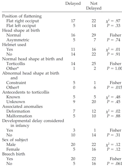
Table 2 compares participants in the telephone interview who reported any delays in school with those without any reported delays or need for special services. Delays could not be anticipated by any simple set of early factors. However, patients at high-est risk included those born with an abnormal head-shape at birth secondary to constraint (P⫽ .015) of which all were males. Breech placement was not statistically significant (P⫽.061). In addition, helmet therapy was not a risk factor for a subsequent history of delays (P⫽ .91).
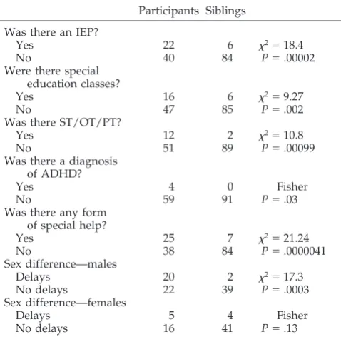
Table 3 compares participants in the telephone interview with their siblings in regards to history of need for special services during the school-age years. The need for any special services frequently docu-mented on an IEP to included special education as-sistance, speech therapy, physical therapy, or occu-pational therapy was dramatically different compared with siblings (P ⫽ .0000041). Controlling for gender, a significant difference was found for males (P ⫽ .0003) but not for females (P ⫽ .13).
Questions about attention deficit hyperactivity dis-order were not specifically asked during the tele-phone interview; however, parents provided this in-formation when discussing services on the IEP for 4 of the patients previously diagnosed with plagio-cephaly and in none of the siblings (P ⫽ .03). In regards to the type of therapy required for the pa-tients with plagiocephaly, 10 received speech ther-apy, 1 occupational therther-apy, and 1 physical therapy. The 2 siblings who required therapy services both received speech therapy. The percentage of patients on an IEP with plagiocephaly was 34.9% and their siblings 6.6%. The percentage of children qualifying for special services on an IEP in the state of Wash-ington for 1997, as per the Individuals with Disabil-ities Education Act, Part B, was 11.9%.
Strabismus was not found to be statistically signif-icant in this study, with only 1 patient with plagio-cephaly and 1 sibling with a history of strabismus resulting in evaluation and treatment by an ophthal-mologist.
DISCUSSION
The population of children with deformational plagiocephaly evaluated at CHRMC from 1980 through 1991 was consistent with previous studies in regards to position of plagiocephaly. Right-sided oc-cipital flattening was more common as previously reported with percentages ranging from 61% to 73%,
TABLE 1. Comparison Between 63 Participants and 191
Non-participants Based on Chart Review Data Participants
Non-participants Position of flattening
Flat right occiput 39 122 2⫽.04 Flat left occiput 19 53 P⫽.85 Head shape at birth
Normal 45 129 2⫽.00
Asymmetric 12 36 P⫽.95
Helmet used
Yes 27 64 2⫽1.42
No 36 127 P⫽.23
Normal head shape at birth and
Torticollis 39 108 Fisher
Other* 3 6 P⫽.70
Abnormal head shape at birth and
Constraint 6 14 2⫽.11
Other† 6 22 P⫽.74
Antecedents to torticollis
Known 10 37 2⫽.62
Unknown 29 71 P⫽.43
Associated anomalies
Deformation 19 54 2⫽.24
Malformation 15 32 P⫽.62
Developmental delay considered in infancy
Yes 4 29 2⫽2.06
No 24 67 P⫽.15
Sex of subject
Male 42 110 2⫽1.27
Female 21 81 P⫽.26
Breech birth
Yes 7 8 Fisher
No 56 183 P⫽.061
Area of residence
West Washington 54 108 2⫽.92
Other 9 10 P⫽.34
* Forced sleep position, hemivertebrae, neuromotor dysfunction, unknown.
† Asymmetric brain, unknown.
TABLE 2. Comparison Between 25 Delayed Participants and
36 Not Delayed Participants Based on Interview Delayed Not
Delayed Position of flattening
Flat right occiput 17 22 2⫽.97 Flat left occiput 5 14 P⫽.33 Head shape at birth
Normal 16 29 Fisher
Asymmetric 5 7 P⫽.74
Helmet used
Yes 11 16 2⫽.01
No 14 22 P⫽.91
Normal head shape at birth and
Torticollis 14 25 Fisher
Other* 1 2 P⫽1.00
Abnormal head shape at birth and
Constraint 5 1 Fisher
Other† 0 6 P⫽.015
Antecedents to torticollis
Known 5 5 2⫽.48
Unknown 9 20 P⫽.45
Associated anomalies
Deformation 7 12 2⫽.02
Malformation 5 10 P⫽.88
Developmental delay considered in infancy
Yes 3 1 Fisher
No 10 14 P⫽.31
Sex of subject
Male 20 22 2⫽.12
Female 5 16 P⫽.12
Breech birth
Yes 20 22 Fisher
No 5 16 P⫽.061
* Forced sleep position, hemivertebrae, neuromotor dysfunction, unknown.
compared with 63% in this study.2,10,11Bruneteau and Mulliken2reported torticollis as an associated find-ing in 64% of infants with deformational plagioceph-aly; however, our population had a higher associa-tion with torticollis in 84%. Bruneteau and Mulliken also noted other congenital anomalies in 43% of pa-tients, which was similar to 47% of patients in this sample with other deformations or malformations. Demographics were similar to the results reported by Hellbusch et al9 with the age of deformity first being noted at an average age of 1.6 months and the age that the patient was first seen in a doctor’s office reported to be an average of 7.3 months. Results from the CHRMC population was an average age of 1.6 months and 6.5 months, respectively.
Concern about developmental delay in children who have a history of plagiocephaly secondary to deformation has not previously been evaluated in the literature. There is reportedly no increased risk of functional neurologic disability in patients with pla-giocephaly without synostosis.12 Rekate8 stated that there were few reports documenting any late effects of occipital plagiocephaly in his comprehensive crit-ical review of the literature. Late effects discussed by Rekate included visual disturbances, orthodontic/ oral surgical issues, cosmetic concerns, and psycho-social issues. A statement was made in that review that the possibility that cranial distortion without increased intracranial pressure or definable damage being done to the distorted underlying areas of the brain could lead to overt or subtle neuropsycholog-ical problems, had not been reported to that point. Although this study does not attempt to explain an underlying mechanism, there clearly seems to be a difference in children with plagiocephaly, compared with their siblings in regards to need for special
services during the school-age years. Problems noted related to subtle problems of cerebral dysfunction, which were manifest during the school-age years, involving language disorders, learning disability, and attention deficits. These are clearly issues that primary care providers need to be aware of, because they may care for children in the high-risk group.
If others can confirm that deformational plagio-cephaly is associated with increased rates of behav-ioral and learning problems during the school-age years, then the question to be asked is: “Is plagio-cephaly an early sign of subtle brain dysfunction or does early brain molding lead to subtle brain dysfunction?” Referrals to craniofacial centers for evaluation of deformational plagiocephaly and brachycephaly are increasing.8 This increase in de-formations has been temporally linked to the Back to Sleep program advanced by the American Academy of Pediatrics in 1992 that advises the avoidance of the prone sleeping position as a method of reducing the rates of sudden infant death syndrome.10,12,13There is a delay in early gross motor milestones in children forced to sleep supine but these delays seem tran-sient and have not been linked as yet to any longer term problems.14 Children who are encouraged to sleep on their backs and develop abnormal head shapes as a result are a different population than children who spontaneously restricted their move-ment in bed for one reason or another. It will be important to follow both groups over the next sev-eral years and document the rates of attentional and cognitive disabilities that arise.
The use of helmet therapy did not seem to affect the rate of developmental delay. A total of 44% of the delayed patients had worn helmets, whereas a total of 42% of the nondelayed patients had worn helmets. The sample seemed large enough to conclude that if the additional pressure on the head from the helmet contributed to the delay, it must be a very small part of the variance.
A limitation of this study was the number of pa-tients included in the telephone interview compared with the chart review because of an inability to con-tact families previously evaluated in the Craniofacial Center. We were unable to contact 73 families be-cause of an inaccurate address and telephone num-ber. Two thirds of the families who presumably re-ceived our letter of introduction and invitation did not respond in any way. This is a typical response rate for a survey and obviously we could not ask the families who did not respond why they did not respond. The families who were eventually con-tacted were shown to be a random and representa-tive sample of the whole, based on comparison data in the charts. The reader might speculate that fami-lies with concerns about their child’s development might be more likely to reply to the letter, but most parents involved with the telephone interview did not have significant complaints about their child’s developmental progress. Further, only 12% of fami-lies who had had an infant noted to have delays, and therefore, at risk for additional neurodevelopmental problems responded to our invitation, compared with 44% of families in which no early delay had
TABLE 3. Comparisons Between 63 Participants and Their 91 Siblings Based on Interviews
Participants Siblings
Was there an IEP?
Yes 22 6 2⫽18.4
No 40 84 P⫽.00002
Were there special education classes?
Yes 16 6 2⫽9.27
No 47 85 P⫽.002
Was there ST/OT/PT?
Yes 12 2 2⫽10.8
No 51 89 P⫽.00099
Was there a diagnosis of ADHD?
Yes 4 0 Fisher
No 59 91 P⫽.03
Was there any form of special help?
Yes 25 7 2⫽21.24
No 38 84 P⫽.0000041
Sex difference—males
Delays 20 2 2⫽17.3
No delays 22 39 P⫽.0003
Sex difference—females
Delays 5 4 Fisher
No delays 16 41 P⫽.13
been found. Although all the interviewees were will-ing to discuss their children’s needs for special ser-vices in the school setting, most parents reported an expectation of being asked questions about cosmetic appearance and social acceptance. Of note, most par-ents reported no significant psychosocial issues re-lated to persistent facial asymmetry. It was the opin-ion of the interviewers that the families involved with the telephone interview were not a group in search of additional services, and they did not asso-ciate their child’s need for special school services with a previous diagnosis of deformational plagio-cephaly.
An additional question considered in this study as part of the telephone interview was whether patients with persistent plagiocephaly have an increased rate of strabismus related to their underlying craniofacial asymmetry. Synostotic plagiocephaly is known to be associated with vertical strabismus and contralateral head tilt.7 The underlying mechanism resulting in strabismus involves traction on the ocular globe in-duced by bone deformation caused by craniosynos-tosis.6Rekate8reports that strabismus, especially in-volving vertical eye movements, is common in the more severe forms of this disorder. A study by Fredrick et al7 evaluated 13 patients with deforma-tional plagiocephaly, of which 9 were found to have ipsilateral torticollis and 1 of these presented with strabismus. The need for early identification of stra-bismus is based on the development of binocular vision by⬃6 months of age, whereby late correction may be an obstacle to the development of normal visual function.6History of strabismus of any type or need for ophthalmology evaluation was found in only 7 patients during the chart review. One of these 7 patients with plagiocephaly was part of the tele-phone interview, and only 1 sibling had a history of strabismus. The results of this study do not suggest a need for increased awareness of this issue by provid-ers caring for children with deformational plagio-cephaly.
Primary care physicians commonly diagnose de-formational plagiocephaly early in its course and offer appropriate recommendations for prevention of additional cranial distortion. However, even with appropriate intervention, there will continue to be a subset of children with persistent cranial asymmetry
in need of additional evaluation and follow-up. Chil-dren with deformation plagiocephaly in the high-risk group, which includes males with abnormal head shapes at birth associated with uterine con-straint, need to be closely followed for potential de-velopmental delay presenting as subtle problems of cerebral dysfunction during the school-age years. If concerns arise, timely evaluation is indicated to en-sure that children in the high-risk group are receiv-ing appropriate services based on their underlyreceiv-ing special needs.
ACKNOWLEDGMENT
We thank Dr Susan J. Astley of CHRMC for her statistical support throughout this study.
REFERENCES
1. Clarren SK, Smight DW, Hanson JW. Helmet treatment for plagioceph-aly and congenital muscular torticollis.J Pediatr. 1979;94:43– 46 2. Bruneteau RJ, Mulliken JB. Frontal plagiocephaly: synostotic,
compen-sational, or deformation.Plast Reconstr Surg. 1992;89:21–31
3. Pollack IF, Losken HW, Fasick P. Diagnosis and management of poste-rior plagiocephaly.Pediatrics. 1997;99:180 –185
4. Dias MS, Klein DM. Occipital plagiocephaly: deformation or lambdoid synostosis?Pediatr Neurosurg. 1996;24:69 –73
5. Clarren SK. Plagiocphaly and torticollis: etiology, natural history, and helmet treatment.J Pediatr. 1981;98:92–95
6. Dennis D, Genitori L, Conrath J, Lena G, Choux M. Ocular findings in children operated on for plagiocephaly and trigonocephaly.Child Nerv Syst. 1996;12:683– 689
7. Fredrick DR, Mulliken JB, Robb RM. Ocular manifestations of deforma-tional frontal plagiocephaly.J Pediatr Opththalmol Strabismus. 1993;93: 92–95
8. Rekate HL. Occipital plagiocephaly: a critical review of the literature.J Online. 1997:1–14
9. Hellbusch JL, Hellbusch LC, Bruneteau RJ. Active counter-positioning treatment of deformation occipital plagiocephaly.Nebr Med J. 1995: 344 –349
10. Turk AE, McCarthy JG, Thorne CHM, Wisoff JH. The “back to sleep campaign” and deformational plagiocephaly: is there cause for con-cern?J Craniofac Surg. 1996;7:12–18
11. Watson GH. Relation between side of plagiocephaly, dislocation of hip, scoliosis, bat ears, and sternomastoid tumors.Arch Dis Child. 1979;46: 203–210
12. Kane AA, Mitchell LE, Craven KP, Marsh JL. Observations on a recent increase in plagiocephaly without synostosis. Pediatrics. 1996;97: 877– 885
13. Argenta LC, David LR, Wilson JA, Bell WO. An increase in infant cranial deformity with supine sleeping position.J Craniofac Surg. 1996; 7:5–11
DOI: 10.1542/peds.105.2.e26
2000;105;e26
Pediatrics
Robert I. Miller and Sterling K. Clarren
Plagiocephaly
Long-Term Developmental Outcomes in Patients With Deformational
Services
Updated Information &
http://pediatrics.aappublications.org/content/105/2/e26
including high resolution figures, can be found at:
References
http://pediatrics.aappublications.org/content/105/2/e26#BIBL
This article cites 12 articles, 2 of which you can access for free at:
Subspecialty Collections
http://www.aappublications.org/cgi/collection/genetics_sub
Genetics
following collection(s):
This article, along with others on similar topics, appears in the
Permissions & Licensing
http://www.aappublications.org/site/misc/Permissions.xhtml
in its entirety can be found online at:
Information about reproducing this article in parts (figures, tables) or
Reprints
http://www.aappublications.org/site/misc/reprints.xhtml
DOI: 10.1542/peds.105.2.e26
2000;105;e26
Pediatrics
Robert I. Miller and Sterling K. Clarren
Plagiocephaly
Long-Term Developmental Outcomes in Patients With Deformational
http://pediatrics.aappublications.org/content/105/2/e26
located on the World Wide Web at:
The online version of this article, along with updated information and services, is
by the American Academy of Pediatrics. All rights reserved. Print ISSN: 1073-0397.