O R I G I N A L R E S E A R C H
Global Sequence Analysis and Expression of
Azurin
Gene in Different Clinical Specimens of Burn
Patients with
Pseudomonas aeruginosa
Infection
This article was published in the following Dove Press journal: Infection and Drug Resistance
Hajar Mohammadi Barzelighi1 Bita Bakhshi 2
Bahram Daraei 3 Hossein Fazeli 1 Bahram Nasr Esfahani1
1Department of Microbiology, School of
Medicine, Isfahan University of Medical
Sciences, Isfahan, Iran;2Department of
Bacteriology, Faculty of Medical Sciences, Tarbiat Modares University, Tehran, Iran;
3Department of Toxicology and
Pharmacology, School of Pharmacy, Shahid Beheshti University of Medical Sciences, Tehran, Iran
Aim:The purpose of this study was to analyze the sequence ofazuringene in relation to its expression inPseudomanas aeruginosastrains isolated from different clinical specimens of burn patients. Moreover, in silico sequence analysis of azuringene using globally reported sequences was intended.
Materials and Methods: Fifty-nine multidrug-resistant P. aeruginosa isolates were selected from different clinical specimens of patients suffering from burn wound infections in two university hospitals and subjected to antibacterial susceptibility testing. The frequency and genetic diversity of theazuringene was determined by polymerase chain reaction (PCR) and Sanger sequencing. The azuringene sequences were compared with the sequence data from other countries. The expression level of azurin gene in P. aeruginosa isolates with differentazurinsequences from different clinical specimens was evaluated by real-time PCR.
Results and Conclusion: About 98%–100% of the isolates were resistant to gentamicin, tobramycin, cefoxitin, ciprofloxacin, amikacin, and imipenem, while 100% and 23.9% of the isolates were susceptible to colistin and ceftazidime, respectively. Only eight point mutations were detected with amino acid substitutions in only two positions (81 and 102). In global analysis, 93% of strains showed missense mutation at positions 81 (alanine to threonine). The majority (81%) of Iranian strains were allocated to two major clusters distinct from the rest of world, which may suggest that strains from Iran have made a distinct genetic stockpile through point mutations which has established them separate from the other counties. However, 19% were distributed in different clusters together with the strains from different countries of North and South America, Europe, South and East Asia. The expression level of the azurin gene was statistically higher in the isolates collected from the blood of burns patients with systemic infection compared to the isolates collected from other speci-mens (wound, catheter and tissue), which shows a positive correlation betweenazuringene expression and increased pathogenicity and capability for dissemination. This study may open new insight about azurin genetic variation and significance in P. aeruginosa
pathogenesis.
Keywords:azurin, pathogenicity,Pseudomonas aeruginosa, burn, sequence analysis
Highlights
● The azurin gene was highly conserved and present in all clinical strains of P. aeruginosa.
● Global analysis ofazuringene sequence indicated 97.3% similarity among the 168 sequences.
Correspondence: Bita Bakhshi Department of Bacteriology, Faculty of Medical Sciences, Tarbiat Modares University, Jalal-Ale-Ahmad Ave, Tehran 14117-13116, Iran
Email [email protected]
Infection and Drug Resistance
Dove
press
open access to scientific and medical research
Open Access Full Text Article
Infection and Drug Resistance downloaded from https://www.dovepress.com/ by 118.70.13.36 on 27-Aug-2020
● The expression of the azurin gene was significantly higher in the strains from systemic infections. ● Azurin probably affects the pathogenicity and
cap-ability of the bacterium for dissemination.
Introduction
Pseudomonas aeruginosa is an opportunistic pathogen.1 Clinical infections ofP. aeruginosa are usually related to the immune system compromised situations such as burns, AIDS, cancer and cystic fibrosis.1 P. aeruginosa is fre-quently recognized as an inhabitant of chronic non-healing wound infections which cause high morbidity and mortal-ity, especially in burn patients, despite antibiotic therapy.2 Several virulence factors are involved in P. aeruginosa infection and pathogenesis. Among them, azurin is a cupredox protein called blue-copper protein which is located in the periplasmic and cytoplasmic space and which has proven different biological functions.3Azurin is involved in the electron transfer within denitrification process and anaerobic biofilm formation inP. aeruginosain cysticfi bro-sis patients.4,5This protein probably gives the bacterium the capacity to evade the immune system via induction of p53-mediated apoptosis in macrophages and provides bacterial ability to escape from the immune system.6,7Azurin is also involved in Cu uptake and hemostasis inP. aeruginosa, while the lack of Cu+-ATPase induces an increase in azu transcription.3,8This protein is involved in cytoplasmic oxi-dative stress and anaerobical respiration.6
The azurin protein structure has similarity to the variable domains of the immunoglobulin superfamily members (Ig superfamily).9 This protein has antitumor,10 antiparasitic (against Plasmodium falciparum and Toxoplasma gondii) and anti-HIV9,11properties associated with different domains of the protein. The P18 (amino acids 50–67) is the azurin transport domain (PTD) which is responsible for its penetra-tion into cancerous cells and P28 (amino acids 50–77) is a functional region which interacts with the DNA-binding domain of p53, preventing its proteasomal degradation, enhancing its levels and Bax protein, then resulting in the release of mitochondrial cytochromecinto the cytosol and apoptosis.10Azurin also can bind to Ephreceptor tyrosine kinase and VEGFA (vascular endothelium growth factor A) in cancerous and endothelial cells and inhibits the progres-sion of cell cycle and angiogenesis, respectively.12–15
Azurin and its derivatives can attach to the surface antigen SAG1 inT. gondii, the C-terminal of the merozoite surface protein 1 (MSP1) inP. falciparum, gp120 in HIV-1, and the dendritic cell-specific adhesion receptor DC-SIGN and
mimics the function of the intercellular adhesion molecule ICAM-3 and suppressesT. gondiiadhesion andP. falciparum and HIV-1 growth in peripheral blood mononuclear cells.9,11 The antibacterial properties of azurin (as an inhibitor of growth, biofilm formation, adhesion and invasion) against different epithelial and intestinal bacterial pathogens was deter-mined in our previous studies.16,17Therefore, azurin acts like a scaffold protein and possesses high affinity to interact with different molecules using distinct regions.18These character-istics make it a proper candidate of therapeutic peptides for oncotherapy and antimicrobial purposes. Despite the informa-tion available about the effects of azurin against cancerous cells and different microorganisms, there is limited information on its role inP. aeruginosapathogenesis and virulence.
It was demonstrated that physiological conditions and mutations or deletions in theazuringene lead to loss of azurin production in some P. aeruginosa isolates.18,19 The main objective of the present study was to determine the genetic variation within theazurin gene sequence ofP. aeruginosa from different clinical specimens as a possible factor in the progression of bacterial pathogenesis and to determine the probable correlation betweenazuringene frequency, expres-sion and nucleotide sequence variation with the site of infec-tion and geographical origin of strains. Moreover, nucleotide polymorphism analysis of globally reported azurin gene sequences was also intended.
Materials and Methods
Collection and Identi
fi
cation of
P. aeruginosa
Isolates from Different
Clinical Specimens
In this study, 133P. aeruginosastrains were isolated from burned patients admitted to two major university hospitals in Tehran and Isfahan provinces in Iran from May to October 2016. The strains were isolated from wound, tissue (deep wound), blood (patients with both wound and systemic infections), and genitourinary catheters (patients with both wound and urogenital infections) of burned patients (with Degree III burn) and identified in the hospitals laboratories using biochemical tests. All patients fulfilled the criteria of nosocomial infection with no initial infection prior to admission.
The isolates were transferred to the laboratory of Tarbiat Modares University and subjected to re-identification and confirmation by phenotypic tests including Gram staining, catalase, oxidase, citrate, triple sugar iron agar (TSI), oxida-tive-fermentative test, growth at 42°C, methyl red/Voges
Infection and Drug Resistance downloaded from https://www.dovepress.com/ by 118.70.13.36 on 27-Aug-2020
Proskauer (MRVP), sulfur indole motility test (SIM), and pigment production on Muller Hinton agar (MHA)20 and further confirmed by molecular methods using specific poly-merase chain reaction (PCR) for 16s rDNA ofP. aeruginosa. The primer sequences were species-specific and encom-passed variable regions (V2 and V8) of 16S rDNA.21 All culture media were purchased from Merck, Germany, and in all assays,P. aeruginosaATCC27853 was included as stan-dard control.
Antimicrobial Susceptibility Testing (AST)
Antibiotic susceptibility test was performed by the Kirby– Bauer method on MHA (Merck, Germany), according to the Clinical Laboratory Standard Institute (CLSI) guidelines.22 The antibiotics were chosen from different groups including: ciprofloxacin (5 μg), amikacin (30μg), gentamicin (10 μg), tobramycin (10 μg), colistin (10μg), ceftazidime (30 μg), aztreonam (30 μg), imipenem (10
μg), cefotaxime (30μg), cefoxitin (30 μg), ticarcillin (75
μg), piperacillin (100 μg), and piperacillin/tazobactam (100/10μg) (Mast, England). Resistance to three or more antibiotic groups was considered as multidrug-resistant (MDR).23
Alignment and In Silico Analysis of
Azurin
Sequences from GenBank
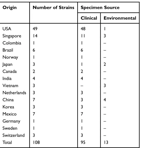
The complete sequences ofazuringene plusflanking regions from whole genome sequence of 108P. aeruginosastrains (95 strains from clinical specimens such as bacteremia, pneu-monia, and burn infections; and 13 strains of environmental origin such as marine, hospital wastewater, and dental clinic wastewater) with defined source and region were selected and acquired from NCBI GenBank (http://www.ncbi.nlm.
nih.gov/) (Table 1). Multiple sequence alignment was carried
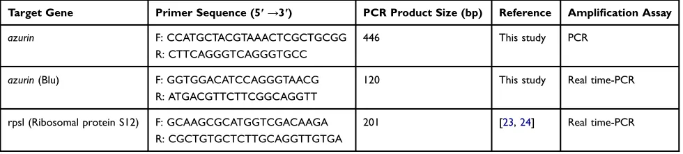
out using CLC Sequence Viewer software Ver. 7.6 (CLC, Denmark). The 3ˈand 5ˈflanking regions were composed of very diverse sequences (with 65% similarity), which made it impossible to achieve a conserved sequence for designing a primer pair which could anneal and amplify wholeazurin gene sequence in all of the isolates. The aligned sequences were used to design a primer pair which could anneal and amplify a conserved region from the very beginning to the end of azurin gene. The primers were designed by Gene Runner and CLC Sequence Viewer software. The designed primers were analyzed by Primer-BLAST on NCBI (http:// www.ncbi.nlm.nih.gov). Schematic representation ofazurin
gene sequence and primers position are depicted inFigure 1, and the primer pairs’sequences are displayed inTable 2.
PCR Ampli
fi
cation and Sequencing of
Azurin
Gene
All genomic DNA was extracted from the isolates by YTA genomic DNA extraction mini kit (Yekta Tajhiz Azma, Iran) according to the manufacturer’s instructions. DNA isolation procedure was performed in a room physically separated from the room applied for nucleic acid amplifi -cation reaction and also from the post-PCR room in order to inhibit or minimize contamination and false positive results. PCR amplification was performed using primers specifically designed forazuringene (Table 2).
The amplification assay was performed in a total volume of 12.5μL containing 1μL of purified DNA (20 ng), 6.5 μL of PCR Master-mix (Ampliqon, Denmark), and 0.5 μL of each primer (10 pM) using Bio Rad Thermal Cycler, Germany. The PCR steps included: an initial denaturation at 94°C for 5 min, followed by 30 cycles of denaturation at 94°C for 40 s, annealing at 63° C for 45 s, extension at 72°C for 60 s, and afinal extension at 72°C for 3 min. The products of PCR were run on electrophoresis by 1% agarose gel and visualized under UV doc apparatus. The amplified fragments of 59 MDR strains were selected (according to the isolate geographical
Table 1GenBbank Extracted AzurinGene Sequences Analyzed in This Study
Origin Number of Strains Specimen Source
Clinical Environmental
USA 49 48 1
Singapore 14 11 3
Colombia 1 1 –
Brazil 6 6 –
Norway 1 1 –
Japan 3 1 2
Canada 2 2 –
India 4 4 –
Vietnam 3 – 3
Netherlands 3 3 –
China 7 3 4
Korea 3 3 –
Mexico 7 7 –
Germany 1 1 –
Sweden 1 1 –
Switzerland 3 3 –
Total 108 95 13
Infection and Drug Resistance downloaded from https://www.dovepress.com/ by 118.70.13.36 on 27-Aug-2020
region and clinical specimen type) and subjected to direct sequencing using Applied Biosystems (ABI) capillary sequencer (Macrogen, Korea).
Alignment and Sequence Analysis of
Azurin
Gene from
P. aeruginosa
Isolates
Sequences ofazuringene of 59P. aeruginosastrains and P. aeruginosaATCC27853 were delivered as Chromas for-mat (Technelysium, Australia). The nucleotide sequence of each strain was deposited in the GenBank database and assigned a GenBank accession number (MK276543– MK276602). The sequences were analyzed and aligned by CLC Sequence Viewer 7 (Qiagen, Denmark) and Gene Runner 6.5.46 (Softpedia, Romania) software. The phylo-genetic tree was constructed for the azurin gene using UPGMA (unweighted pair group method with arithmetic mean) algorithm. P. aeruginosa strain PAO1 sequence, which was recorded in GeneBank (accession number AE004091.2), was included in alignment analysis as con-trol. Diversity index was calculated by DI = 1–n(n–1)/ƩN (N–1).
Global Nucleotide Polymorphism of
Azurin
Gene
A total of 168 azurin gene sequences (59 P. aeruginosa isolates and standardP. aeruginosaATCC27853 from this study and 108P. aeruginosastrains from Genbank) (Table 1) were compared and aligned by BioNumerics Ver. 7.6 (Applied Maths company, Belgium). The nucleotide changes, consensus blocks, similarities, amino acid trans-lations and mutations were determined. The circular den-drogram and multidimensional layout were constructed using UPGMA method.
RNA Extraction and cDNA Synthesis
In total, 40 isolates (including 39 clinical isolates and P. aeruginosa ATCC27853 as control) were subjected to total RNA extraction and cDNA synthesis based on the sequence analysis (Sequence Type), isolation location, and clinical sample type (blood, wound, tissue, and catheter). Total RNA was extracted from the isolates by YTA Total RNA Purification Mini kit (Yekta Tajhiz Azma, Iran) according to the manufacturer’s protocol. The removal of 1
50 100 150 200 250 300 350 446
1
60 120 148
azuringene (Accesion number: AE004091.2)
Azurin protein
Forward primer: 5522089-5522112 Reverse primer: 5521667-5521689
5' flanking region 3' flanking region
400
FP
RP
Figure 1The schematic representation ofazuringene andflanking regions. Position of primers for PCR amplification ofazuringene.
Table 2Primer Sequences Used in This Study
Target Gene Primer Sequence (5ʹ→3ʹ) PCR Product Size (bp) Reference Amplification Assay
azurin F: CCATGCTACGTAAACTCGCTGCGG
R: CTTCAGGGTCAGGGTGCC
446 This study PCR
azurin(Blu) F: GGTGGACATCCAGGGTAACG
R: ATGACGTTCTTCGGCAGGTT
120 This study Real time-PCR
rpsl (Ribosomal protein S12) F: GCAAGCGCATGGTCGACAAGA
R: CGCTGTGCTCTTGCAGGTTGTGA
201 [23,24] Real time-PCR
Infection and Drug Resistance downloaded from https://www.dovepress.com/ by 118.70.13.36 on 27-Aug-2020
genomic DNA was performed by DNase I- RNase free kit (Thermo Scientific, USA). The purity and quality of pre-pared RNA were assessed by measuring the optical density in 260/280 nm values and electrophoresis on agarose gel. The cDNA synthesis was performed using cDNA synthesis kit (Yekta Tajhiz Azma, Iran). For this purpose, 100 ng of total RNA, 1 µL oligo (dT) 18 primer (50 µM), 1 µL random hexamer primer (50 µM), and DEPC-treated water up to 13.4 µL were added into a sterile, nuclease-free tube on ice. The mixture was centrifuged briefly and incubated at 70°C for 5 min. In the following cDNA Synthesis Mix, 4 µL 5x-first strand buffer, 1 µL dNTP (10 mM), 0.5 µL RNasin (40 U/µL), and 1 µL M-MLV were added into the microtube, centrifuged, and incubated at 37°C for 60 min. Finally, the reaction was terminated by heating at 70°C for 5 min. The quality of synthesized cDNA was evaluated by gel electrophoresis and reverse transcription PCR. The synthesized cDNAs were subjected to real-time PCR assay.
Real-Time PCR Analysis of
Azurin
Gene
Expression
Relative-real-time PCR was performed to determine the expression level of azurin gene in different isolates of P. aeruginosa using the Light Cycler 96 Real-Time PCR system (Roche Life Science, Germany). Each PCR reac-tion was performed in a total reacreac-tion volume of 20 μL containing 12 μL of real-time PCR Master Mix (Amplliqon, Denmark), 1 μL cDNA template, 1 μL of each primer (Blu), and 5 μL distilled water. Primer desig-nation and sequences are depicted in Table 2. The qPCR was performed according to the following conditions: pre-incubation at 95°C for 5 min, 45 cycles of denaturation at 95°C for 30 s, annealing at 60°C for 45 s, and extension at 72°C for 30 s. The analysis of melting pick was performed at 95°C for 5 min. In each sample, the same amount of RNA was used, which were converted into the cDNA and pipetted. Ribosomal protein S12 (Rpsl) mRNA expression was applied as the internal control for each sample (Dumas et al., 2006), and the ΔCT(CTtarget − CT
refer-ence) and expression fold change were calculated for each sample according to the comparative CT method (Pfaffl
formula).25Each real-time PCR reaction was performed in duplicate, and the standard deviation was calculated. The efficiency of real-time PCR was determined by amplifying a serial dilution of the template cDNA (10 folds) and calculatingE= −1+10(−1/slope).
Statistical Analysis
The quantity ofazuringene expression in all clinical strains of different locations (Tehran or Isfahan), different clinical samples (wound, blood, tissue and catheter), and different sequence clusters was compared by one-way analysis of variance (ANOVA) statistical analysis test. Thet-test analy-sis was used to compare azurin expression level between the clinical isolates andP. aeruginosaATCC 27853. Ap-value <0.05 was considered as significant in all statistical analysis. The correlation between the sequence type and expression level ofazuringene was analyzed by Pearson test.
Results
Isolates Collection and Identi
fi
cation
In this cross-sectional study, 133 isolates were collected from burned patients, among which 57 and 43% were from the major centers of burn patients in Tehran and Isfahan, respec-tively. The bacterial strains were isolated from blood [7 (5.3%): 4 (5%) + 3 (5%)], wound [117 (88%): 67 (50%) + 50 (50%)], tissue [1 (0.7%): 1 (1%) + 0], and catheter [8 (6%): 4 (5%) + 4 (7%)] of patients admitted to Tehran and Isfahan university hospitals, respectively. The blood speci-mens were obtained from patients with both wound and systemic infections, and catheter specimens were from patients with both wound and genitourinary urogenital infec-tions due P. aeruginosa. All isolates (100%) were re-identified asP. aeruginosaby phenotypic tests and confirmed by species-specific 16s rDNA PCR amplification assay.
Antimicrobial Susceptibility Testing
About 100% of the isolates were resistant to gentamicin, tobramycin, and cefoxitin. About 98.5% were resistant to ciprofloxacin, amikacin, and imipenem while 100% and 23.9% of the isolates were susceptible to colistin and ceftazidime, respectively. All strains were determined as MDR according to the criteria determined by CLSI.23
Alignment and In Silico Analysis of
Azurin
Sequences from GenBank
According to the results of the multiple sequence alignment method, primer pairs were designed to cover the whole azuringene sequence at the position 5521667–5522112 in P. aeruginosaPAO1 (accession number: AE004091.2). This primer pair amplified the 446 bp amplicon, and the primer blast in NCBI indicated that the primers were specific for P. aeruginosa azuringene.
Infection and Drug Resistance downloaded from https://www.dovepress.com/ by 118.70.13.36 on 27-Aug-2020
Ampli
fi
cation and Sequencing of
Azurin
Gene
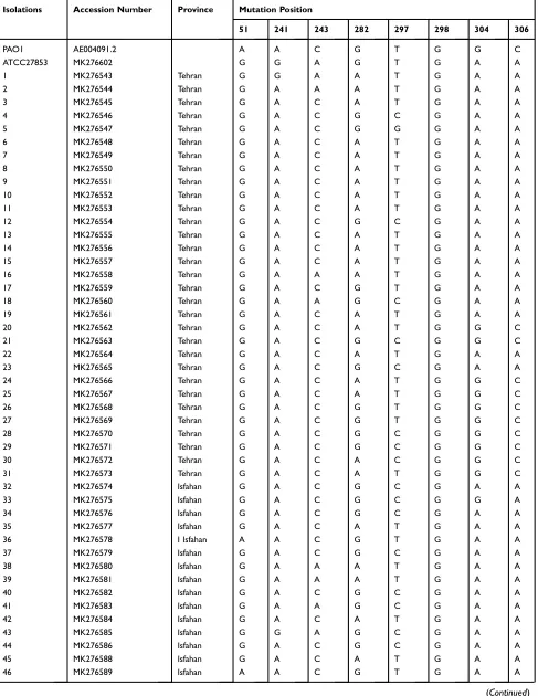
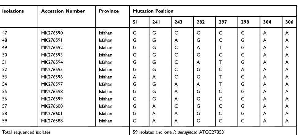
Total isolates (100%) under study containedazuringene, and it was possible to amplify 446 bp of theazuringene (Figure 2). In this study, 59 strains isolated from blood (6, 10%), wound (47, 80%), tissue (1, 2%), and catheter (5, 8%) of patients were sequenced. After trimming the low-quality sequences at the ends, a region of 406 bp (which covered 5521700–5522105) was subjected to analysis. The sequence analysis of azurin gene revealed point mutations at eight positions located in nucleotides 51, 241, 243, 282, 297, 298, 304, and 306
(Table 3). Mutation at four locations (241, 243, 304, and
306) resulted in different amino acid substitutions in two positions (threonine to alanine at position 81 and alanine to threonine at position 102 of amino acid sequences) (Table 4). About 62.8% of the isolates from Tehran and Isfahan harbored mutations which resulted in amino acid substitution in two positions (81 and 102). Also, 37.2% of the strains contained mutations in nucleotides 241, 243, 304, and 306, resulting in amino acid substitution at two positions 81 and 102 (missense mutation): A and T at position 81 and T and A at position 102 (Table 4). Generally, in addition, the amino acid sequence type (ST) to P. aeruginosa ATCC27853 and PAO1 there are 3 amino acid STs in our strains (Table 4).
The diversity index was calculated as 0.9 for the popula-tion. A dendrogram was constructed based on the sequences of azurin gene from 59 isolated strains, P. aeruginosa ATCC27853, and P. aeruginosa PAO1. The strains were spread in 19 STs (17 STs for Iranian strains and two STs for P. aeruginosaATCC27853 and PAO1). It was shown that 15 (25%) and nine (15%) clinical isolates allocated in common sequence types (A and B, respectively) (Figure 3). Also, 58 and 50% of the strains from Tehran and Isfahan were distrib-uted in common clusters (A, B, D, and E), while clusters C, G and F, H were specific for the isolates of Isfahan and Tehran, respectively. The strains distributed in cluster A were obtained
from the wound (86%), blood (7%), and catheter (7%) sam-ples. On the other hand, 80% and 20% of cluster A strains were collected from Tehran and Isfahan hospitals, respec-tively. Cluster B included the strains isolated from patients with wound infection (100%). This cluster's strains were iso-lated in greater numbers from Isfahan (67%) hospital.
Nucleotide Polymorphism Analysis of
Globally Reported
Azurin
Gene Sequences
The alignment of 168azurinsequences (59 Iranian clinical strains, 108 strains from GenBank, and P. aeruginosa ATCC27853) displayed 97.3% similarity using BioNumerics Ver. 7.6 (free trial version, Applied Maths Company, Belgium). The point mutations were shown in 23 nucleotides at different positions (51, 81, 114, 117, 124, 126, 169, 201, 241, 243, 255, 273, 282, 297, 298, 304, 306, 336, 342, 378, 418, 432, and 433). The nucleotide sequences were comple-tely conserved in other parts. Only one nucleotide deletion was detected at position 170 in the BH9 strain from India.
The amino acid substitutions (missense mutation) were determined at positions 81 (alanine to threonine) in 93% of isolates, position 99 (valine to isoleucine) in 1% of isolates and position 102 (threonine to alanine or threonine to serine) in 68% and 3% of isolates, respectively. The dele-tion of nucleotide in BH9 resulted in frameshift and unde-termined protein sequence.
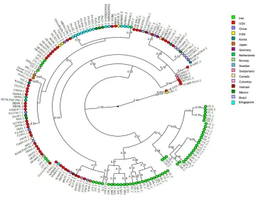
Global analysis of nucleotide polymorphism of azurin gene revealed a total of 26 STs among the P. aeruginosa strains according to the circular dendrogram (Figure 4). The results showed that 59 strains from Iran were distrib-uted in 17 STs, 65 strains from America (USA, Canada, Mexico Brazil, and Colombia) were spread in nine STs, 29 strains from Asia (China, Japan, Korea, Singapore, Vietnam, and India) belonged to seven STs, and nine strains from Europe (Norway, Netherlands, Sweden, Switzerland, and Germany) were distributed in four STs.
Among the Iranian strains, 48 (81%) strains belonged to two major clusters, and 11 (~19%) strains were distrib-uted in different clusters together with the strains from different countries of North and South America, Europe, South and East Asia (Figure 4). Also, 13 environmental strains were located in five STs within the same clusters where the clinical strains were located (Figure 4).
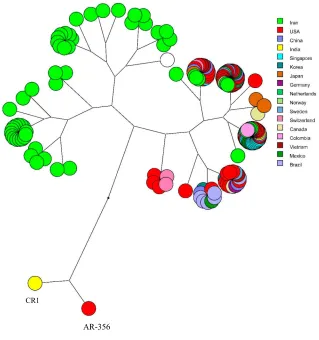
Multidimensional layout of global analysis of azurin gene in different isolates ofP. aeruginosashowed that all the isolates were originated from a common ancestor and branched in two main stems, while two strains (CR1 from Figure 2PCR assay ofazuringene. Lanes 1–5:P. aeruginosaisolates, Lanes N and P:
representatives of negative and positive controls. M: 100 bp DNA size marker.
Infection and Drug Resistance downloaded from https://www.dovepress.com/ by 118.70.13.36 on 27-Aug-2020
Table 3Point Mutations Within the 446-Bp Segment ofAzurinGene in IranianP. Aeruginosa Isolates in Relation to the Location of Isolation and Accession Numbers
Isolations Accession Number Province Mutation Position
51 241 243 282 297 298 304 306
PAO1 AE004091.2 A A C G T G G C
ATCC27853 MK276602 G G A G T G A A
1 MK276543 Tehran G G A A T G A A
2 MK276544 Tehran G A A A T G A A
3 MK276545 Tehran G A C A T G A A
4 MK276546 Tehran G A C G C G A A
5 MK276547 Tehran G A C G G G A A
6 MK276548 Tehran G A C A T G A A
7 MK276549 Tehran G A C A T G A A
8 MK276550 Tehran G A C A T G A A
9 MK276551 Tehran G A C A T G A A
10 MK276552 Tehran G A C A T G A A
11 MK276553 Tehran G A C A T G A A
12 MK276554 Tehran G A C G C G A A
13 MK276555 Tehran G A C A T G A A
14 MK276556 Tehran G A C A T G A A
15 MK276557 Tehran G A C A T G A A
16 MK276558 Tehran G A A A T G A A
17 MK276559 Tehran G A C G T G A A
18 MK276560 Tehran G A A G C G A A
19 MK276561 Tehran G A C A T G A A
20 MK276562 Tehran G A C A T G G C
21 MK276563 Tehran G A C G C G G C
22 MK276564 Tehran G A C A T G A A
23 MK276565 Tehran G A C G C G A A
24 MK276566 Tehran G A C A T G G C
25 MK276567 Tehran G A C A T G G C
26 MK276568 Tehran G A C G T G G C
27 MK276569 Tehran G A C G T G G C
28 MK276570 Tehran G A C G C G G C
29 MK276571 Tehran G A C G C G G C
30 MK276572 Tehran G A C A C G G C
31 MK276573 Tehran G A C A T G G C
32 MK276574 Isfahan G A C G C G A A
33 MK276575 Isfahan G A C G C G G A
34 MK276576 Isfahan G A C G C G A A
35 MK276577 Isfahan G A C A T G A A
36 MK276578 I Isfahan A A C G T G A A
37 MK276579 Isfahan G A C G C G A A
38 MK276580 Isfahan G A A A T G A A
39 MK276581 Isfahan G A A A T G A A
40 MK276582 Isfahan G A C G C G A A
41 MK276583 Isfahan G A A G C G A A
42 MK276584 Isfahan G A C A T G A A
43 MK276585 Isfahan G G A G C G A A
44 MK276586 Isfahan G A C G C G A A
45 MK276588 Isfahan G A C A T G A A
46 MK276589 Isfahan A A C G T G A A
(Continued)
Infection and Drug Resistance downloaded from https://www.dovepress.com/ by 118.70.13.36 on 27-Aug-2020
USA and AR-356 from India) were completely separated from the others (Figure 5).
Real-Time PCR Analysis
The purity of each extracted RNA was determined as 1.80–2 by calculating OD260/280 nm, and its quality was confirmed by gel electrophoresis and RT-PCR (data not shown). The amplification picks and melting curve analysis of products in real-time PCR are depicted inFigure S1aandS1b, respec-tively (supplementary data). No non-specific product and primer dimer were detected at melting temperature of approximately 84°C. Primer efficiency was calculated as 1.96 (Figure 1Sc in supplementary data).
The expression level ofazuringene in different isolates is presented inFigures 3and6. The expression level ofazurin gene in different Iranian clinical strains was statistically
different from its expression level in P. aeruginosa ATCC27853 (p< 0.001) and was 0.015–4.3-fold higher. The azurin gene expression level in the strains isolated from blood was 1.28–4.3-fold higher than in the standard strain, while the expression level in the strains isolated from wound, tissue, and catheter was 0.015–0.91-fold higher
(Figure 3). These results showed that the expression level
ofazuringene was statistically higher in the strains isolated from blood, compared with the strains of other clinical origin (p< 0.001). There was no significant difference regarding the expression level of azurin between the strains of two pro-vinces (Tehran and Isfahan) (p> 0.05).
The Pearson correlation test displayed that the expres-sion level of azuringene was not affected by the nucleo-tide sequence changes in this gene.
Discussion
In this study, 100% of isolates were susceptible to colistin, which means that this antibiotic is effective for treatment of infections caused by MDRP. aeruginosa. According to CLSI guidelines colistin could be used in antimicrobial assay and probably treatment of MDR P. aeruginosa.22 Moreover, colistin has been reported to be effective against MDR infections in several literatures in this
field,26–28although the use of polymyxins (colistin) should be optimized for dosage administration and indications, in order to maximize effectiveness, prevent the emergence of further polymyxin resistance and reduce adverse effects.29 In the study performed by Sabuda et al. (2008), the
Table 3(Continued).
Isolations Accession Number Province Mutation Position
51 241 243 282 297 298 304 306
47 MK276590 Isfahan G G C G C G A A
48 MK276591 Isfahan G G A G C G A A
49 MK276592 Isfahan G G C A T G A A
50 MK276593 Isfahan G G C G C G A A
51 MK276594 Isfahan G G C A T G A A
52 MK276595 Isfahan G G C G C A A A
53 MK276596 Isfahan A A C G T G A A
54 MK276597 Isfahan G G A A T G A A
55 MK276598 Isfahan G G A G C G A A
56 MK276599 Isfahan G G A G C G A A
57 MK276600 Isfahan G A C G C G A A
58 MK276601 Isfahan G A A G C G A A
59 MK276588 Isfahan G A A G C G A A
Total sequenced isolates 59 isolates and oneP. aeruginosaATCC27853
Table 4 The Frequency of Amino Acid Substitutions Among Iranian Azurin Protein Sequences
Strains Amino Acids Substitution Site
81 102
PAO1 . . T . . . . A . .
ATCC 27853 . . A . . . . T . .
37 strains (62.8%) . . T . . . . T . .
11 strains (18.6%) . . A . . . . T . .
11 strains (18.6%) . . T . . . . A . .
Total 59 isolates (100%)
Infection and Drug Resistance downloaded from https://www.dovepress.com/ by 118.70.13.36 on 27-Aug-2020
Isolates Province Patient sample Azurin expression (fold change)*
27 T Wound 0.53
26 T Blood 3.7
PAO1
31 T Wound
25 T Wound 0.56
24 T Wound
20 T Wound 0.75
28 T Wound 0.69
21 T Wound 0.91
29 T Wound
30 T Wound
42 I Wound 0.90
35 I Wound 0.47
22 T Wound 0.24
19 T Wound 0.02
15 T Wound
14 T Wound
13 T Wound
11 T Wound
10 T Blood 2.15
9 T Wound
8 T Wound 0.69
7 T Wound
6 T Wound 0.47
3 T Catheter 0.015
45 I Wound
51 I Wound 0.24
49 I Wound
39 I Blood 1.49
38 I Wound 0.24
16 T Wound 0.15
2 T Wound
54 I Wound
1 T Blood 4.3
ATCC 1
46 I Wound 0.59
36 I Wound 0.26
53 I Blood 1.28
12 T Wound 0.21
4 T Wound 0.37
57 I Wound
44 I Wound 0.94
40 I Wound 0.46
37 I Wound 0.87
34 I Wound 0.46
32 I Wound
23 T Wound
33 I Catheter 0.87
17 T Tissue 0.55
5 T Wound 0.55
50 I Wound
47 I Catheter 0.53
52 I Catheter 0.21
59 I Blood 4.28
58 I Catheter 0.60
41 I Wound 0.21
18 T Wound 0.75
56 I Wound
55 I Wound 0.61
48 I Wound 0.48
43 I Wound
C (6.6%)
G (5%)
A (25%)
D (6.6%)
H (5%)
B (15%)
E (6.6%)
F (6.6%)
Figure 3UPGMA dendrogram ofP. aeruginosaclinical strains based on the difference in the nucleotides ofazuringene sequences.P. aeruginosaATCC27853 and PAO1 were used as control. Each strain is presented based on the geographical region of its isolation, clinical sample, and gene expression fold change. T: Tehran, I: Isfahan.
Infection and Drug Resistance downloaded from https://www.dovepress.com/ by 118.70.13.36 on 27-Aug-2020
intravenous and nebulized colistin was effective for treat-ment of MDRP. aeruginosa, with mild toxicity in kidneys of two-third of patients.26
In this study,azuringene was detected in 100% of 133 clinical strains under study, while Sereena and colleagues (2016) reported the presence of this gene in 80% of the strains of environmental origin.14 In another study by Sarwar et al. (2018), the frequency ofazuringene in envir-onmental strains was determined as 25%.30These findings signify its putative role in the biology of clinical strains and probably disease development in clinical strains.
Sequence alignment and azurin gene analysis in P. aeruginosastrains in this study displayed the presence of eight point mutation sites, of which four resulted in amino acid substitution at two locations (81 alanine to threonine and 102 threonine to alanine) (missense mutation) (Tables 3
and 4). Similarly, the amino acid substitution in azurin sequence were detected in positions 81 and 102 in studies reported from different parts of the world.33–35Alanine and threonine are structurally different with an extra OH group in threonine (polar amino acid). This is compatible with the preference of alanine (as a hydrophobic amino acid) to form a helical structure and the preference of threonine (as a polar amino acid) to support beta-sheet structures.31Of course, as
the structure of azurin is a complex of alpha helixes and beta sheets, it seems that these amino acid substitutions have no great impact on protein structure and function. In the same study by Nguyen et al. (2019), point mutation and amino acid substitutions were detected, none of which affected the azurin function.32
In the constructed dendrogram based on the sequence analysis of azuringene in clinical isolates (Figure 3), the isolates were classified in 19 STs and nine (A–H) clusters, considering P. aeruginosa PAO1 and P. aeruginosa ATCC27853 as standard strains. Also, 25% and 15% of the isolates belonged to clusters A and B, respectively. The Diversity Index was calculated as 0.9 for total population. In addition, 58% and 50% of the strains from Tehran and Isfahan were included in common clusters (A, B, D and E), while clusters C, G, and F, H were restricted for Isfahan and Tehran strains, respectively, which may show the dissemination of specificazurin sequence types in each province. According to the distribution of the strains from different clinical specimens (blood, wound, catheter and tissue) in common clusters such as A and B, it was assumed thatazurinST type has no effect on the site of infection. About 80% and 20% of cluster A strains were collected from Tehran and Isfahan hospitals, respectively, Figure 4Circular dendrogram according to the global analysis of nucleotide polymorphism ofazuringene amongP. aeruginosastrains. C and E next to the ID of the isolates determine the clinical and environmental sources of isolates. The geographical origin of the isolates is displayed using different colors.
Infection and Drug Resistance downloaded from https://www.dovepress.com/ by 118.70.13.36 on 27-Aug-2020
and 67% of cluster B strains were isolated from Isfahan. The frequency of distribution of ST types based on the geographical region in different clusters demonstrates the possible role of geographical area on the ST distribution.
Due to limited point mutations and considering the fact that only purine:purine and pyrimidine:pyrimidine conver-sions were observed in nucleotides, it seems thatazuringene is structurally and functionally conserved in clinical strains and may be considered a useful tool for the detection of clinical isolates ofP. aeruginosa. In the study performed by Nguyen et al. (2019), the sequences ofazuringene from the metagenomic DNA sample andP. aeruginosaisolates were analyzed andfive point mutations at (51, 282, 297, 305, and 432) were determined. Thefirst mutation was located in the signal peptide region and the other mutations did not affect the three main domains of azurin protein. They concluded that mutations in the mature peptide did not interfere with azurin protein structural properties.32
Kamalakannan et al. (2011) examined the primary and secondary structure of azurin protein and its phylogenetic relatedness in various species ofPseudomonas. Their results showed differences in molecular weight and amino acid composition of azurin in different species; however, phylo-genetic analysis indicated that azurin protein of various Pseudomonas species originated from a common ancestor.36 Their study results strengthened our hypothesis aboutazuringene conservation inP. aeruginosaspecies.
The global analysis ofazuringene sequences among the 168 strains of P. aeruginosa (59 sequences from Iranian clinical strains and 108 Genbank registered sequences) dis-played point mutations in 22 nucleotides, resulting in amino acid substitution (missense mutations) at three positions: 81 (alanine to threonine), 99 (valine to isoleucine), and 102 (threonine to alanine/serine). The sequence alignment and analysis indicated 97.3% similarity among the 168 sequences with a clear distinct lineage for Iranian sequences (Figure 4).
CR1
AR-356
Figure 5The multidimensional layout of global analysis ofazuringene in different isolates ofP. aeruginosaaccording to UPGMA algorithms.
Infection and Drug Resistance downloaded from https://www.dovepress.com/ by 118.70.13.36 on 27-Aug-2020
The analysis results are re-emphasizing that theazuringene is conserved and it may be related the important function of this protein.
The circular dendrogram was constructed according to the sequences alignment and divided the sequences into 26 STs, in which the sequences from Iranian strains were distributed in 17 STs, and the sequences reported from all
other countries were spread over nine STs. Among the Iranian sequences, 48 (81%) sequences belonged to two main clusters, but 11 sequences were distributed in other clusters together with the sequences from different coun-tries (Figure 4), emphasizing the probable role of the iso-lates’ geographical location on azurin gene nucleotide sequence variation. This may suggest that somehow related
0 0.5 1 1.5 2 2.5 3 3.5 4 4.5 5
27 20 25 28 21 42 35 22 19 8 6 51 38 16 46
AT
C
C 36 12 4 40 37 34 44 5 41 18 55 48 26 59 53 1 39 10 3 33 47 52 58 17
Fo
ld
c
h
an
g
e
o
f
az
ur
in
expr
es
si
o
n
Strains
Wound
Blood
Catheter
Tissue
*
*
* *
* *
Figure 6The fold change ofazuringene expression in differentP. aeruginosaclinical strains isolated from different specimens (wound, blood, catheter, and tissue). *p< 0.001.
Infection and Drug Resistance downloaded from https://www.dovepress.com/ by 118.70.13.36 on 27-Aug-2020
strains from Iran have made a genetic stockpile through point mutations, which has established them distinct from strains of rest of world.
The sequences of 13 environmental strains (from hos-pital or dental clinic sewage and marine) were distributed in five STs together with the clinical strains (Figure 4), which may be due to (i) the contamination of hospital or dental clinic sewage by clinical strains or (ii) inefficient disinfection of hospital or dental clinic sewage, which might have caused the contamination to be disseminated to clinics and caused infections.
The multidimensional layout of global analysis of azuringene in different strains ofP. aeruginosa(Figure 5) determined that all isolates were originated from a common ancestor and divided the isolates into two main branches, one of which contained Iranian isolates. Two clinical strains from India and USA (CR1 and AR-356) were absolutely fell apart.
The present study results demonstrated that the strains isolated from blood samples of patients with systemic infections belonged to several clusters and STs; thus, it seems that the azuringene sequence does not have effect on the infection type.
The results of azurin gene expression among the sequenced isolates demonstrated that the expression of azuringene was significantly higher in the strains isolated from blood than in those isolated from wound, catheter, and tissue (p< 0.001;Figures 3and6). The former isolates were obtained from patients with systemic infections which ultimately resulted in their deaths. This is probably related to the contribution of azurin protein to the patho-genicity and the possibility of bacterial attachment, inva-sion (our previous study)16,17 and survival in blood. According to the results, it was assumed that the interfer-ence or inhibition of azurin function probably led to the destruction of invasion or systematic pathogenicity of P. aeruginosa.
Overall, there are limited studies concerning azurin expression. It was found that azurin is highly expressed in anaerobically constructed biofilms in cystic fibrosis patients.5 This implies the significant role of azurin in P. aeruginosa infection, especially in cystic fibrosis patients. Furthermore, it was previously determined that azurin protein induces apoptosis in macrophages involved in phagocytosis.7 It seems that a high level of azurin expression in blood isolate strains may correlate with its role as an apoptosis inducer in macrophages, which helps its dissemination and increased pathogenesis.
It is worth noting that in this study, no obvious correlation was detected between theazurinsequence type and the level of expression, signifying the importance of clinical source of isolation as the main crucial factor affectingazurin expres-sion level. This increased expresexpres-sion may be due to the presence of strongerazurinpromoters in these strains, indis-putably explaining why these strains escaped the host immune system and disseminated into the bloodstream.
Conclusion
This study revealed the presence of azurin gene in all clinical isolates ofP. aeruginosa, which was highly con-served with point mutations occurring in only eight posi-tions, resulting in only two amino acid substitutions (missense mutation) at positions 81 and 102 (alanine to threonine). It seems that the point mutations have no effect on the virulence potency of P. aeruginosa. The multidi-mensional layout of global analysis of azurin gene revealed that the majority of Iranian sequences (81%) were allocated in two main clusters distinct from the rest of world, which suggests that Iranian strains have made a distinct genetic stockpile through point mutations which has established them separate from the other counties. However, 19% of Iranian strains were distributed in simi-lar clusters together with strains from different countries of North and South America, Europe, South and East Asia.
The expression ofazuringene was significantly higher in the strains isolated from blood of patients with systemic infections, which may demonstrate their increased patho-genicity and ability to escape the host immune system and disseminate into the bloodstream. This finding may open new insight aboutazuringenetic variation and significance inP. aeruginosa pathogenesis.
Data Sharing Statement
The data sets of the current study are available within article/its supplementary files or can be obtained from corresponding upon request. DNA sequences of genes that have been deposited in GenBank are available in
https://pubmed.ncbi.nlm.nih.gov/.
Ethics Approval and Consent to
Participate
This study was approved by Medical Ethics Committee of Isfahan University of Medical Sciences Code: 3.094 before the study began. All research was performed in accordance with relevant guidelines/regulations. The consent to
Infection and Drug Resistance downloaded from https://www.dovepress.com/ by 118.70.13.36 on 27-Aug-2020
participate was obtained from the patients or parents/guar-dians of the minors included in this study and the data were analyzed anonymously.
Acknowledgments
I would like to express my special thanks to the laboratory staff of two burns center hospitals, Imam Musa kazem in Isfahan and Shahid Motahhari in Tehran.
Author Contributions
All authors contributed to data analysis, drafting or revising the article, gavefinal approval of the version to be published, and agree to be accountable for all aspects of the work.
Disclosure
The authors report no conflicts of interest in this work.
References
1. Japoni A, Farshad S, Alborzi A. Pseudomonas aeruginosa: burn infection, treatment and antibacterial resistance.Iran Red Crescent Med J IRCMJ ©iranian Red Crescent Med J.2009;11(3):244–253. 2. Lau GW, Hassett DJ, Britigan BE. Modulation of lung epithelial
functions by Pseudomonas aeruginosa. Trends Microbiol. 2005;13 (8):389–397. doi:10.1016/j.tim.2005.05.011
3. Han Y, Wang T, Chen G, et al. APseudomonas aeruginosatype VI secretion system regulated by CueR facilitates copper acquisition.
PLoS Pathog.2019;15(12):e1008198. doi:10.1371/journal
4. Yamada T, Mehta RR, Lekmine F, et al. A peptide fragment of azurin induces a p53-mediated cell cycle arrest in human breast cancer cells.
Mol Cancer Ther. 2009;8(10):2947–2958. doi:10.1158/1535-7163. MCT-09-0444
5. Yoon SS, Hennigan RF, Hilliard GM, et al. Pseudomonas aeruginosa anaerobic respiration in biofilms. Dev Cell. 2002;3(4):593–603. doi:10.1016/S1534-5807(02)00295-2
6. Kamp M, Silvestrini MC, Brunori M, Beeumen J, Hali FC, Canters GW. Involvement of the hydrophobic patch of azurin in the electron-transfer reactions with cytochrome c551 and nitrite reductase. Eur J Biochem. 1990;194(1):109–118. doi:10.1111/ j.1432-1033.1990.tb19434.x
7. Yamada T, Goto M, Punj V, et al. The bacterial redox protein azurin induces apoptosis in J774 macrophages through complex formation and stabilization of the tumor suppressor protein p53.Infect Immun.
2002;70(12):7054–7062. doi:10.1128/IAI.70.12.7054–7062.2002 8. Wright BW, Kamath KS, Krisp C, et al. Proteome profiling of
Pseudomonas aeruginosa PAO1 identifies novel responders to copper stress. BMC Microbiol. 2019;19(1):1–19. doi:10.1186/s12866-019-1441-7
9. Chaudhari A, Fialho AM, Ratner D, et al. Azurin, Plasmodium falciparum malaria and HIV/AIDS: inhibition of parasitic and viral growth by azurin.Cell Cycle.2006;5(15):1642–1648. doi:10.4161/ cc.5.15.2992
10. Das Gupta TK, Green A, Shilkaitis A, et al. Noncationic peptides obtained from azurin preferentially enter cancer cells.Cancer Res.
2009;69(2):537–546. doi:10.1158/0008-5472.can-08-2932
11. Naguleswaran A, Fialho AM, Chaudhari A, et al. Azurin-like protein blocks invasion of toxoplasma gondii through potential interactions with parasite surface antigen SAG1.Antimicrob Agents Chemother.
2008;52(2):402–408. doi:10.1128/AAC.01005-07
12. Apiyo D, Wittung-Stafshede P. Unique complex between bacterial azurin and tumor-suppressor protein p53. Biochem Biophys Res Commun.2005;332(4):965–968. doi:10.1016/j.bbrc.2005.05.038 13. Fialho A, Das Gupta T, Chakrabarty A. Designing promiscuous
drugs? Look at what nature made!Lett Drug Des Discov. 2006;4 (1):40–43. doi:10.2174/157018007778992946
14. Sereena M, Sebastian D. Molecular detection of azurin: a powerful anticancer protein from native pseudomonas isolates. J Adv Med Pharm Sci.2015;5(1):1–7. doi:10.9734/jamps/2016/20557
15. Chen J, Zhuang G, Frieden L, Debinski W. Eph receptors and ephrins in cancer: common themes and controversies.Cancer Res.2008;68 (24):10031–10033. doi:10.1158/0008-5472.CAN-08-3010
16. Mohammadi Barzelighi H, Nasr-Esfahani B, Bakhshi B, et al. Analysis of antibacterial and antibiofilm activity of purified recombi-nant Azurin from Pseudomonas aeruginosa. Iran J Microbiol.
2019;11(2):166–176. doi:10.18502/ijm.v11i2.1083
17. Mohammadi Barzelighi H, Esfahani BN, Bakhshi B, et al. Influence of heterologously expressed azurin from pseudomonas aeruginosa on the adhesion and invasion of pathogenic bacteria to the Caco-2 Cell Line.Probiotics Antimicrob Proteins.2019. doi:10.1007/s12602-019-09573-2
18. Ramachandran S, Sarkar S, Mazumadar A, Mandal M. Azurin synth-esis from Pseudomonas aeruginosa MTCC 2453, properties, induc-tion of reactive oxygen species, and p53 stimulated apoptosis in breast carcinoma cells. Journal of Cancer Science & Therapy.
2011;3(5):104–111. doi:10.4172/1948-5956.1000069
19. Osman YA, El-Deep D, Younis SA. Azurin: A Powerful Anticancer from“A”Local Pseudomonas aeruginosa Isolate.J Am Sci.2013;9 (12):755–764.
20. Nanvazadeh F, Khosravi AD, Zolfaghari MR, et al. Genotyping of Pseudomonas aeruginosa strains isolated from burn patients by RAPD-PCR. Burns. 2013;39(7):1409–1413. doi:10.1016/j. burns.2013.03.008
21. Spilker T, Coenye T, Vandamme P, et al. PCR-based assay for differentiation of pseudomonas aeruginosa from other pseudomonas species recovered from cystic fibrosis patients. J Clin Microbiol.
2004;42(5):2074–2079. doi:10.1128/JCM.42.5.2074-2079.2004 22. Clinical and Laboratory Standards Institute.M07-A10: Methods for
Dilution Antimicrobial Susceptibility Tests for Bacteria That Grow Aerobically; Approved Standard—Tenth Edition. January2015. 23. Falagas ME, Koletsi PK, Bliziotis IA. The diversity of definitions of
multidrug-resistant (MDR) and pandrug-resistant (PDR) Acinetobacter baumannii and Pseudomonas aeruginosa. J Med Microbiol.2006;55(12):1619–1629. doi:10.1099/jmm.0.46747-0 24. Dumas JL, Van Delden C, Perron K, et al. Analysis of antibiotic
resistance gene expression in Pseudomonas aeruginosa by quantita-tive real-time-PCR. FEMS Microbiol Lett. 2006;254(2):217–225. doi:10.1111/j.1574-6968.2005.00008.x
25. Schmittgen TD, Livak KJ. Analyzing real-time PCR data by the comparative Ct method. Nat Prod Rep. 2008;3(6):1101–1108. doi:10.1038/nprot.2008.73
26. Sabuda DM, Laupland K, Pitout J, et al. Utilization of colistin for treatment of multidrug-resistant Pseudomonas aeruginosa. Can J Infect Dis Med Microbio.2008;19(6):413–418. doi:10.1155/2008/ 743197
27. Martis N, Leroy S, Blanc V. Colistin in multi-drug resistant
Pseudomonas aeruginosablood-stream infections.J Infect.2014;69 (1):1–12. doi:10.1016/j.jinf.2014.03.001
28. Cai Y, Yang D, Wang J, Wang R. Activity of colistin alone or in combination with rifampicin or meropenem in a carbapenem-resistant bioluminescent Pseudomonas aeruginosa intraperitoneal murine infection model. J Antimicrob Chemother. 2018;73(2):456–461. doi:10.1093/jac/dkx399
29. Bassetti M, Peghin M, Vena A, Giacobbe DR. Treatment of Infections Due to MDR Gram-Negative Bacteria. Front Med.
2019;6:1–10. doi:10.3389/fmed.2019.00074
Infection and Drug Resistance downloaded from https://www.dovepress.com/ by 118.70.13.36 on 27-Aug-2020
30. Sarwar F. Molecular Detection of Anticancer Protein Azurin in Native Pseudomonas Isolates. BRAC University;2018:31–36. 31. Malkov S, Zivkovic MV, Beljanski MV, Zaric SD. Correlations of
amino acids with secondary structure types: connection with amino acid structure.arxiv.org.2005;34:1–13.
32. Nguyen VD, Nguyen TT, Pham TT, Packianather M, Le CH. Molecular screening and genetic diversity analysis of anticancer Azurin-encoding and Azurin-like genes in human gut microbiome deduced through cultivation-dependent and cultivation-independent studies.
Int Microbiol.2019;22(4):437–449. doi:10.1007/s10123-019-00070-8 33. Ichise Y, Kosuge T, Uwate M, Nakae T, Maseda H. Complete
genome sequence ofpseudomonas aeruginosastrain 8380, isolated from the human gut.GenomeAnnounc.2015;3(3):e00520–e005215. doi:10.1128/genomeA.00520-15
34. Espinosa-Camacho LF, Delgado G, Miranda-Novales G, Soberón-Chávez G, Alcaraz LD, Morales-Espinosa R. Complete genome sequences of twoPseudomonas aeruginosastrains isolated from children with bacteremia. Genome Announc. 2017;5(35): e00927–e00917. doi:10.1128/genomeA.00927-17
35. Magalhães B, Senn L, Blanc DS. High-quality complete genome sequences of three Pseudomonas aeruginosaisolates retrieved from patients hospitalized in intensive care units. Microbiol Resour Announc.2019;8(9):e01624–18. doi:10.1128/MRA.01624-18 36. Kamalakannan T, Rajagopal K, Jothi A, Nirai ST. Analysis of Azurin
Protein from Different Pseudomonas sp Using Proteomic Tools.
J Commun Comput.2011;8:727–730.
Infection and Drug Resistance
Dove
press
Publish your work in this journal
Infection and Drug Resistance is an international, peer-reviewed open-access journal that focuses on the optimal treatment of infection (bacterial, fungal and viral) and the development and institution of preventive strategies to minimize the development and spread of resis-tance. The journal is specifically concerned with the epidemiology of
antibiotic resistance and the mechanisms of resistance development and diffusion in both hospitals and the community. The manuscript manage-ment system is completely online and includes a very quick and fair peer-review system, which is all easy to use. Visit http://www.dovepress.com/ testimonials.php to read real quotes from published authors.
Submit your manuscript here:https://www.dovepress.com/infection-and-drug-resistance-journal
Infection and Drug Resistance downloaded from https://www.dovepress.com/ by 118.70.13.36 on 27-Aug-2020