Changes in Upper Extremity Position Cause Migration of
Peripherally Inserted Central Catheters in Neonates
Ali M. Nadroo, MD*; Ronald B. Glass, MD‡; Jing Lin, MD*; Robert S. Green, MD*; and Ian R. Holzman, MD*
ABSTRACT. Objective. The migration of peripherally inserted central catheters (PICCs) from the superior or inferior vena cava into the right atrium can pose a sig-nificant risk of lethal pericardial effusion and tampon-ade secondary to myocardial perforation. Arm movement has been reported to cause displacement of the catheter tip toward the heart and lead to ventricular tachycardia in adults. The objective of this study was to investigate whether adduction or abduction at the shoulder and flex-ion or extensflex-ion at the elbow affect the positflex-ion of PICCs placed via upper limb veins. We also hypothesized that arm movements can be used to reposition malpositioned catheters.
Methods. A total of 280 radiographs of 60 neonates with PICCs inserted via upper limb veins from July 2000 through June 2001 were reviewed. Differences in catheter tip position as a result of abduction versus adduction at the shoulder, flexion versus extension at the elbow, and combination changes in arm posture were determined by measurements in paired radiographs. Correction of mal-positioned catheters was attempted in 10 patients by using arm movements without any alterations at the site of insertion.
Results. Arm movements were associated with sig-nificant displacement of catheters. Catheters that were placed via the basilic or axillary vein migrated toward the heart with adduction of the arm, whereas those that were placed via the cephalic vein moved away from the heart with adduction. Flexion of the elbow displaced catheters that were placed in the basilic or cephalic vein below the elbow toward the heart but did not have any effect on catheters that were placed via the axillary vein. For cath-eters that were placed in the basilic vein, simultaneous shoulder adduction and elbow flexion caused the great-est movement toward the heart (15.11 ⴞ 1.22 mm). We were able to reposition correctly inappropriately placed catheters in 9 of 10 patients by using arm movements.
Conclusions. Arm movements significantly affect the position of the tip of the PICCs. Prevention of catheter migration into the right atrium requires radiographic determination of vein of insertion and monitoring of catheter tip position with upper extremity in position of maximum inward movement of catheter for that vein. Arm movements can be used to correct the malpositioned catheters. Pediatrics 2002;110:131–136; catheters, migra-tion, arm movements, neonates.
ABBREVIATIONS. PICC, peripherally inserted central catheter; SEM, standard error of the mean; ANOVA, analysis of variance.
P
eripherally inserted central catheters (PICCs) have become increasingly popular in the treat-ment of neonates in the intensive care unit. We recently reported that migration of these catheters is a common problem, experienced by as many as 82% of neonatologists who responded to a national sur-vey.1Lethal pericardial effusion and tamponade sec-ondary to myocardial perforation is a known com-plication of PICCs.2– 4 Our recent survey and previous case reports suggest that this complication can result from either placement of the catheter tip in the right atrium or migration of the catheter tip into the right atrium from a proximal site, such as the superior vena cava. For preventing myocardial per-foration, it is recommended that the tip of the PICC not be placed in the right atrium or the intrapericar-dial portion of the inferior or superior vena cava.5 However, migration of catheters still may pose a significant risk for cardiac tamponade.1 It also has been reported in adults that adduction of the arm can both displace catheters medially (toward the heart) by 5 to 8 cm and lead to ventricular tachycardia.6,7 The present study was conducted to examine the effects of arm movements on the position of PICCs placed through the upper limb veins in neonates. We hypothesized that arm movements, such as adduc-tion or abducadduc-tion at the shoulder and flexion or extension at the elbow, affect position of the catheters placed via upper extremity veins. We also tested the hypothesis that knowledge of catheter tip movement with changes in upper extremity posture might al-low correction of malpositioned catheters (eg, coiled catheters).METHODS
The study was approved by the Institutional Review Board of the Mount Sinai Medical Center. The radiographs taken as part of routine management in our neonatal intensive care unit based on our previous study of all neonates with PICCs inserted via upper limb veins from July 2000 through June 2001 were reviewed in this study. Catheters (1.9 French) from a single manufacturer (Becton-Dickinson, Sandy, UT) were used in all patients. Before insertion, catheters were prepared according to the suggestions of the man-ufacturer by cutting to a length equal to the distance from the point of insertion to the medial end of the right clavicle and then down to the second intercostal space. The measurement was taken with the arm abducted 90 degrees at the shoulder. After insertion, an initial chest radiograph was taken, and, if necessary, the cath-eter was adjusted so that the cathcath-eter tip was in the superior vena
From the *Department of Pediatrics, Division of Newborn Medicine, and ‡Department of Radiology, Mount Sinai School of Medicine, New York, New York.
Received for publication Jul 10, 2001; accepted Feb 8, 2002.
Reprint requests to (A.M.N.) Department of Pediatrics, Division of New-born Medicine, Box 1508, Mount Sinai School of Medicine, One Gustave L Levy Pl, New York, NY 10029. E-mail: [email protected]
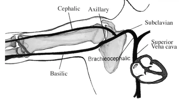
cava or subclavian vein as confirmed by a repeat radiograph. Subsequent radiographs were performed twice a week to monitor the position of the catheter as part of our existing neonatal inten-sive care unit policy. Sequential radiographs were taken with the arm and forearm in a variety of positions. A pediatric radiologist examined all of the radiographs. The reference points by which the position of the tip of the catheter was determined were the vertebral bodies, the midline, the outer margin of the first rib, and the medial end of the clavicle. The length of the catheter left outside the body at the site of insertion was documented daily. Radiographs of any patient whose catheter length outside the body had changed from that of initial insertion were excluded from the study. Radiographs also were excluded when the tip of the catheter was not clearly visible or the radiograph was rotated, making it difficult to interpret accurately the catheter tip position. PICCs were inserted into 1 of 3 different vessels: cephalic vein, basilic vein, or axillary vein (Fig 1). Insertion in the cephalic and basilic veins was below the elbow. Axillary vein insertion was in the axilla. Depending on the duration of catheter insertion and position of the arm and forearm in various radiographs, different comparisons were available for different infants. The change in the catheter tip position was measured between paired radiographs of the same catheter with the upper extremity in various combina-tions of adduction/abduction at the shoulder and flexion/exten-sion at the elbow. Observations of paired radiographs for catheter tip migration with change in arm position were divided into 3 groups, each with 2 subgroups. The numbers indicated for each subgroup are the numbers of paired radiographs available for the specific comparisons.
Groups
Group 1: Effect of Shoulder Adduction
In group 1, catheter tip position in a radiograph with the arm adducted at the shoulder was compared with the catheter tip position in a paired radiograph with the arm abducted at the shoulder. In group 1A (n⫽24), the elbow was in flexion in both radiographs. In group 1B (n⫽47), the elbow was in extension in both radiographs.
Group 2: Effect of Elbow Flexion
In group 2, catheter tip position in a radiograph with the forearm flexed at the elbow was compared with the catheter tip position in a paired radiograph with the forearm extended at the elbow. In group 2A (n⫽37), the shoulder was in adduction in both radiographs. In group 2B (n ⫽ 24), the shoulder was in abduction in both radiographs.
Group 3: Effect of Shoulder Adduction Combined With Simultaneous Elbow Flexion or Extension
In group 3A (n⫽31), catheter tip position in a radiograph with the arm adducted at the shoulder and the forearm flexed at the elbow was compared with the position of the catheter tip in a
paired radiograph with shoulder abducted and elbow extended. In group 3B (n⫽25), catheter tip position in a radiograph with the arm adducted at the shoulder and the forearm extended at the elbow was compared with the catheter tip position in a paired radiograph with the shoulder abducted and elbow flexed. Thus, in groups 1 and 2, the effects of difference in position at 1 joint with the other joint fixed are examined; in group 3, the effects of simultaneous change in position of both the shoulder and elbow joints are examined.
Statistical Analysis
For statistical analysis, the measurements of the movements of the catheters were normalized to the individual infant’s body length at birth and expressed as millimeters per centimeter. Re-sults are reported as the mean⫾standard error of the mean (SEM) for the respective group. Catheter tip movement between paired radiographs for each group was analyzed by 2-way analysis of variance (ANOVA) with arm/forearm position subgroup and the vein of insertion being the main effects. When the ANOVA showed a significant difference, Tukey’s test for multiple compar-isons was performed. Subsequent to partial compilation of the information acquired from comparing paired radiographs, the knowledge of catheter tip movement as a function of arm position was used to attempt correction of the position of malpositioned catheters in 10 patients.
RESULTS
A total of 280 radiographs from 60 patients (mean birth weight: 1283 g; range: 480-4570 g; mean gesta-tional age: 29 weeks; range: 23– 41 weeks) with PICCs were reviewed in the study. Thirty-three catheters were inserted via basilic veins, 18 through cephalic veins, and 9 through axillary veins. In Fig 2, the actual catheter tip movement in millimeters⫾SEM, as a result of various changes in position, as de-scribed below is illustrated. The displacement nor-malized to the body length expressed as millimeters per centimeter is shown in parentheses.
Group 1: Effect of Adduction at Shoulder With Fixed Forearm Position
Catheters that were inserted in the basilic and axillary veins moved toward the heart with shoulder adduction, but those that were placed in cephalic veins moved away from the heart. The effect of shoulder adduction was not altered by the fixed elbow position in the ANOVA; therefore, actual shoulder adduction movement in millimeters of
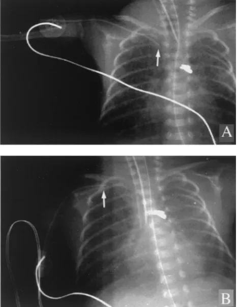
catheters placed in a given vein regardless of elbow position were averaged, and the results are illus-trated in the first panel of Fig 2. Catheters that were placed via the basilic and axillary veins were dis-placed toward the heart with shoulder adduction 8.95⫾0.85 mm (0.24 ⫾0.02 mm/cm;P⬍.001) and 7.70 ⫾ 1.31 mm (0.21 ⫾ 0.04 mm/cm, P ⬍ .001), respectively. Catheters that were placed via the ce-phalic vein moved significantly away from the heart as indicated by the negative number ⫺5.17 ⫾ 0.53 mm (0.14⫾ 0.01 mm/cm;P⬍.001). An example is shown in Fig 3.
Group 2: Effect of Flexion at the Elbow With Fixed Shoulder Position
Catheters that were inserted in the basilic and cephalic veins moved toward the heart an equivalent distance with elbow flexion, but those that were placed in axillary veins did not move. Because fixed shoulder position had no effect on elbow flexion-induced catheter movement in the ANOVA, actual movement in millimeters of catheters placed in a given vein regardless of shoulder position was aver-aged, and the results are illustrated in the second panel of Fig 2. Catheters that were placed via the basilic and cephalic veins were displaced toward the heart with elbow flexion 7.35⫾0.62 mm (0.20⫾0.02 mm/cm;P⬍.001) and 5.71⫾ 0.80 mm (0.15⫾0.02 mm/cm;P⬍.001), respectively. Catheters that were placed via the axillary veins had no detectable
mo-Fig 2. Catheter tip movements induced by changes in upper extremity position. Group 1, adduction of the arm with fixed elbow position; group 2, flexion of the elbow with fixed shoulder position; group 3A, adduction of the arm and simultaneous flexion of the elbow; group 3B, adduction of the arm and simultaneous extension of the elbow. Vein of insertion indicated by the legend. Values shown are mean⫾ SEM. *P⬍.05 versus the other 2 veins in the respective group; #P⬍.05 versus same vein in group 3A.
tion with elbow flexion in the paired chest radio-graphs.
Group 3: Effect of Shoulder Adduction and Simultaneous Elbow Flexion or Extension
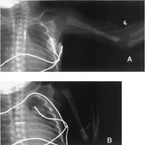
For each vein separately, we performed an analy-sis of the effect on catheter tip motion of flexion versus extension at the elbow simultaneous with shoulder adduction. The actual changes in position for catheters in groups 3A (adduction plus flexion) and 3B (adduction plus extension) are illustrated for catheters that were placed in each of the 3 veins in the last two panels of Fig 2, respectively. For cathe-ters that were placed in the basilic vein, simultaneous shoulder adduction and elbow flexion (group 3A) caused movement of 15.11 ⫾ 1.22 mm (0.41 ⫾ 0.02 mm/cm; P ⬍ .001) toward the heart, the greatest movement observed in this study; an example is shown in Fig 4. The shoulder adduction-induced inward movement was blunted by the elbow exten-sion-induced outward movement of the catheters in the basilic vein (4.8⫾2.07 mm; 0.126⫾0.04 mm/cm; P⬍.001; group 3B). For catheters that were placed in the cephalic vein, the effects of shoulder adduction and elbow flexion counteracted one another; thus, there was minimal motion in group 3A (⫺0.38⫾0.0 mm;⫺0.014⫾0.03 mm/cm; NS), whereas shoulder adduction and elbow extension both tended to pull the catheter outward in group 3B (⫺8.36⫾1.66 mm; 0.23 ⫾ 0.04 mm/cm; P ⬍ .001). For catheters that were placed in the axillary vein, catheter tip move-ment as a result of adduction at the shoulder was the same regardless of the elbow motion.
Table 1 summarizes the direction of the move-ments of the catheter tips caused by changes in the arm or forearm position in paired chest radiographs. There was no difference in the direction or the extent
of the catheter migration between the catheters placed on the right or left arm.
Correction of Catheter Malposition
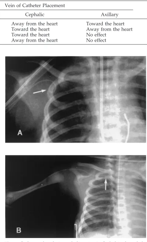
On the basis of the above observations, we used arm movements to attempt to correct the radiograph-ically demonstrated malposition of 10 PICC tips. One catheter, inserted via a cephalic vein, was coiled in the subclavian vein at the outer edge of the second rib, and the other 9, inserted via basilic veins, were looped into the neck veins. The following maneuver was performed to correct the malposition of cathe-ters that were inserted via basilic veins: The arm was abducted at the shoulder and extended at the elbow as far as possible, thereby pulling the catheter tip peripherally; then adduction at the shoulder and flexion at the elbow were performed to advance the catheter tip centrally. After this maneuver, a repeat radiograph was performed. An example of success-ful repositioning of the tip of a PICC inserted in a basilic vein is shown in Fig 5. These maneuvers were successful in correcting the catheter tip malposition in 9 of the 10 PICCs placed in basilic veins. For the malpositioned catheter in the cephalic vein, the arm was adducted at the shoulder and extended at the elbow as far as possible to pull the catheter tip pe-ripherally; the shoulder was then abducted and the elbow was flexed to advance the catheter tip cen-trally. The radiograph showing correction of this malposition was then repeated and is illustrated in Fig 6.
DISCUSSION
Extravasation of intravenous fluid into the pericar-dial sac either secondary to perforation of the myo-cardium directly by the catheter tip or tissue necrosis caused by the hyperosmolar fluid can lead to cardiac tamponade and sudden death.2–5To avoid this com-plication, we believe that the tip of PICCs should not be placed in the right atrium.1,8 Even adherence to this precaution is not entirely adequate because mi-gration of PICCs is common. Catheters have also been reported to migrate from the superior vena cava into the neck veins or conversely to move to an acceptable site from an initially malpositioned one.9,10The mechanism of these migrations is poorly understood.
The present study indicates that upper extremity movements can significantly displace PICCs in neo-nates. These movements alter the length of the soft tissue segments of the arm that include the veins. Because in all of the observations in this study the length of catheter outside the body remained con-stant, it can be assumed that the length of catheter inside the veins also remained constant as it seems unlikely that the catheter material stretched or con-tracted. Thus, compression of soft tissue around a catheter of fixed length could lead to catheter tip displacement centrally and restretching peripherally. PICCs that are inserted in the basilic vein move toward the heart with both shoulder adduction and elbow flexion; the combination of these 2 move-ments, ie, flexion at the elbow and adduction at the shoulder of the previously extended, abducted arm,
results in the greatest inward movement observed in this study. PICCs that are inserted in the cephalic vein also move inward with elbow flexion; however, they move away from the heart as the abducted arm is adducted at the shoulder. Therefore, cephalic vein PICCs have their greatest inward motion with the combination of flexion at the elbow and abduction at the shoulder of the previously extended, adducted arm. PICCs that are inserted in the axillary vein move inward with adduction at the shoulder and are
unaffected by elbow motion. It is important to em-phasize that all of these observations were made in patients in whom the length of the catheter outside the body and, by inference, that inside the body were documented to remain constant.
There are several implications of these findings. First, the outward movement of a catheter can bring the tip of the catheter from the superior vena cava or subclavian vein to the origin of a more distal vein, for example, 1 of the jugular veins, and lead to its mi-gration into that vessel on reversal of the initial arm movement. Second, estimation of the length of the catheter to be inserted is usually done by measuring the distance from the site of insertion to the point of desired placement with the forearm extended and the arm abducted 90 degree ; this is likely to be an overestimate for catheters placed in the basilic vein because infants generally maintain some degree of shoulder adduction and elbow flexion.11 Third, when interpreting radiographs, it is important to keep in mind that the maximum movement toward the heart for catheters inserted in the basilic vein is when the arm is adducted and the elbow is flexed;
Fig 5. Catheter placed in basilic vein. A, Tip looped into the neck; B, movement of the tip to the medial end of the clavicle with abduction of arm and extension of elbow; C, reposition to junction of superior vena cava and subclavian vein with shoulder adduc-tion and elbow flexion.
Fig 6. Catheter placed via cephalic vein. A, Coiled in the subcla-vian vein; B, repositioned to desired site after arm movement maneuver.
TABLE 1. Direction of the Displacement of the PICCs Related to Various Movements of the Upper Extremity Shoulder and
Elbow Movement
Vein of Catheter Placement
Basilic Cephalic Axillary
Shoulder adduction Toward the heart Away from the heart Toward the heart
Shoulder abduction Away from the heart Toward the heart Away from the heart
Elbow flexion Toward the heart Toward the heart No effect
for the cephalic vein, when the arm is abducted and the elbow is flexed; and for the axillary vein, when the arm is adducted regardless of elbow position. For ensuring that the PICC cannot enter the right atrium with arm position changed and to prevent the sub-sequent complications, radiographs taken to confirm the position of the catheter tip should be performed with the arm in these particular positions. Fourth, a similar mechanism could operate in spontaneous correction of malpositioned catheters. Finally, knowledge of the effects of upper extremity position on catheter tip movements can be used to reposition malpositioned catheters without pulling the catheter back at the site of insertion in some cases in which the tip has not migrated too far into an unacceptable vessel.
REFERENCES
1. Nadroo AM, Lin J, Green RS, Magid MS, Holzman IR. Death as a complication of peripherally inserted central catheters in neonates.J Pe-diatr.2001;138:599 – 601
2. Rogers BB, Berns SD, Maynard EC, Hansen TW. Pericardial tamponade secondary to central venous catheterization and hyperalimentation in a very low birth infant.Pediatr Pathol.1990;10:819 – 823
3. Byard RW, Bourne AJ, Moore L, Little KE. Sudden death in early infancy due to delayed cardiac tamponade complicating central venous line insertion and cardiac catheterization.Arch Pathol Lab Med.1992;116: 654 – 656
4. Goutail-Flaud MF, Sfez M, Berg A, et al. Central venous catheter-related complications in newborns and infants: a 578-case survey.J Pediatr Surg.
1991;26:645– 650
5. Pesce C, Mercurella A, Musi L, Campobasso P, Negri M. Fatal cardiac tamponade as a late complication of central venous catheterization: a case report.Eur J Pediatr Surg.1999;9:113–115
6. Kalso E, Rosenberg PH, Vuorialho M, Pietila K. How much do arm movements displace cubital central venous catheter tip?Acta Anaesthe-siol Scand.1982;26:354 –356
7. Kasten GW, Owens E, Kennedy D. Ventricular tachycardia resulting from central venous catheter tip migration due to arm position changes: report of two cases.Anesthesiology.1985;62:185–187
8. Bar-Joseph G, Galvis AG. Perforation of the heart by central venous catheters in infants: guidelines to diagnosis and management.J Pediatr Surg.1983;18:284 –287
9. Sigurdsson J, Riba P, Sigurdsson SS. The wandering central venous catheter. An unusual case of catheter displacement.Intensive Care Med.
1985;11:263–264
10. Rastogi S, Bhutada A, Sahini R, Berdon WE, Wung JT. Spontaneous correction of the malpositioned percutaneous central venous line in infants.Pediatr Radiol.1998;28:694 – 696
11. Peripherally Inserted Central Catheter: The Selection, Insertion, Use and Care of the First PICC Catheter: Clinical Education Class Manual. Sandy, UT: Becton Dickinson and Company; 1998:29
BIRTH OF THE WORD “NEONATOLOGY”
“On page vi of the preface of his book, the author uses the term ‘clinical neonatology’ to describe his intention ie, to write an ‘atlas of diseases of the newborn.’ ”
Schaffer AJ.Diseases of the Newborn.Philadelphia, PA: WB Saunders; 1960
DOI: 10.1542/peds.110.1.131
2002;110;131
Pediatrics
Ali M. Nadroo, Ronald B. Glass, Jing Lin, Robert S. Green and Ian R. Holzman
Central Catheters in Neonates
Changes in Upper Extremity Position Cause Migration of Peripherally Inserted
Services
Updated Information &
http://pediatrics.aappublications.org/content/110/1/131
including high resolution figures, can be found at:
References
http://pediatrics.aappublications.org/content/110/1/131#BIBL
This article cites 10 articles, 0 of which you can access for free at:
Subspecialty Collections
sub
http://www.aappublications.org/cgi/collection/fetus:newborn_infant_
Fetus/Newborn Infant
following collection(s):
This article, along with others on similar topics, appears in the
Permissions & Licensing
http://www.aappublications.org/site/misc/Permissions.xhtml
in its entirety can be found online at:
Information about reproducing this article in parts (figures, tables) or
Reprints
http://www.aappublications.org/site/misc/reprints.xhtml
DOI: 10.1542/peds.110.1.131
2002;110;131
Pediatrics
Ali M. Nadroo, Ronald B. Glass, Jing Lin, Robert S. Green and Ian R. Holzman
Central Catheters in Neonates
Changes in Upper Extremity Position Cause Migration of Peripherally Inserted
http://pediatrics.aappublications.org/content/110/1/131
located on the World Wide Web at:
The online version of this article, along with updated information and services, is
by the American Academy of Pediatrics. All rights reserved. Print ISSN: 1073-0397.