How you became you
Life of a cell from the time it is first formed until its own division into two daughter cells
The Cell Cycle
Reproduce by cell division
Unicellular organisms
100 µm
(a) Reproduction. An amoeba, a single-celled eukaryote, is dividing into two cells. Each new cell will be an individual organism (LM).
Reproduce by a type of cell division known as Binary Fission
Bacterial chromosome replicates and two daughter chromosomes actively move apart
Binary Fission
Origin of replication
E. coli cell
Bacterial Chromosome Cell wall Plasma Membrane Two copies of origin Origin Origin
Chromosome replication begins. Soon thereafter, one copy of the origin moves rapidly toward the other end of the cell.
1
Replication continues. One copy of the origin is now at each end of the cell.
2
Replication finishes. The plasma membrane grows inward, and new cell wall is deposited.
3
Two daughter cells result.
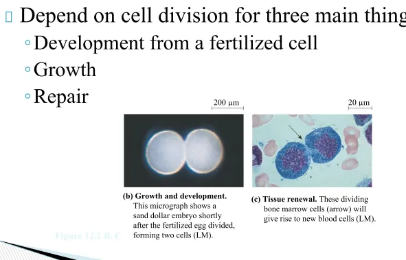
Depend on cell division for three main things:
◦
Development from a fertilized cell
◦
Growth
◦
Repair
Multicellular Organisms
20 µm 200 µm
(b) Growth and development. This micrograph shows a sand dollar embryo shortly after the fertilized egg divided, forming two cells (LM).
(c) Tissue renewal. These dividing bone marrow cells (arrow) will give rise to new blood cells (LM).
Results in two genetically identical daughter cells
Before division occurs, daughter cells need to
have an exact copy of the genetic material (DNA)
DNA contains the blueprint (genome)
DNA molecules are packaged into chromosomes
◦ Eukaryotes: Chromatin {DNA + Protein (histones)}
◦ Animals: Somatic Cells have 2 sets of chromosomes
◦ Gametes: Have 1 set of chromosomes
What is a gene?
A sequence of nucleotides that provides the cell with instructions to make a RNA or a protein
◦ Average is 1000-4000 nucleotides per gene
Genes influence how cells, tissues and organs appear
What is a chromosome?
Chromosome: where cells package DNA
Chromatin: Strings of DNA and associated proteins called histones
◦ State of the DNA inside the nucleus when the cell is not dividing
How many chromosomes do you have?
Most humans have 23 pairs of chromosomes
◦ Called a homologous pairs
◦ Autosomes: 1-22
◦ Sex Chromosomes: X and Y
Chromosome Structure
Consist of two thin rod-like structures of DNA called sister chromatids
◦ Exact replicas of each other copied during DNA Replication
Before Cell Division
DNA is replicated and the chromosomes condense Each duplicated chromosome has two sister
Before Cell Division
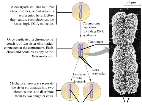
0.5 µm Chromosome duplication (including DNA synthesis) Centromere Separation of sister chromatids Sister chromatidsCentromeres Sister chromatids
A eukaryotic cell has multiple chromosomes, one of which is
represented here. Before duplication, each chromosome
has a single DNA molecule.
Once duplicated, a chromosome consists of two sister chromatids connected at the centromere. Each chromatid contains a copy of the
DNA molecule.
Mechanical processes separate the sister chromatids into two
chromosomes and distribute them to two daughter cells.
Eukaryotic Cell Division consists of:
◦ Mitosis: Division of the Nucleus
◦ Cytokinesis: Division of the Cytoplasm
In Meiosis
◦ Sex cells are produced after a reduction in chromosome number
● But…that’s next semester…
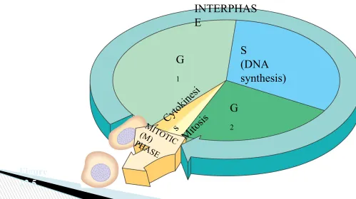
Regulation system for the cell division process Consists of the Mitotic phase and Interphase
The Cell Cycle
The cell spends most of the time in this phase
“Living” phase of the cell
Three main stages:
◦
G
1phase
◦
S phase
◦
G
2phase
Chromosomes are indistinct
Nucleolus may be visible
Centrioles are present
The cell grows and functions normally Protein synthesis occurs
More organelles are produced
Until the cell divides it will stay in this phase
The cell duplicates its DNA
Cell resumes its growth in preparation for division Checks for any errors in the duplicated DNA
Mitosis and Cytokinesis occur
Consists of five phases:
◦ Prophase
◦ Prometaphase
◦ Metaphase
◦ Anaphase
◦ Telophase
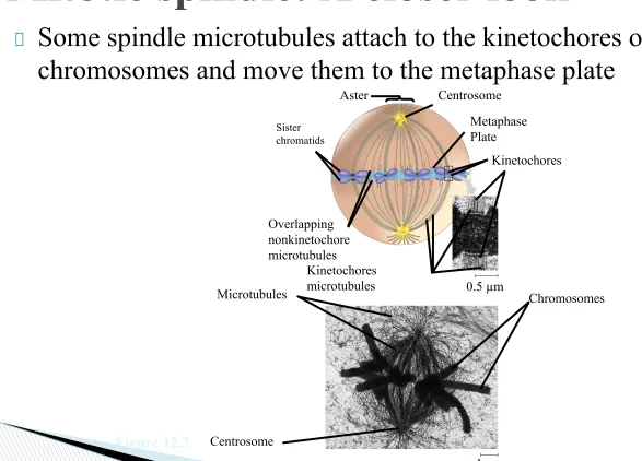
Centrosomes
◦ Where the spindle arises from
Centrioles
◦ Responsible for organization of the microtubules that form the
spindle
Asters
◦ Star shaped system of microtubules formed around each centrosome
Kinetochores
◦ Responsible for attachment of the chromosomes and the
microtubules that are part of the spindle
Mitotic Spindle
◦ Microtubules that control chromosome movement during Mitosis
Phases of Mitosis
G2 OF INTERPHASE PROPHASE PROMETAPHASE Centrosomes
(with centriole pairs) Chromatin (duplicated) Early mitotic spindle Aster Centromere Fragments of nuclear envelope Kinetochore Nucleolus Nuclear envelope Plasma membrane Chromosome, consisting of two sister chromatids
Kinetochore microtubule
Figure 12.6
Phases of Mitosis
Centrosome at one spindle pole
Daughter chromosomes
METAPHASE ANAPHASE TELOPHASE AND CYTOKINESIS
Some spindle microtubules attach to the kinetochores of chromosomes and move them to the metaphase plate
Mitotic spindle: A closer look
Centrosome Aster Sister chromatids Metaphase Plate Kinetochores Overlapping nonkinetochore microtubules Kinetochores microtubules Centrosome Chromosomes Microtubules 0.5 µm
1 µm
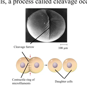
In animal cells, a process called cleavage occurs.
Cytokinesis: A Closer look
Cleavage furrow
Contractile ring of
microfilaments Daughter cells 100 µm
(a) Cleavage of an animal cell (SEM)
Frequency of division varies with the type of cell The cell cycle is regulated by a molecular control system
How do we know this?
Molecules in the cytoplasm regulate progress through the cell cycle
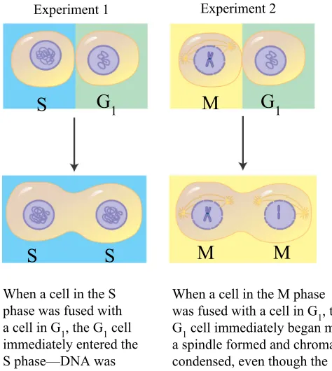
Experimental Evidence
In each experiment, cultured mammalian cells at two different phases of the cell cycle were induced to fuse.
When a cell in the M phase was fused with a cell in G1, the G1 cell immediately began mitosis— a spindle formed and chromatin condensed, even though the
chromosome had not been duplicated.
EXPERIMENT S
RESULTS
CONCLUSION The results of fusing cells at two different phases of the cell cycle suggest that molecules present in the cytoplasm of cells in the S or M phase control the progression of phases.
When a cell in the S phase was fused with a cell in G1, the G1 cell immediately entered the S phase—DNA was synthesized.
S
S S M M
M
G1 G1
Experiment 1 Experiment 2
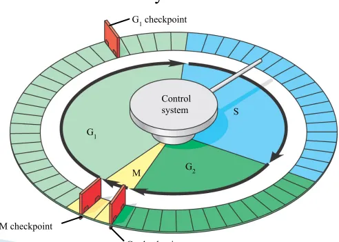
Sequential events of the cell cycle
Checkpoints
Figure 12.14
Control system
G2 checkpoint M checkpoint
G1 checkpoint
G1
S
The cell cycle stops until it receives a go-ahead signal
Red Light; Green Light
G1 checkpoint
G1 G1
G0
(a) If a cell receives a go-ahead signal at the G1 checkpoint, the cell continues on in the cell cycle.
(b) If a cell does not receive a go-ahead signal at the G1checkpoint, the cell exits the cell cycle and goes into G0, a nondividing state.
Located at the end of the cell cycle’s G1 phase
◦ Choices:
● Division
● Delay of Division
● Enter a resting stage (G0 phase)
Located at the end of G2 phase
Checks the success of DNA Replication
Makes the decision if the cell is ready for Mitosis
◦ Yes Goes through to the M phase
◦ No The cell stops and performs the necessary repairs
Metaphase checkpoint
◦ Is the Mitotic spindle formed properly?
◦ Are the chromosomes lined up on the plate correctly?
◦ Are the chromosomes attached to the microtubules?
● Yes Go onto Anaphase
● No Fix the problem or start the cell process of apoptosis (Cell
Death)
Cell Cycle Regulation
Late S phase: cyclins are synthesized and it
continues through G2
Cyclin combines with Cdk to form MPF Promotes mitosis by
phosphorylating various protein
During Anaphase, the cyclin component of MPF is
Internal and External signal control the cell cycle checkpoint
Growth factors play a large part in stimulating cell division
◦ Molecular peer pressure
Normal Cells
Density-Dependent Inhibition
Anchorage Dependence
Crowded cells stop dividing
Cells need to be
attached to a surface to divide
Cells anchor to dish surface and divide (anchorage dependence).
When cells have formed a complete single layer, they stop dividing
(density-dependent inhibition).
Abnormal Cells
Cancer Cells
Do not exhibit Anchorage or Density
Dependent Inhibition
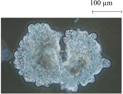
25 µm
Cancer cells do not exhibit anchorage dependence or density-dependent inhibition.
Cancer cells. Cancer cells usually continue to divide well beyond a single layer, forming a clump of overlapping cells.
(b)
Transformation: conversion of a normal cell to a cancer cell
Benign tumor: remain at the original site and do not move
Malignant tumor: have the ability to move to other locations
They do not respond normally to the body’s control mechanisms
Form Tumors
◦ Malignant tumors invade surrounding tissues and can metastasize
● Exportation cancer cells to other parts of the body where they can
form secondary tumors
Cancer Cells
Chemotherapy drugs interfere with specific steps of the cell cycle
◦ Taxol
● Freezes the mitotic spindle by preventing it from shortening
Radiation
● Destroys the DNA of cancer cells
● Most cancer cells have lost the ability to repair their DNA when it is
destroyed