Amit Levy
1*, Choaa El‑Mochtar
1, Chunxia Wang
2, Michael Goodin
3and Vladimir Orbovic
2Abstract
Background: Transient gene expression is a powerful tool to study gene function in plants. In citrus, Agrobacterium transformation is the method of choice for transient expression studies, but this method does not work efficiently with many gene constructs, and there is a need for a more robust transient expression system in citrus leaves. Biolistic particle delivery is an alternative to Agrobacterium transformation, and in some plants, such as Arabidopsis, gives higher transformation rates in leaf tissues than Agrobacterium.
Results: Here we describe an improved method for gene expression in epidermal cells of citrus leaves, using the Bio‑Rad Helios gene‑gun. Gene‑gun bombardment of GFP‑HDEL produced highly efficient gene expression in large number of cells and in different citrus varieties. We show here that transiently expressed proteins have maintained their functions in plants, and this is demonstrated by the subcellular localization of different organelle markers, and by a functional assay of Xanthomonas citri effector AvrGF1. To further expand the available tools for subcellular localiza‑ tion studies in citrus, we also generated a new set of transgenic citrus plants that contain organelle markers labelling the nuclei, actin and endoplasmic reticulum. Using these new tools, we were able to show that the coat protein of citrus tristeza virus localizes to the cytoplasm and nuclei when expressed in epidermal cells fused to GFP.
Conclusion: We have optimized a new method for transient expression in citrus leaves, to give highly reproducible and efficient transformation without producing a high level of injury or artifacts to the bombarded tissue. We also generated the first set organelle markers for use in citrus. These fluorescent protein markers label the nucleus and the actin. With these new resources, protein activity and subcellular localization can be studied in citrus rapidly and in high throughput. The handheld gene‑gun device can also be used in the grove to deliver therapies for citrus diseases, such as canker and Huanglongbing, into trees.
Keywords: Citrus, Transient expression, Transformation, Cell biology, Citrus tristeza virus
© The Author(s) 2018. This article is distributed under the terms of the Creative Commons Attribution 4.0 International License (http://creativecommons.org/licenses/by/4.0/), which permits unrestricted use, distribution, and reproduction in any medium, provided you give appropriate credit to the original author(s) and the source, provide a link to the Creative Commons license, and indicate if changes were made. The Creative Commons Public Domain Dedication waiver (http://creativecommons.org/ publicdomain/zero/1.0/) applies to the data made available in this article, unless otherwise stated.
Background
Genetic engineering is becoming widely accepted as a solution for disease control in plants, including diseases in citrus. This is especially true for the expression of plant-derived genes, and for new methods of gene edit-ing. Recent studies in genomics and plant pathology studies using citrus and other, especially model, plants have identified many more gene candidates that can be
incorporated into other species to generate pathogen-resistant plants. For example, it was recently shown that the expression of NPR1 in citrus trees resulted in partial resistance/tolerance to diseases such as citrus canker and greening under field conditions [1]. When working with perennials such as citrus, generation of a genetically engi-neered plant is a long and laborious process [2, 3]. For this reason, there is a need to develop an efficient, repro-ducible and easy methodology for transient genetic trans-formation of citrus tissues in order to easily screen these candidates and identify genes that perform well and can be used to produce stably transformed plants. Moreover, *Correspondence: [email protected]
1 Department of Plant Pathology, Citrus Research and Education Center,
University of Florida, Gainesville, FL, USA
tools to study citrus proteins properties, such as intracel-lular localization, are lacking. Transient gene expression can provide valuable data about various characteristics of these proteins, such as subcellular localization and intra/ inter cellular trafficking, expression levels, stability and degradation, interactions with other proteins, and activ-ity [for example, induction of hypersensitive response (HR)]. The development of an easy and efficient transient expression system in citrus will therefore undoubtedly speed up our investigations of gene function in this host plant [4].
In citrus, transient expression is carried out by two methods. In the first method, gene expression is delivered by Agrobacterium infection. Agrobacterium transforma-tion was successfully applied to various citrus cultivars, and this method has been employed to study the type III effector gene avrGf1 in grapefruit and pepper Bs2 gene in Citrus limon [5, 6]. However, Agrobacterium -medi-ated transient expression has been especially difficult to use [7, 8]. Recently, a modification for this technique was described, in which Xanthomonas citri subsp. citri
(Xcc) pre-treatment before agro infiltration dramatically enhanced transient β-glucuronidase (GUS) expression in leaves of six citrus varieties—Valencia sweet orange (Citrus sinensis var. Valencia), Duncan grapefruit ( Cit-rus paradisi), Key lime (C. aurantifolia L.), Carrizo cit-range (C. sinensis × Poncirus trifoliata), Sour orange (C. aurantium), and Meiwa kumquat (Fortunella crassifo-lia) [9]. Agrobacterium infection may carry a limitation by generating potentially unpredictable effects of bacte-rial effector proteins known to be exported into the plant cells together with the transforming T-DNA [10]. Add-ing Xcc pretreatment might even further complicate this undesirable effect, and influence the interpretation of the obtained results.
A second transient expression method is based on delivery of DNA by particle bombardment. Using this method with Carrizo citrange thin epicotyl segments, efficiencies of up to 93% of the bombarded plants, with an average of 102 GUS spots per tissue, have been reported [8]. Still, no efficient bombardment method has been described for citrus leaves, which is the preferable tissue for transient expression since it is represented by organs that are the largest and easiest to harvest, manipulate and observe [4].
Fluorescent proteins (FPs) tags are suitable for plant research especially for those integrated functional genomics projects where intracellular localization of pro-teins synthesized in response to infection by the patho-gen is followed in respect to changes in the host patho-gene expression. In this work, we describe a highly efficient method for the delivery of plasmid DNA into the epider-mis of young citrus leaves, using the Bio-Rad Helios gene
gun system [4]. This technique displayed a highly robust, efficient and reproducible transient expression of a vari-ety of functional proteins with diverse biological activi-ties and subcellular localizations. We also describe a new set of transgenic citrus plants that allow visualization of actin and the nuclei in citrus, and we demonstrate how these new lines can be combined with transient trans-formation for functional studies in citrus. Using these new tools, we were able to demonstrate that the coat protein (CP) of—citrus tristeza virus (CTV) co-localized with Histone-RFP in the nuclei of citrus cells. These new resources will provide an efficient new toolset for study-ing function and localization of proteins in citrus, and will enable more rapid citrus gene studies.
Results
Bio‑Rad Helios gene gun system for robust transient expression in citrus
We used the Helios gene-gun in order to develop an effi-cient transient-expression system for citrus leaves. In this technique, DNA-coated gold particles are precipitated on a plastic tube and then fired into the plant by helium pressure. Bombardment of an endoplasmic reticulum (ER) GFP marker (GFP-HDEL) into detached leaves of
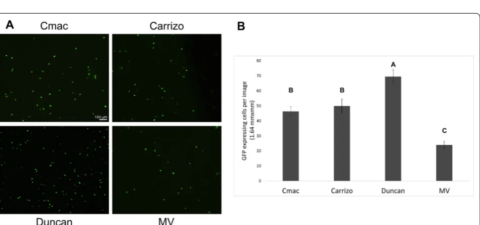
Citrus macrophylla (C-mac) resulted in up to ~ 700 GFP expressing cells in a typical 10 × magnification micro-scope field (Fig. 1). This highly efficient level of expression was obtained when young (2nd–3rd from the branch tip) leaves were used. In older leaves with better-developed cuticle, expression rate decreased dramatically. Bom-bardment was carried out most efficiently with high con-centrations of both DNA and 0.6 µm gold particles (5 µg/ shot and 0.5 mg/shot, respectively). Lowering the con-centration of both DNA and gold particles decreased the average number of expressing cells in both C-mac; how-ever, these differences were not statistically significant (Additional file 1). Surprisingly, lower gold concentration resulted in a statistically significant reduction of expres-sion rate in Madam Vinous sweet orange (Citrus sinen-sis (L.) Osbeck) (Additional file 1), highlighting the level of variability found with particle bombardment between different varieties, and different bombardment events, which is a limitation of the bombardment method.
orange. The number of fluorescent cells in a 10 × image in C-mac and Carrizo citrange, was identical. In Madam Vinous, expression levels were about half of C-mac and Carrizo levels, and in Duncan grapefruit about 50% higher (Fig. 2).
Localization and activity of transiently expressed proteins in citrus
We determined the effectiveness of transient expression method in order to conduct functional studies of pro-teins with diverse biological activities and subcellular localization specificities. First, we expressed three differ-ent proteins that localize in differdiffer-ent cellular organelles:
(1) ER protein GFP-HDEL, (2) actin protein UtrCH-GFP and (3) plasmodesmata protein AtBG_ppap. GFP-HDEL is retained in the ER lumen [11]. UtrCH-GFP binds to actin through the calponin binding domain of utro-phin (UtrCH) a probe reported to mark F-actin without altering the balance of actin assembly/disassembly [11]. AtBG_ppap is a beta-1,3-glucanase that targets the plas-modesmata after attachment to a glycosylphosphatidylin-ositol (GPI) plasma membrane anchor [12]. Forty hours after bombardment into C-mac leaves, these three mark-ers were clearly localized at their expected target orga-nelles (Fig. 3). Actin filaments labelled with UtrCH:GFP appeared highly bundled (Fig. 3). These results indicate Fig. 1 CLSM‑projected Z‑series of C‑mac abaxial leaf section after bombardment with ER‑GFP. Image was taken 40 h after bombardment. Green fluorescence (a), brightfield (b) and merged image (c) are shown. White areas in the brightfield image are oil cavities
that basic function of studied proteins (such actin bind-ing, GPI anchoring etc.) are well preserved when tran-siently expressed in citrus by bombardment (Fig. 3).
To further determine the biological functionality of
genes introduced via bombardment, we used AvrGF1
from Xanthomonas citri. AvrGF1 protein is a bacte-rial effector that was shown to induce an hypersensitive response (HR) when expressed in leaves of grapefruit (Citrus paradisi) by Agrobacterium inoculation [5]. In this assay, we bombarded young leaves of intact plants. Seven days after bombardment into leaves of C-mac, AvrGF1 expression resulted in the appearance of clear HR cell-death symptoms in the inoculated areas.
Bombardment of yellow fluorescent protein (YFP) gene, on the other hand, resulted in minimal bombardment-associated damage, but did not result in HR phenotype (Fig. 4). Altogether, our results show that protein activity is maintained with this transient expression assay.
Generation of transgenic citrus with labelled organelles
incorporation of any of the four genes into the genomes of citrus plants did not induce any phenotypic changes, as transgenic plants looked similar to wild-type plants for the duration of this study when some of them were already 3 years old. Actin was labeled with Talin:GFP, ER was labelled with RFP-KDEL and two different AFP variants (CFP and RFP) fused to Histone2B were used to label nuclei [13]. Nuclear markers clearly displayed fluo-rescence of the fusion protein in the expected subcellular compartment (Fig. 5). Similar to the transiently expressed actin marker UtrCH:GFP (Fig. 3), Talin:GFP stably labelled actin which also appeared in a bundled form, and was extremely condensed in the oil cavities (Fig. 5). RFP-ER stable lines clearly labelled the nuclear enve-lope and high amounts at the cell periphery. However, the polygonal network of the ER did not display clearly, probably because of RFP low expression levels of the ER (and lower quantum yield of RFP compared to GFP). As a result, we were unable to expose the intricate structure of ER within the cells, and could not use these transgenic plants as accurate ER marker lines (data not shown).
The coat protein of CTV localizes to nucleus
We used these new resources in order to study the sub-cellular localization of the coat protein (CP) of CTV
fused to GFP (CPCTV–GFP) in citrus. First, we bom-barded CPCTV–GFP fusion under the control of a 35S promoter into citrus using the gene-gun. Forty hours
after CPCTV–GFP gene was bombarded into C-mac,
we could detect the protein in the cytoplasm of epider-mal cells, and in what looked like the nuclei of the cells (Additional file 2). This expression pattern was similar to that of unfused YFP (Additional file 2). Next, we used the transgenic Histone 2B lines in order to verify the nuclear localization. We bombarded CPCTV–GFP gene into transgenic Duncan grapefruit plants that express Histone2B:RFP fusion. In these plants, CPCTV–GFP co-localized with Histone2B-RFP, indicating that CPCTV– GFP was indeed in the nucleus (Fig. 6). To further verify that nuclear localization fluorescence is not the result of GFP cleavage, we used the bimolecular fluorescence complementation (BiFC) method, in which CPCTV is not fused to full-length YFP. Instead, fusions of CPCTV with nEYFP and cEYFP [14] were co expressed from a single plasmid. Bombardment of this construct into C-mac resulted in the same expression cytoplasmic and nuclear pattern that was seen for CPCTV–GFP (Fig. 6). These results suggest that beyond its structural role in the for-mation of CTV capsids, CPCTV may have another activity that involves an uncharacterized nuclear stage.
Discussion
Here we describe a method for the efficient delivery of plasmid DNA into the epidermis of citrus leaves by microparticle bombardment, using the Bio-Rad Helios gene gun system. The main advantages of this system are: (1) it gives a high transformation efficiency (high number of transformed cells); (2) it is highly reproducible; (3) it does not produce significant damage or artifacts; (4) it is broadly applicable to different types of functional assays; and (5) it is broadly applicable to different citrus varie-ties. In addition, the gene gun has an important advan-tage over the traditional bombardment methods in the fact that it does not need any vacuum applied. The gun can be carried and used with any target material in the plate, greenhouse or field, with or without detaching the leaves, and this can be employed in order to deliver ther-apies directly into leaves of citrus plants in the grove. The
technique described here, having all these characteristics, will therefore be suitable for functional studies of various citrus proteins with diverse biological activities and sub-cellular localizations, and for gene delivery in the field.
The main limitation of this method is that it is relatively expensive. Both the gene-gun and the gold particles are expensive, compared to the inexpensive Agrobacterium
inoculation procedure. In addition, the bombardment does cause some damage to the bombarded tissue (see Fig. 4), although by avoiding the damaged area, thou-sands of undamaged transformed cells can be observed/ used for intended purposes. A third limitation is that this method works much more efficiently with very young leaves, at least under the conditions described here. When older, but still not fully developed leaves were used, efficiency decreased dramatically, probably because the conditions were not suitable for sufficient Fig. 5 CLSM‑projected Z series of abaxial transgenic Duncan grapefruit leaf epidermal cells stably expressing either Histone2B‑RFP (a–c),
particle penetration into the leaf. The last limitation is that there is variability between the experiments, which is characteristic of all bombardment methods. Despite all above-mentioned limitations, this method still generates reliable and robust transient expression in citrus, result-ing in extremely high level of transformation, and there-fore should be the method of choice when possible.
A major importance of transient expression systems is that it enables us to study protein function in transgenic or mutant citrus backgrounds. For example—studying protein localization in a transgenic plant where known organelle markers are expressed fused to FPs. To dem-onstrate this possibility, we generated three new citrus transgenic lines that express markers for nuclei and actin filaments. A forth marker for the ER did not display the expected results. Using these lines in combination with the gene-gun delivery method, we were able to carry out
and MFB1 [16, 17], and the one from Turnip vein clearing virus (TVCV) associated with nuclear F-actin-contain-ing filaments [11] that were hypothesized to be involved in transcription regulation [18]. The crucifer strain of tobacco mosaic virus (TMV-cg) has been shown to modulate the expression of a WRKY8 transcription fac-tor [19]. Recently it was also shown that TMV enhances its access to the phloem of mature plant tissues through the targeted disruption of auxin/indole acetic acid (Aux/ IAA) transcriptional regulators that control expression of host genes involved in virus cell-to-cell movement, plas-modesmata gating, and defense [20]. The possible biolog-ical importance of CPCTV in the nuclei is still unknown, but may involve interaction with host transcription fac-tor. CPCTV does not contain any nuclear localization sig-nals in its sequence, and its entry into the nuclei may be by diffusion as a result of its small size. We used BiFC in order to demonstrate that nuclear localization is not the result of a cleavage in the fluorescent protein. However, we cannot completely rule out the rare scenario in which both nEYFP and cEYFP are cleaved from the fusions, and bind each other. Nevertheless, our BiFC expression strongly suggest CPCTV self-interacts inside the nuclei, and thus points to a functional activity of CPCTV in the nuclei. Future experiments will need to address this questions.
Finally, the possibilities to conduct functional studies to test candidate genes in citrus are very limited, while gen-erating transgenic plants is an extremely long and labori-ous process [3]. One of the major promises of the method described here is that it will enable researchers to meet the challenges facing the citrus industry from citrus can-ker to citrus greening by conducting rapid functional studies in citrus to test protein expression and activ-ity, before generating transgenic plants. In addition, the handheld gun can be used in order to deliver therapies directly to the plant leaves in the grove.
Conclusions
We have optimized a new method for transient expres-sion in citrus leaves, which is based on the Bio-Rad Helios gene-gun bombardment. This method is highly reproducible, gives a high transformation efficiency, and does not produce a high level of injury or artifacts to the bombarded tissue. This method routinely resulted in transformation of hundreds of cells in a typical micro-scope field at 10 × magnification. We have used subcel-lular localization studies, and HR induction to show that proteins that are expressed using this method maintain their activities in planta. To further expand the toolset for functional assays in citrus, we also generated a new set of transgenic citrus organelle marker lines, with markers to the nucleus, actin and ER. Bombardment of the CP of
CTV into the nucleus marker line determined that CPCTV localizes in the cytoplasm and nucleus when expressed fused to GFP. Diseases such as citrus canker and citrus greening are threatening citrus production worldwide. The idea of using genetic engineering in order to provide the needed solutions is becoming more acceptable, but progress is slowed down by the inability to conduct quick functional studies in citrus to test candidate genes when generating transgenic plants is an extremely long and laborious process. The new tools and resources described here can help conduct functional assays with diverse pro-teins and citrus varieties, and can speed up the race for greening and canker solutions.
Methods
Plant material and plasmid constructs
All plants used in this study were grown in the Citrus Research and Education Center (lake Alfred, FL), and maintained under greenhouse conditions. Construc-tion of GFP-HDEL, RFP-HDEL, UtrCH-GFP, AtBG_ pap-GFP, Talin-GFP, Histone-RFP and Histone-CFP have been described previously [12, 13, 21]. To express AvRGF1 under the 35S promoter, the entire coding sequence of AvrGF1 was PCR amplified directly from
X. citri cells grown in liquid culture overnight, using the
primers 5′-GGGGACAAGTTTGTACAAAAAAGCAGG
CTTCATGGCTCCGAGCATGCATTC-3′ and 5′-GGGG
ACCACTTTGTACAAGAAAGTGGGTCTTAGTCGC
TGCTGGTCATTG-3′. PCR product was purified,
cloned into pDONR207, and then into pSITE0A destina-tion vector [22].
For generating CPCTV–GFP, we used overlap extension PCR [23] to build a hybrid gene consisting of a 5′UTR of tobacco etch virus as a translational enhancer fused to the 5′ end of the CTV T36 strain coat protein ORF fol-lowed by a linker sequence and the 5′ end of EYFP ORF,
using primers C-1728: 5′-AACACAACATATACAAAAC
AAACGAAT-3′, C-1863: 5′-CACCATTTACGAACGAT
AGCAATGGACGACGAAACAAAGAAATT-3′, C-1864:
5′-AATTTCTTTGTTTCGTCGTCCATTGCTAT
CGTTCGTAAATGGTG-3′, C-1856: 5′-TAT CCATGGT
TAGTACAGCTCGTCCATGCCGAGAGTGAT-3′,
C-1858: 5′-AGATCAATAGCCACG ATGGTGAGCAAG
GGCGAGGAGCTGTT-3′ and C-1866: 5′-TCACCATCG
TGGCTATTGATCTACGTGTGTTGAATTTCCCA A-3′. Cloning was done into pCASS4N (a variant of pCASS2 that contains NcoI [24]) between the StuI and NcoI restriction sites.
plasmids with I-SceI. pPZP-RCS1 was alkaline phos-phatase treated to prevent self-ligation. The third step was to use the plasmid generated in step 2 to introduce the gene cassette of CP in pSAT1A-nEYFP-N1 to pPZP-RCS1 carrying the expression cassette of pSAT4-cEYFP-C1(B). Both plasmids were digested with AscI, and the appropriate fragments were ligated.
Particle bombardment into citrus leaves
For bombardment, all plasmids were first purified from
E. coli with the Qiagen plasmid maxi-kit (Qiagen, Hilden, Germany). Particle bombardment was carried out as described in [4], with few modifications. Gold particles coating with DNA was performed as described in [4], but only 0.6 µm particles were used, mixed with 150 µg of DNA for the preparation, to give ~ 5 µg of DNA per bullet. Gold concentration was 0.5 mg/shot, unless noted otherwise. Bombardment was carried out on the abaxial side of the leaf, using the 2nd and 3rd newest leaves of the citrus plants. Finally, delivery into the plants was carried out with a helium pressure of 260–280 psi. After bom-bardment, leaves were kept in the dark inside a petri dish with wet filter paper, to maintain humid conditions. In all the experiments described here, we used a single cartridge per leaf. However, the size of citrus leaves is usually big enough for multiple shots if needed for functional studies.
Transformation
Transgenic plants that belong to Carrizo citrange root-stock cultivar and Duncan grapefruit cultivar were produced according to well established protocol that employed Agrobacterium tumefaciens [3]. Starting mate-rial in transformation experiments were juvenile explants cut from stems of germinated seedlings. All transgenic shoots were micro-grafted onto Carrizo rootstock and allowed to grow in vitro for 4–6 weeks. The plantlets con-sisting of growing transgenic shoots and Carrizo roots were moved from tubes to 2′ × 2′ × 2′ pots kept under plastic dome on the light bench in the laboratory for additional 2 months. Subsequently, plants were moved to greenhouse and re-planted into bigger pots.
Selection of transgenic shoots carrying gene for GFP-Talin fusion was done by visualizing GFP. Transgenic
Selection of transgenic shoots carrying gene for GFP-Talin fusion was done using a Leica Wild M3Z stereomi-croscope (Leica Microsystems Inc., Buffalo Grove, IL, USA) outfitted with a NIGHTSEA™ Stereo Microscope Fluorescent Adaptor (NIGHTSEA, Lexington, MA, USA) and the filter set for Royal Blue (excitation 400–415 nm with a 460 nm longpass emission filter). GFP express-ing cells on different citrus varieties were imaged usexpress-ing an Olympus BX61 epi-fluorescence microscope and an OMAX A35140U 14MP CMOS camera. Fluorescence was captured using a FITCI dichroic cube. All other CLSM images were collected using a Leica SP8 laser-scanning confocal microscope (Leica Microsystems Inc., Buffalo Grove, IL, USA). Samples were imaged using lasers with excitation wavelengths for their respective flu-orescent reporting range: GFP fluorescence was excited with a 488-nm argon laser, and emission was detected at 500–530 nm. RFP fluorescence was excited with a 561-nm diode-pumped solid-state (DPSS) laser, and emission was detected at 590–630 nm. Cyan fluorescent protein (CFP) was excited with near UV diode 405-nm laser, and emission was detected at 475–501 nm. Leica Application Suite—LAS X (Leica Microsystems Inc., Buffalo Grove, IL, USA) was used to collect z-stacks composed of opti-cal sections at a 1024 × 1024 resolution. Images were exported as TIFF files to produce photographs for publi-cation using Adobe Photoshop software.
Statistical analysis
Statistical analysis of images was preformed using the JMP Pro 13.2 Statistical program (SAS, Cary, NC). Differ-ences in gold concentration, plasmid concentration and
Table 1 The sequences of PCR primers used for selection of transgenic shoots
Primer Sequence
CFP‑Histone 2B (f ) AGCTGACCCTGAAGTTCATCTG
CFP‑Histone 2B (r) GATATAGACGTTGTGGCTGATGTAG
RFP‑Histone 2B (f ) CGTAATGCAGAAGAAGACCA
RFP‑Histone 2B (r) CTTGATGTCGGTCTTGTAGG
RFP‑ER (f ) CCGACTACTTGAAGCTGTCCTTC
AvrGF1 HR response were analyzed using T-test, while the difference between varieties was analyzed using the Tukey HSD.
Authors’ contributions
AL, VO and CE‑M and MG conceived and designed the experiments. AL, CW and VO performed the experiments. AL performed data analysis. AL prepared and wrote the manuscript with help from VO and CE‑M. All authors read and approved the final manuscript.
Author details
1 Department of Plant Pathology, Citrus Research and Education Center, Uni‑
versity of Florida, Gainesville, FL, USA. 2 Citrus Research and Education Center,
University of Florida, Lake Alfred, FL, USA. 3 Department of Plant Pathology,
University of Kentucky, Lexington, KY, USA.
Acknowledgements
pSAT vectors were kindly provided by Dr. Stanton Gelvin and Dr. Lan‑Ying Lee (Purdue university). We would like to thank Daniel Stanton (Citrus Research and Education microscope facility) for technical help and for critically review‑ ing the manuscript. We also thank Dr. Bill Dawson and Cecile J Robertson (University of Florida) for providing us citrus plants.
Competing interests
The authors declare that they have no competing interests.
Availability of data and material
All data generated in this study are included in this article and additional files. Material is available from the corresponding author on reasonable request.
Ethics approval and consent to participate Not applicable.
Consent for publication Not applicable.
Funding
This work was supported by the IFAS Citrus Initiative Research fund for the years 2016–2017 to AL.
Use of plant material
Genetically modified citrus plants, and plants for transient expression were generated and used in accordance with the State of Florida regulations.
Publisher’s Note
Springer Nature remains neutral with regard to jurisdictional claims in pub‑ lished maps and institutional affiliations.
Received: 20 June 2017 Accepted: 19 December 2017
Additional files
Additional file 1. Relation between expression efficiency and DNA/Gold particle concentration. Graphs of the average number of fluorescent cells in a 10 × image area after bombardments with different gold particle concentrations and plasmid DNA concentrations.
Additional file 2. CLSM images of CPCTV–GFP and free YFP after bom‑
bardment into epidermal cells of C‑mac. (A‑C) CLSM image of CPCTV–GFP
(A, green), brightfield image (B) and superimposed image (C). (D‑F) CLSM image of unfused YFP (A, yellow), with brightfield image (B), Chlorophyll channel (C, Blue) and superimposed image (D).
References
1. Dutt M, Barthe G, Irey M, Grosser J. Transgenic citrus expressing an Arabi‑ dopsis NPR1 gene exhibit enhanced resistance against Huanglongbing (HLB; Citrus Greening). PLoS ONE. 2015;10:e0137134.
2. Donmez D, Simsek O, Izgu T, Aka Kacar Y, Yalcin Mendi Y. Genetic transfor‑ mation in citrus. Sci World J. 2013;2013:491207.
3. Orbović V, Grosser JW. Citrus transformation using juvenile tissue explants. In: Wang K, editor. Agrobacterium protocols, vol. 2. New York: Springer; 2015. p. 245–57.
4. Ueki S, Lacroix B, Krichevsky A, Lazarowitz SG, Citovsky V. Functional tran‑ sient genetic transformation of Arabidopsis leaves by biolistic bombard‑ ment. Nat Protoc. 2009;4:71–7.
5. Figueiredo JFL, Römer P, Lahaye T, Graham JH, White FF, Jones JB. Agrobacterium‑mediated transient expression in citrus leaves: a rapid tool for gene expression and functional gene assay. Plant Cell Rep. 2011;30:1339–45.
6. Sendín LN, Filippone MP, Orce IG, Rigano L, Enrique R, Peña L, Vojnov AA, Marano MR, Castagnaro AP. Transient expression of pepper Bs2 gene in Citrus limon as an approach to evaluate its utility for management of citrus canker disease. Plant Pathol. 2012;61:648–57.
7. Ahmad M, Mirza B. An efficient protocol for transient transformation of intact fruit and transgene expression inCitrus. Plant Mol Biol Rep. 2005;23:419–20.
8. Bespalhok Filho JC, Kobayashi AK, Pereira LFP, Galvao RM, Vieira LGE. Transient gene expression of b‑glucuronidase in citrus thin epicotyl transversal sections using particle bombardment. Braz Arch Biol Technol. 2003;46:1–6.
9. Jia H, Wang N. Xcc‑facilitated agroinfiltration of citrus leaves: a tool for rapid functional analysis of transgenes in citrus leaves. Plant Cell Rep. 2014;33:1993–2001.
10. Lacroix B, Vaidya M, Tzfira T, Citovsky V. The VirE3 protein of Agrobacterium mimics a host cell function required for plant genetic transformation. EMBO J. 2005;24:428–37.
11. Levy A, Zheng JY, Lazarowitz SG. The tobamovirus turnip vein clearing virus 30‑kilodalton movement protein localizes to novel nuclear filaments to enhance virus infection. J Virol. 2013;87:6428–40.
12. Levy A, Erlanger M, Rosenthal M, Epel BL. A plasmodesmata‑associated beta‑1,3‑glucanase in Arabidopsis. Plant J. 2007;49:669–82.
13. Martin K, Kopperud K, Chakrabarty R, Banerjee R, Brooks R, Goodin MM. Transient expression in Nicotiana benthamiana fluorescent marker lines provides enhanced definition of protein localization, movement and interactions in planta. Plant J. 2009;59:150–62.
14. Citovsky V, Lee L‑Y, Vyas S, Glick E, Chen M‑H, Vainstein A, Gafni Y, Gelvin SB, Tzfira T. Subcellular localization of interacting proteins by bimolecular fluorescence complementation in Planta. J Mol Biol. 2006;362:1120–31. 15. Solovyev AG, Savenkov EI. Factors involved in the systemic transport
of plant RNA viruses: the emerging role of the nucleus. J Exp Bot. 2014;65:1689–97.
16. Matsushita Y, Deguchi M, Youda M, Nishiguchi M, Nyunoya H. The tomato mosaic tobamovirus movement protein interacts with a putative tran‑ scriptional coactivator KELP. Mol Cells. 2001;12:57–66.
17. Matsushita Y, Miyakawa O, Deguchi M, Nishiguchi M, Nyunoya H. Cloning of a tobacco cDNA coding for a putative transcriptional coactivator MBF1 that interacts with the tomato mosaic virus movement protein. J Exp Bot. 2002;53:1531–2.
18. Levy A. Turnip vein clearing virus movement protein nuclear activity: Do Tobamovirus movement proteins play a role in immune response sup‑ pression? Plant Signal Behav. 2015;10:e1066951.
19. Chen L, Zhang L, Li D, Wang F, Yu D. WRKY8 transcription factor functions in the TMV‑cg defense response by mediating both abscisic acid and ethylene signaling in Arabidopsis. Proc Natl Acad Sci. 2013;110:E1963–71. 20. Collum TD, Padmanabhan MS, Hsieh Y‑C, Culver JN. Tobacco mosaic
virus‑directed reprogramming of auxin/indole acetic acid protein tran‑ scriptional responses enhances virus phloem loading. Proc Natl Acad Sci. 2016;113:E2740–9.
• We accept pre-submission inquiries
• Our selector tool helps you to find the most relevant journal • We provide round the clock customer support
• Convenient online submission • Thorough peer review
• Inclusion in PubMed and all major indexing services • Maximum visibility for your research
Submit your manuscript at www.biomedcentral.com/submit