Dr. Nasreen Laiq (Corresponding Author) Department of Anaesthesia, (MTI),
Lady Reading Hospital, Peshawar - Pakistan Cell: +92-333-9590610
Email: [email protected]
Date Received: February 2, 2017 Date Revised: March 20, 2017 Date Accepted: May 10, 2017
EFFECTIVENESS OF CONTINUOUS POSITIVE PRESSURE
VENTILATION IN REDUCING THE LENGTH OF STAY IN POST
CARDIAC BYPASS SURGERY PATIENTS
Nasreen Laiq, Shahid Khan, Nasir Islam, Muhammad Naeem Khan
Department of Anaesthesia, Medical Teaching Institute, Lady Reading Hospital, Peshawar - Pakistan
ABSTRACT
Objectives: To asses the length of stay using Continuous Positive pressure ventilation (CPAP) during weaning in post bypass patients in cardiac ICU and compared this procedure with MV (Mechanical ventilation) by analyzing cardiac and respiratory parameters and complications.
Material and Methods: A randomized clinical trial was conducted from June 2011 to July 2012 with patients in the cardiac ICU of Lady Reading Hospital Peshawar Pakistan. Patients of both sexes and ages between 45-70 years, post bypass surgeries and were on mechanical ventilation for more than 48 hours, who failed at 30 minutes of spontaneous breathing trial (SBT) were included in the study. Sealed envelopes were used for random assignment. The established weaning criteria was routinely followed in the ICU. Before SBT, the following measurements were carried out: arterial blood gases; parameters of ventilation such as f (frequency of breaths),VT (Tidal volume), PEEP (Positive end expiratory
pressure), FiO2(Fraction of inspired oxygen), HR(Heart rate), systolic (SBP) diastolic (DBP) blood pressure and SpO2 (Oxygen saturation).SBT , 30 minutes was given,If failure occurred before the 30th minute ,he/she was included in the group previously defined by random assignment. Patients in the experimental group were extubated and placed on CPAP, whereas the other patients (the control group) returned to MV, which was classified as the conventional treat-ment. Spontaneous ventilation mode using a bi-levelCPAP support was used in experimental group immediately after trachealextubation,. The interface chosen was facemask. Daily SBT was carried out thereafter in order to evaluate the possibility of extubation in control group.
Results: Eighty patients who failed T-piece trials ventilation, 40 were placed on CPAP and 40,on intermittent mandatory ventilation or mechanical ventilation (IMV). The ages of patients in the CPAP and IMV groups were 45.7± 18.12 and 47.10 ± 18.44 years respectively. In both groups, ventilation time before T-piece trial was 2 days or 48 hours. Heart and respiratory parameters were similar for the two groups at 30 minutes of T-piece trial. The percentage of minor complica-tions in both groups were lower. The comparisons of gas measurements between the CPAP and IMV groups showed no significant differences . Patients of the CPAP group had a shorter stay in the ICU and in the hospital i-e 2.95 ±0.78 days versus 7.44+1.12 days for IMV group (Table No 2). Mortality was similar in the two groups. Of the 80 patients in both groups no serious complications were seen on ventilator support while discharged from the ICU.
Conclusion: The combination of early extubation and CPAP is a good alternative for ventilation in a group of patients who initially failed weaning. The length of stay is significantly reduced in Cardiac ICU ,compared to Mechanical ven-tilation. Therefore, CPAPis a useful and safe strategy that may be considered during mechanical ventilation weaning. Key words: CPAP, IMV,Mechanical ventilation.
This article may be cited as: Laiq N, Khan S, Islam N, Khan MN. Effectiveness of continuous positive pressure ventilation in reducing the length of stay in post cardiac bypass suegery patients. J Med Sci 2017; 25: (2) 257-261.
INTRODUCTION
including ,Pleural opening, phrenic nerve damage and postoperative pain. Prolong atelectasis may lead to decreased oxygenation, increase in shunting, altered work of breathing, and pulmonary complications post-operatively. Different maneouvers for recruitment of the alveoli are adopted. Deep breaths, chest physiotherapy and high positive end-expiratory pressure (PEEP) and CPAP are the methods used to increase lung functions and to prevent postoperative lung collapse.3
Continuous Positive pressure ventilation (CPAP) has been found to improve oxygenation equally as effective as conventional mechanical ventilation in im-proving pulmonary gas exchange in case of respiratory failure. It has been reported that length of stay is shorter in the intensive care unit (ICU) compared to mechan-ical ventilation.4 Respiratory failure needs mechanical ventilation to rest, respiratory muscles and to improve gas exchange in case of respiratory failure. Prolonged Ventilation is usually associated with ventilator induced pneumonia, and hence increased morbidity and mor-tality.
Patient’s are ventilated by CPAP mode with pressure support by means of oronasal, nasal, or total face mask at the patient-ventilator interface. Patient also maintains the ability to speak and cough6. CPAP has been reported to reduce tachypnea,, provides rest to the respiratory muscles,improves oxygenation and hence improves clinical outcomes in desired patients.7
A number of studies have been done, which supports CPAP ventilation. Nava and colleagues8, in a randomized clinical trial, found lower mortality with the application of CPAPin 50 patients having failed trials for spontaneous ventilation compared with Mechanical ventilation. Girault and colleagues9 compared Positive pressure ventilation with mechanical ventilation in 33 patients after a failed trials for spontaneous ventilation for 2-hour, and found a reduction in total mechanical ventilation time in the CPAP group. Ferrer and col-leagues10 in one of his study suggested that Positive pressure ventilation be assessed as a means to facil-itate weaning from mechanical ventilation in patients having failed trials for spontaneous ventilation. Later a meta-analysis documented that non invasive positive pressure ventilation (CPAP) reduces mortality and facilitates early weaning compared to intermittened mandatory ventilation.11
Our study was to asses and compare the length of stay by the application of CPAP during weaning from mechanical ventilation, in post bypass patients in an ICU in our own setup. and to analyze the outcome in terms of cardiac and respiratory parameters, clinical course, and complications in both groups.
MATERIAL AND METHODS
This study was approved by the Institutional Research and Ethics Committee of Lady Reading Hospital Peshawar Pakistan. A randomized clinical
trial was conducted from June 2011 to July 2012 with patients in the cardiac ICU of Lady Reading Hospital Peshawar Pakistan. Patients of both sexes and ages between 35-70 years, post bypass surgeries and were on Mechanical ventilation for more than 48 hours, who failed at 30 minutes of spontaneous breathing trial (SBT) were included in the study.
The established weaning criteria was routinely followed in the ICU i-e improvement of the cause of Acute respiratory failure that led to the use of ventilation support, correction of arterial hypoxemia (arterial partial pressure of oxygen (PaO2 ) of greater than 60 mm Hg, fraction of inspired oxygen (FiO2) of less than or equal to 0.4, and positive end-expiratory pressure (PEEP) of less than or equal to 5 cm H2O during pressure support ventilation. All patients were breathing at low levels of pressure support ventilation (less than 15 cm H2O). Fully consciousness level having Glasgow coma score of greater than or equal to 13, not on vasoactive drug and have an adequate cough reflex are included in the study .
Failure or intolerance at 30 minutes of SBT was defined according to one of the following criteria: pe-ripheral oxygen saturation (SpO2) measured by pulse oximetry of less than 90% (80% in chronic respiratory failure), respiratory rate (f) of greater than 35 respirations per minute, heart rate (HR) of greater than 140 or less than 50 beats per minute (bpm) or increase or decrease of greater than 20% in previous mechanical ventilation, and systolic arterial blood pressure of greater than 180 mm Hg or less than 70 mm Hg or increase or decrease of greater than 20% in previous mechanical ventilation. Agitated or non-cooperative behavior patients and hav-ing tracheotomy, excessive respiratory secretion, and unwilling for study,were excluded from the study.
Data collection
Patients were included in the study after an informed consent form, signed by a family member or guardian. Patients considered apt to undergo the weaning procedure were given spontaneous breathing trail (SBT) for at least 30 minutes.At that moment, if the patients had failed SBT,they will be put into one of the two studied groups i-e either CPAP or IMV(Mechanical ventilation). Sealed envelopes were used for random assignment.
was extubated after having rested in the mechanical ventilation for 30 minutes in the experimental group. Immediately after tracheal extubation, spontaneous ventilation mode using a bi-level CPAP support was used. Inspiratory positive airway pressure was delivered according to patient tolerance and varied from 10 to 30 cm H2O.
FiO2 was set according to an SpO2 of greater than 90%, as measured by pulse oximetry. Expiratory positive airway pressure was set to provide adequate gas exchange. CPAP mask is chosen as the interface. Weaning from CPAP was performed on a daily basis by gradually reducing pressure levels and FiO2 until ade-quate tidal volumelevels could be reached and proper alveolar ventilation could be established. In the control group, mechanical ventilation followed the previously administrated ICU ventilation support routinely. Daily SBT was carried out thereafter in order to evaluate the possibility of extubation. X-Ray chest were carried out daily in order to compare improvement in two groups and to exclude any lung collapse during ICU stay.
RESULTS
Of 80 patients who failedspontaneous breathing trail, 40 were placed on CPAP and 40 were placed on IMV. The ages of patients in the CPAP and IMV groups were 45.7± 18.12 and 47.10 ± 18.44 years respectively. In both groups, ventilation time before T-piece trial was 2 days. Heart and respiratory parameters were similar
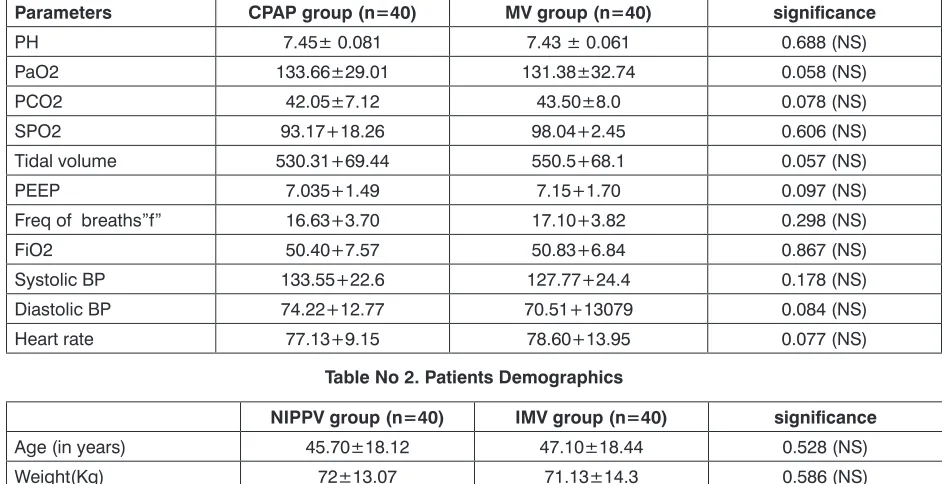
Table No 1. At the end of ventilation support
Parameters CPAP group (n=40) MV group (n=40) significance
PH 7.45± 0.081 7.43 ± 0.061 0.688 (NS)
PaO2 133.66±29.01 131.38±32.74 0.058 (NS)
PCO2 42.05±7.12 43.50±8.0 0.078 (NS)
SPO2 93.17+18.26 98.04+2.45 0.606 (NS)
Tidal volume 530.31+69.44 550.5+68.1 0.057 (NS)
PEEP 7.035+1.49 7.15+1.70 0.097 (NS)
Freq of breaths”f” 16.63+3.70 17.10+3.82 0.298 (NS)
FiO2 50.40+7.57 50.83+6.84 0.867 (NS)
Systolic BP 133.55+22.6 127.77+24.4 0.178 (NS)
Diastolic BP 74.22+12.77 70.51+13079 0.084 (NS)
Heart rate 77.13+9.15 78.60+13.95 0.077 (NS)
for the two groups at 30 minutes of spontaneous breath-ing trail. The percentage of complications in the CPAP group was lower in both the groups. Length of stay in the intensive care unit was statistically significant when comparing the two groups.
The comparisons of gas measurements between the CPAP and mechanical ventilation (IMV) groups showed no significant differences. The blood gas val-ues at the end of ventilation support removal were as follows PH; 7.45 ± 0.081 verses 7.43 ± 0.061 , PaCO2 42.05±7.12 versus 43.50±8.0, Arterial Oxygen Pres-sure; 133.66± 29.01 verses 131.38 ±32.74 and Oxygen saturation was 93.17 ± 18.26 versus 98.04 ± 2.95 for CPAP and 7.44 for IMV groups respectively.,similarly tidal volume, Systolic BP,Diastolic BP,Frequency of breaths (f),Heart rate,FiO2 and PEEP were statistically not significant in two groups. Patients of the CPAP group had a shorter stay in the ICU (statistically significant ) compare to mechanical ventilation. Mortality was similar in the two groups. Of the 40 patients in the CPAP group 36 had no serious complications while only 4 patients had mild nausea and soreness on face which were treated with routine medication. No marked atelactasis were seen in both studied groups on X-ray findings.
DISCUSSION
The main findings of this study were that CPAP compared to MV (mechanical ventilation) resulted in better early postoperative blood gases,less atelectasis
Table No 2. Patients Demographics
NIPPV group (n=40) IMV group (n=40) significance
Age (in years) 45.70±18.12 47.10±18.44 0.528 (NS)
Weight(Kg) 72±13.07 71.13±14.3 0.586 (NS)
Male / Female 25 / 15 30 / 10
and a shorter duration of hospitalization and ICU stay. Patients usuallysuffer from atelectasis after by-pass surgery. The incidence of atelectasis has been seen in the first postoperative days of cardiac operations to be found as 87.7%. Although atelectasis decreases with time, the incidence has been shown to be high (30%) on the 6th postoperative day. Lung collapse has been shown to be associated with impaired gas exchange and increased shunt fraction, and therefore with reduced arterial oxygenation postoperatively.12 Lung oedema due to Increased extravascular water, in addition to atelectasis may aggravate the shunt.13 Pulmonary function tests obtained 06 days after open heart surgery have revealed a 26% decrease in FEV1 and forced vital capacity (FVC) in patients with a normal chest X-rays, this may reach upto 42% when atelectasis was present. PEEP( Peak end-inspiratory pressures) or CPAP have been shown to provide lung recruitment using high inspiratory pressures of about 35 to 40 cm H2O.14
Radiological improvement of atelectasis has been seen after open heart surgery with CPAP compared to control groups. Higher PEEP or sustained inflation may reduce cardiac output and left ventricular end-diastolic Pressure in hemody namically stable patients after car-diac surgery15 Atelectasis can be prevented by taking deep breaths and incentive spirometry. CPAP is used to prevent and treat atelectasis in the postoperative period. It has been shown to reduce venous admixture, shunt fraction and restrictive lung pattern after cardiac surgery.16 CPAP by means of increasing intrathoracic pressure can restore decreased FRC(Fractional re-sidual capacity) and prevent lung collapse,improving hypoxemia and decreases work of breathing, therefore there is recommendation for early application of CPAP in high-risk patients, because a decrease in FRC and deterioration of pulmonary function develop rapidly after extubation.17 Early treatment with CPAP has been shown to reduce the need for intubation, ICU length of stay, and the incidence of ventilator induced pneumonia.Pro-long ventilation can lead toinfection ,resulting in acute hypoxemia after elective major abdominal surgery.18 In some studies pressure support ventilation( PSV) seems to provide greater patient comfort than CPAP due to better alveolar opening and a decrease in work of breathing.16,19 But CPAP is still preferred because of its beneficial effects.
Prophylactic use of Nasal CPAP is recommend-ed by some authors, as a preventive measure against postoperative atelectasis, because it is simple method, well-tolerated and improves pulmonary functions,hence reducing morbidity, mortality and length of stay in ICU and hospitalization as well.20
The reason for the intermittent CPAP application in our study was to avoid nausea, vomiting, gastric distention and restricted oral intake. We applied CPAP 02 hourly with 30 minutes gap if the patients conditions
allows, airway pressure was kept at a Pressure support of 15mm H2O,with a PEEP of 07 cm H2O through a tight fitting CPAP mask, better blood gas levels were obtained at that inspiratory and expiratory pressure levels. All the patients were hemodynamically stable at thisairway pressure, in our study.
CPAP provides more advantages in selected group of patients However, there may be some prob-lems limiting the treatment such as patient adaptation, atalectasy and facial ulcers caused by mask pressure. Inspite of the above mentionedlimiting factors, CPAPis easily tolerable,lessens the need for sedation, protects the airway swallowing and speech mechanisms,pro-vides opportunity for early mobilization.21
The contraindication for CPAP is in patients with risk for aspiration or excessive secretion, upper airway obstruction, loss of preventive airway reflex and those who are candidates for intubation, it might not be con-sidered in acute respiratory distress syndrome with severe hypoxemy. Some of the side effects are disten-sion of stomach, some ledisten-sions on the skin, facial ache, sense of drying in the nose, eye irritation (conjunctivity), clostrophobia, sleep disorders and mask leakage22. CPAP should not be used in patients who must not be resuscitated or uncooperative, in cases where secre-tions cannot be removed, systolic blood pressure is lower than 90 mmHg or where there is severe acidoses, shock or arrhythmias that cannot be controlled. Also not suitable for obstruction of upper respiratory system.23 We don’t have come across any major complications in any of our studied groups,minor complications such as nausea and retching were noticed in few patients in CPAP group.
CONCLUSION
The results of this study suggests that the com-bination of early extubation and CPAP is a good alter-native for ventilation in a group of patients who initially failed weaning. Useof CPAP resulted in efficient gas exchange, improved lung functions and decrease ICU and hospital stay, when compared with conventional mechanical ventilation weaning. Therefore, CPAP is a useful and safe strategy that might be considered during mechanical ventilation weaning.
REFERENCES
1. Hedenstierna G, Rothen HU. Atelectasis formation during anesthesia: causes and measures to prevent it. J ClinMonitComput 2000;16:329–35
2. Kotani N, Hashimoto H, Sessler DI, Muraoka M, Wang JS, O’Connor MF, Matsuki A. Cardiopulmonary bypass produces greater pulmonary than systemic proinflam-matory cytokines. AnesthAnalg 2000;90:1039 –45. 3. Matte P, Jacquet L, Van Dyck, Goenen M. Effects
artery bypass grafting.ActaAnaesthesiolScand 2000;44:75–81.
4. Non-Invasive Positive Pressure Vantilation facilitates early extubation in post operative cardiac patients, Laiq N, Khan R A, Malik A, JPMI October - Dec 2013. 27(4): 361-65.
5. Silver MR. BIPAP: useful new modality or confusing acronym? Crit Care Med. 1998;26:1473-74.
6. Hormann C, Baum M, Putensen C, Mutz NJ, Benzer H. Biphasic positive airway pressure (BIPAP) - a new mode of ventilatory support. Eur JAnaesthesiol. 1994;11:37–42.
7. Rathgeber J. Ventilation modes and strategies in intensive care medicine. AnaesthesiolReanim. 1997;22:4–14.
8. Nava S, Ambrosino N, Clini E, Orlando M, Vitacea G, Fracchia C, Rubini F. Noninvasive mechanical ventila-tion in the weaning of patients with respiratory failure
due to chronic obstructive pulmonary disease. Ann
Intern Med. 1998;128:721-28.
9. Girault C, Daudenthun I, Chevron V, Tamion F, Leroy J, Bonmarchand G. Noninvasive ventilation as a sys-tematic extubation and weaning technique in
acute-on-chronic respiratory failure. Am J RespirCrit Care
Med. 1999;160:86–92.
10. Ferrer M, Esquinas A, Arancibia F, Bauer TT, Gonzalez G, Carillo A, Rodriguez-Roisin R, Torres A. Noninva-sive ventilation during persistent weaning failure: a
randomized controlled trial. Am J RespirCrit Care
Med. 2003;168:70–76.
11. Burns KEA, Adhikari NKJ, Meade MO. A meta-analysis of noninvasive weaning to facilitate liberation from me-chanical ventilation. Can J Anesth. 2006;53:305–315. 12. Lindberg P, Gunnarsson L, Tokics L, Secher E, Lund-quist H, Brismar B, Hedenstierna G. Atelectasis and lung function in the post-operative period. ActaAnaes-thesiolScand 1992;36:546–53
13. Hachenberg T, Tenling A, Nystrom SO, Tyden H, Hedenstierna G. Ventilation-perfusion inequality in patients undergoing cardiac surgery. Anesthesiology 1994;80:509–19.
14. Dyhr T, Laursen N, Larsson A. Effects of lung recruit-ment maneuver and positive end-expiratory pressure on lung volume, respiratory mechanics and alveolar
gas mixing in patients ventilated after cardiac surgery. ActaAnaesthesiolScand 2002;46:717–25
15. Nielsen J, Ostergaard M, Kjaergaard J, Tingleff J, Berthelsen PG, Nygard E, Larsson A. Lung recruit-ment maneuver depresses central hemodynamics in patients following cardiac surgery. Intensive Care Med 2005;31:1189–94
16. Matte P, Jacquet L, Van Dyck, Goenen M. Effects of conventional physiotherapy, continuous positive airway pressure and non-invasive ventilatory support with bilevel positive airway pressure after coronary artery bypass grafting. ActaAnaesthesiolScand 2000;44:75–81.
17. Miranda DR, Struijs A, Koetsier P, van Thiel R, Schepp R, Hop W, Klein J, Lachmann B, Bogers AJJC, Gom-mers D. Open lung ventilation improves functional residual capacity after extubation in cardiac surgery. Crit Care Med 2005;33:2253-58.
18. Squadrone V, Coha M, Cerutti E, Schellino MM, Biolino P, Occella P, Belloni G, Vilianis G, Fiore G, Cavallo F, Ranieri VM. Continuous positive airway pressure for treatment of postoperative hypoxemia: a randomized controlled trial. JAMA 2005; 293:589–95.
19. Appendini L, Patessio A, Zanaboni S, Carone M, Gukov B, Donner CF. Physiologic effects of PEEP and mask pressure support during exacerbation of chronic obstructive pulmonary disease. Am J RespirCrit Care Med 1994;149:1069–76
20. Kindgen-Milles D, Mu¨ller E, Buhl R, Bo¨hner H, Ritter D, Sandmann W, Tarnow J. Nasal-continuous positive airway pressure reduces pulmonary morbidity and length of hospital stay following thoracoabdominal aortic surgery. Chest 2005; 128:821–28.
21. Eren NT, Batislam Y. Noninvasive positve pressure ventilation. J. Ankara Medical School.1996;18:49-51. 22. Pennock BE, Crawshaw L, Kaplan PD. Noninvasive
mask ventilation for acute respiratory failure: instution of a new therapeutic technology for routine use in pa-tients with respiratory failure. Chest 1994;105:441-50. 23. Marco C, Alfredo P, Giorgio C, et al. Acute respiratory failure in patients with severe community acquired pneumonia. Am J RespirCrit Care ed.1999;160: 1585-91.
AUTHOR’S CONTRIBUTION
Following authors have made substantial contributions to the manuscript as under: Laiq N: Study design, collection data, Paper Writing
Khan S: Paper writing, finding out references editing, etc
Islam N: Cardiovascular surgeon perfumed cardiac surgeries included in study Khan MN: statistical analysis, typing, editing, finding out references, etc
Authors agree to be accountable for all aspects of the work in ensuring that questions related to the accuracy or integrity of any part of the work are appropriately investigated and resolved.