BIOSCIENCES BIOTECHNOLOGY RESEARCH ASIA,December 2016. Vol. 13(4), 2019-2024
* To whom all correspondence should be addressed. E-mail: [email protected]
Features of the Uterine Cavity in Patients with Myoma
Irina Arkadievna Ozerskaya1, Mikhail V. Medvedev1,
Natalia Anatolyevna Altynnik1, Yakov Aleksandrovich Lobachev1
and Alexander A. Devitskiy2
1Federal State Budgetary Educational Institution of Additional Professional Education
“The Institute of advanced training of Federal medical-biological Agency” Volokolamskoe sh. 91, Moscow, 125371, Russia.
2First Moscow State MedicalUniversity named after I. M. Sechenov,
Malaya Trubetskaya str. 8-2, Moscow, 119991, Russia. http://dx.doi.org/10.13005/bbra/2359
(Received: 25 July 2016; accepted: 21 September 2016)
Was carried out a comparison of the thickness and volume of the endometrium in healthy women and patients with uterine myoma.According to the age composition allgroups were similar and consisted of patients of reproductive age and of premenopausal age.Was revealed a significant (p<0.05) excess of the volume of the uterine cavity in both phases of the menstrual cycle in women suffering from uterine fibroids, in comparison with the control group.Among patients with fibroids along with the growth of the volume of the uterus was detected a significant (p<0.05) increase in endometrium, while the thickness of the median complex was not changed.
Key words: ultrasound, uterine myomas, endometrium.
The problem of ultrasound diagnosis of uterine fibroids is a subject of many scientific papers,and currently it is generally accepted that sonography is a leading instrumental method of identifying the disease.In the scientific literature arediscussed the issues of localization, size, structure and blood supply of the myomatous nodes (Demidov, Zykin, 1990; Zykin, Medvedev, 2000; Fedorova, Lipman, 2002; Ozerskaya, 2013; Fascilla, Cramarossa, Cannone, 2016).Data were presented regarding the ultrasonic criteria of the complications of fibroids and principles of differential diagnosis with benign and malignant diseases (Chekalova, Zuev, 2004; Ozerskaya, Ageyeva, 2009; Medvedev, Rud’ko, 2010; Bulanov, 2014; Takeda, Nakamura, 2016).However, it was not given enough attention for the condition of the
uterus in patients with fibroids. The purpose of the present study was to change this situation.
METHODS
Were examined 72 patients with uterine fibroids in reproductive and premenopausal periods, whose average age was 42 + 7.9 years (ranging from 23 to 56 years).The control group consisted of 73 women with a mean age of 39+6.8 years (from 26 to 54 years),with no gynecological pathology. The distribution of patients by age and phases of the menstrual cycle are presented in table 1.
(70,5%) female patient,for the irregular menstruation – 16 (27.5 per cent), for the secondary dysmenorrhea in 9 women (20,5%).Hypomenstrual syndrome was noted in 5 (6,9%) women. The chronic pelvic pain (not related to the menstrual cycle) – in 15 (20,8%). Scrapping of the uterine cavitybecause of the uterine bleeding and (or) hyperplastic process of the endometrium underwent 11 (15,3%) patients.
Ultrasound examination was carried out on the apparatus Voluson 730 Expert (GE, Austria) by the multifrequency transducers with the transabdominal and transvaginal access in a traditional method. When measuring the volume of the uterus and endometrium was used a software, with the corresponding function of the device with
the inherent formula of ovoid: V=ÀõÂõÑõ0,523, where A, B and C – are the three mutually perpendicular linear dimensions (figure 1, 2).In connection with a high measurement error of the endometriumvolumein patients with submucosal location of the node, such patients were not includedin the study.
The obtained results were processed by standard statistical methods. All quantitative data were obeying to a normal distribution, presented as M±s. In those cases, when data did not obey a normal distribution, they were presented as the median (med) and as 5 and 95 percentiles. To identify differences in quantitative traits between the groups we used univariate analysis of variance and bilateral Student t-test.Differences were considered significant at p < 0.05.
Table 3. The volume of the body of the uterus, the thickness and volume of the endometrium in patients with hysteromyoma
The volume of The thickness of σ The volume of σ between the uterus the endometrium between the endometrium groups (med, 5 and 95 (med, 5 and 95 groups (med, 5 and 95
percentile) percentile) percentile)
Early proliferative 87,0 (47,5-312,9) 5 (3-9) >0,05 3,9 (1,1–10,4) <0,05 Late proliferative 102,2 (62,8-189,8)* 6 (3-11)* >0,05 6,6 (2,3–9,3)* <0,05 Secretory 98,1 (48,7-204,1)* 7 (3,5-14,8)* <0,05 7,9 (2,1–16,4)* <0,05
Note: *– reliability of differences of indicators between cycle phases >0,05.
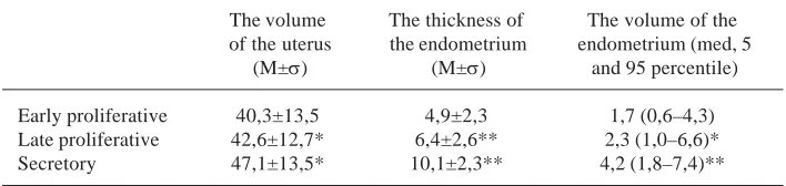
Table 2. The volume of the body of the uterus, the thickness and volume of the endometrium in patients of the control group
The volume The thickness of The volume of the of the uterus the endometrium endometrium (med, 5
(M±σ) (M±σ) and 95 percentile)
Early proliferative 40,3±13,5 4,9±2,3 1,7 (0,6–4,3) Late proliferative 42,6±12,7* 6,4±2,6** 2,3 (1,0–6,6)* Secretory 47,1±13,5* 10,1±2,3** 4,2 (1,8–7,4)**
Note: *– reliability of differences of indicators between the cycle phases >0,05; **– reliability of differences of indicators between cycle phases <0,05.
Table 1. The distribution of patients by age and phases of the menstrual cycle
The phase of the cycle Uterine myoma Control group
n Age (M±σ) n Age (M±σ)
The early proliferative (day 4-9) 36 40.5±8.2 22 41.1±8.0 Late proliferative (day 10-13) 15 42.7±6.5 16 39.9±5.6
The results of the study
At the studying of a control group the volume of the uterine body was in the range of 22.7–72.6 cm3 with an average value of 44.2+13,9
cm3. Any significant differences between phases
of the menstrual cycle were not obtained (p>0.05). The thickness of the endometrium in the early proliferative phase ranged from 2 to 10 mm (4.9+2.3 mm), in the late proliferative phase – from 2 to 11 mm or 6.3+2.6 mm), increasing in the secretory phase from 7 to 15 mm (10,1+2.3 mm). Differences between phases were significant (p<0.05).
The volume of the endometrium in the early proliferative phase ranged from 0.4 to 4.6 cm3
(med 1,7 cm3) and in the late proliferative – from 0.8
to 7.0 CC (med 2,3 cm3) without significant
differences in the rate (p>0.05).In the secretory phase of the cycle the endometrial volume increased from 1.7 to 9.1 cm3 (med 4,2 cm3)
(p<0,5).Volume changes of endometrial thickness and endometrial volume, depending on the phase of the cycle, are presented in table 2.
Pathology of the myometrium, endometrium and (or) ovaries of patients of a control group was not revealed.
The volume of the uterus affected by fibroids, ranged from 25,2 to 450,0 cm3, it was met
among women in the early proliferative phase (med 87,0 cm3).In the late proliferative and secretory
phase fluctuations between the minimum and maximum values of the volume of the uterus were less significant and amounted to 49.5–203,4 cm3and 42.5–242,4 cm3, respectively (med 102,2 and
98,1 cm3).Any significant differences between the
cycle phases were not obtained (p>0.05). A single myomatous node was detected in 27 (37,5%) women, two nodes – in 10 (13.9%) and multiple in 35 (48.6 percent).Isolated lesions of posterior wall were observed in 20 (27.8 percent) cases,the front – 12 (16,7%), the bottom – 7 (9,7%), the nodes at both walls – 33 (45,8%).The combination of myoma in the body of the uterus and cervical-isthmical region were observed in 8 (11,1%) patients.Interstitial and (or)
interstitial-Fig. 1. The measurement of the length and anteroposterior size of the uterus and endometrium in the sagittal plane scanning
Fig. 2. Measurement of the transverse size of the uterus and endometrium while a transverse scanning
Fig. 4. Comparison of the thickness and volume of the endometrium in patients with hysteromyoma
according to the phases of the cycle Fig. 3. The ratio of the thickness and volume of the
subserous location wereidentified in 58 (80,6%) cases,centripetally growth – in 8 (11,1%), subserous fibroids on a wide or narrow base – in 7 (9.7 per cent).The minimum average diameter of the tumor was 6 mm, maximum – 55mm (med 20 mm).
Among patients with myoma the uterine endometrial thickness in the I phase of the cycle ranged from 2 to 14 mm, from 4-th to 9-th day of cycle the median was 5 mm, andfrom 10-th to 14-th day– 6 mm (p>0.05).In the second phase the endometrial thickness ranged from 2 to 17 mm (måd 7 mm) (p>0.05).Any significant difference in endometrial volume trough the different phases of the cycle were not obtained.So, in the early proliferative phase the fluctuations were between 0.8 and 12.2 cm3 (m∑d of 3.9 cm3), in late – from 1.9
to 9.4 cm3 (måd of 6.6 cm3) and secretory – between
1.7 and 16.7 cm3 (måd of 7.9 cm3).The structure and
echogenicity of the endometrium in 47 (65,3%) patients was consistentwith the phase of the menstrual cycle,while in 19 (26,4%) cases was observed lag of a secretory transformation as in I and the II phases of the cycle, and in 6 (8.3%) was observed hyperechogenic endometrium in the first phase.Thebasallayerin 38 (27,4%) women was indistinct and uneven.
The volume of the body of the uterus, the thickness and volume of the endometrium in patients with myoma with accuracy rates compared to the control group are presented in table 3.
The thicknesses of the endometrium among patients with fibroids did not depend on the volume of the uterus,while the volume of the endometrium in thesepatientsincreased with the increasing of a size of the uterus affected by fibroids.
Was made an analysis of the distribution of patients undergoing curettage of the uterus because of the uterine bleeding,depending on the volume of the uterus.It was found that among women with uterine volume of up to 75 cm3there
were no intrauterine interventions on this occasion.Among 17 patients in whom the uterus was from 75 to 100 cm3, 3 patients (17,6%)
underwent the curettage of the uterus.In 18 patients with uterine volume from 100 to 150 cm3anuterine bleeding was observed in 6 (33,5%)
cases.Women with uterine volume of more than 150 cm3were 14 people, among them 2 patients t
(14,3%) – underwent dilatation and curettage of the cavity.
Any structural pathology of the ovaries in patients with uterine myoma was not detected.
DISCUSSION
Uterine fibroids are the most common benign tumor of the myometrium, which affects about 20% of women older than 30-35 years (Vikhlyaeva, 2004; Tikhomirov, Lubnin, 2006; Borgfeldt, Andolf, 2000).In younger women fibroids are found much less frequently, and in children and adolescents – it is an exclusive disease (Borgfeldt, Andolf, 2000; Moroni, Vieira, Ferriani, et al., 2015).A. L. Tikhomirov and D. M. Lubnin (2006) noted that in patients at the age of 22-23 Fig. 5. Comparison of the thickness of
the endometrium in women of the control group and patients with uterine myoma
Fig. 6. Comparison of the volume of the endometrium in women of the control group and patients with uterine myoma
Fig. 7. The dependence of the thickness and volume of the endometrium from the
years are diagnosed predominantly interstitial-subserous fibroids. In the current study these data are confirmed. Thus, among the 8 women whose age was from 23 to 30 years, subserous and interstitial-subserous site location was noted in 6 cases, and interstitial – only in two.
Asymptomatic fibroids are observed in approximately one third of patients with uterine myoma (Vikhlyaeva, 2004; Ishchenko, Botwin, Leninskiy, 2010).Menstrual cycle for the majority of domestic and foreign researchers is the most frequent clinical symptom.
Hypermenstrual syndrome may be associated not only with submucous location of the node, but also with the violation of the contractile activity of the uterus, dilation of the cavity and, consequently, with an increase in the area of the endometrial rejection (Ishchenko, Botwin, Lenchinskiy, 2010; Savitsky, Savitsky, 2003).
The results of the study showed that in the control group, the thickening of the endometriumis synchronouswith an increase of the endometrial volume from the early proliferative to the secretory phase of the menstrual cycle (figure 3).
In patients with uterine myoma the endometrial thickness in the I phase did not differ from the values of healthy women of similar age,but beginning from the late proliferative phasethe increase in thickness significantly (p<0.05) slows down.At the same time, the volume of the endometrium continues to grow (figure 4).
When comparing the values of thickness of the endometrium in patients with myoma and control groups was revealed a significant (p<0.05) reduction only in the secretory phase, whereas in phase I the performance is virtually identical (figure 5).
The volume of the endometrium in patients with myoma in both phases of the cycle was almost in 2 times more than in women in the control group (figure 6).
In addition, noteworthy is the steady increase in the absence of changes in the thickness of the endometrium as the growth of myoma of the uterus (figure 7).
Both facts prove the increase of the uterine cavity.In the work of G. A. Savitsky and A. G. Savitsky (2003) is indicated that the area of the
endometrium in women with fibroids, consistent with 8-9 weeks of pregnancy increases by more than 1.5 times, consistent with 12 weeks – by more than 3.5 times, and 16 weeks – by more than 5.5 times.However, the visual changes of the thickness of the endometrium with hysteroscopy were not detected (Bacsko G. et al., 1997) (Bacsko, Major, Csiszár, Borsos, 1997).
Among clinicians there is no consensus about the tone of the affected uterine myometrium in the mechanism of uterine bleeding.So, A. I. Ischenko et al. (2010) believe that the trigger of menorrhagia and metrorrhagia is a hypotension of the myometrium,and G. A. Savitsky and A. G. Savitsky (2003) – a hypertension.According to I. S. Sidorova (2003) the presence of simple fibroids is much less likely to cause uterine bleeding than proliferating fibroids.Often a combination of fibroids and endometrial hyperplasia can also be the cause of hyperpolymenorrhea(Tikhomirov, Lubnin, 2006; Sidorova, 2003; Lalonde, 1994; Galen, Isaacson, Lee, 2013).Despite the fact that among the surveyed patients with myoma the hyperplastic processes of the endometrium were absent, the combination of these hormone-dependent diseasesdoes not exclude.
In this study is marked a dependence of the frequency of curettage of the uterine cavity due to bleedings on the uterus volume. So, in the group of patients with the uterus, which volume do not exceed 75 cm3, there were no intrauterine
interventions on this occasion. With the increase in the uterus of up to 100 cm3, the curettage was
done in 17.6% of women, in the uterus of up to 150 cm3– in 33,5%. It was probably associated with the
increased area of desquamation. Among women with uterine volume of more than 150 cm3a
frequency of operative hemostasis was 14.3%, which can be associated with a greater frequency of operative treatment of myoma in the early stages and if patients had such enlarged uterus, reaching in the work in maximum of up to 450 cm3,at the
same time the clinical manifestations of the disease were not so significant.In addition, a shown increase in a volume of the endometrium according to the growth of myoma of theanda reducing in the frequency of curettage of the cavity in patients with uterus of more than 150 cm3, confirms
CONCLUSION
According to the conducted research we can draw the following conclusions
1. The thickness of the endometrium in patients with uterine myoma in the I phase of the cycle does not differ from the healthy women. 2. In the second phase of the cycle in patients
with uterine fibroids the processes of secretory transformationare slowing. This contributes to the reducing of thickness of the endometrium. 3. The volume of the endometrium in patients with
myoma is more in both phases of the cycle compared with the control group and it increases at the growth of the uterus.
Thus, not only determining the thickness but also the volume of the endometrium allows usbetter define the condition of the uterine cavity.This method allowsto assess endometrial pathology such as diffuse hyperplasia and polyp, and to conduct differential diagnostics between benign and malignant processes.
REFERENCES
1. Bacsko, G., Major, T., Csiszár, P., Borsos, A.,
Operative hysteroscopy: minimally invasive surgery to control of menorrhagia 1997; 36(1-4): 7-8). Acta Chirurgica Hungarica. https: // www. researchgate. net/ publication/ 13822511_ Operative_ hysteroscopy_ minimally_ invasive_ surgery_ to_ control_ of_ menorrhagia 2. Borgfeldt, C.,Andolf, E., Transvaginal
ultrasonographic findings in the uterus and the endometrium: low prevalence of leiomyoma in a random sample of women age 25-40 years 2000; 79(3): 202-207. Acta Obstetricia et Gynecologica Scandinavica.http://doi/10.1034/j.1600-0412.2000.079003202.x/full
3. Fascilla, F.D.,Cramarossa, P., Cannone, R. et al., Ultrasound diagnosis of uterine myomas 2016; 68(3): 297-312. Minerva Ginecologica. http:// www.minervamedica.it/en/journals/minerva-ginecologica/article.php ? cod= R09Y2016 N03A0297
4. Galen, D.I.,Isaacson, K.B., Lee, B.B., Does menstrual bleeding decrease after ablation of intramural myomas? A retrospective study 2013; 20(6): 830-835. Journal of Minimally Invasive Gynecology.hppt://doi: 10.1016/j.jmig.2013.05. 007.
5. Lalonde, A., Evaluation of surgical options in
menorrhagia 1994; 101(11): 8-14. British Journal of Obstetric and Gynaecology. 6. Moroni, R.M., Vieira, C.S., Ferriani, R.A. et al.,
Presentation and treatment of uterine leiomyoma in adolescence: a systematic review 2015; 15(4): BMCWomen’sHealth.
7. Takeda, A.,Nakamura,H., Torsion of a Subserosal Myoma Managed by Gasless Laparoendoscopic Single-Site Myomectomy with In-Bag Manual Extraction 2016; 783-1270. Case Reports in Obstetric and Gynecology.http://doi: 10.1155/2016/7831270.
8. Bulanov, M.N., Ultrasound gynecology: a course of lectures in two parts. (3rd ed. EXT., part 1). Moscow: Publishing house Vidar-M, 2014. 9. Vikhlyaeva, E. M., Guide to the diagnosis and
treatment of uterine leiomyoma.Moscow, M: Medpress-inform, 2004.
10. Demidov, V. N., Zykin, B. I., Ultrasound diagnosis in gynecology.Moscow, M: Medicine, 1990.
11. Zykin B. I. Medvedev, M. V., Doppler ultrasound in gynecology. Moscow, M: Real time, 2000.
12. Ischenko A. I., Botvin M. A., Lanchinskiy, V. I.,
Uterine fibroids: etiology, pathogenesis, diagnostics, treatment. Moscow: Publishing house Vidar-M, 2010.
13. Medvedev, M. V., Rudko, G. G., Differential diagnostic ultrasound in gynecology (2nd ed., Rev.). Moscow, M: Real Time, 2010.
14. OzerskayaI. A., Sonographyingynecology.(2nd ed., Rev.), M: Publishing house Vidar-M, 2013. 15. Ozerskaya I. A., Ageeva, M. I., Chronic pelvic pain in women of reproductive age. Ultrasound diagnosis. Moscow: Publishing house Vidar-M. 2009.
16. Savitsky, G. A., Savitsky, A. G., Uterine fibroids (problems of pathogenesis and pathogenetic
therapy).(Ed. 3.). Saint Petersburg, St.
Petersburg: ELBI-SPb, 2003.
17. Sidorova, I. S., Hysteromyoma (modern issues of etiology, pathogenesis, diagnosis, and treatment). Moscow, M: MIA, 2003.
18. Tikhomirov A. L., Lubnin D. M., Myoma of the
uterus.Moscow, M: Medical information
Agency, 2006.
19. Fedorova, E. V., Lipman, A. D., The use of color Doppler mapping and Dopplermetry in gynecology. Moscow: Vidar, 2002.