Correspondence: Dr Bijaya Mishra, Department of Biochemistry, BPKIHS, Dharan
Sunsari, Nepal. Email: [email protected], Phone: +977-9849530325.
ABSTRACT
INTRODUCTION
Acute Coronary Syndrome (ACS) occurs when there is acute disruption of coronary blood flow leading to mismatch between myocardial oxygen demand and supply, which results in myocardial ischemia and ultimately infarction.1 Diagnostic approach of ACS is challenging, as most events are clinically not recognizable and biochemically undetectable until onset of myocardial necrosis.2
Cardiac troponins(cTns) and creatine kinase-MB(CK-MB) although sensitive and specific, shows greater rises~3-6
hrs after onset of irreversible myocardial injury, delaying immediate diagnosis of ACS. Ischemia Modified Albumin (IMA),is suggested as useful marker for early detection of myocardial ischemia,3 elevated within 6–10 min after ischemia, remains elevated up to 6–12 hrs, returns to normal within 12–24 hrs.4 Human Serum Albumin(HSA) converts to IMA when convened with ischemic heart tissues.5
The objective of the study was therefore to compare IMA between ACS and healthy control groups, and to evaluate diagnostic performance of IMA compared to cTnI, CK-MB and ECG in ACS patients in our population.
Background:
The diagnosis of acute coronary syndrome remains challenging, as cardiac troponins and creatine
kinase-MB do not detect myocardial ischemia. Ischemia modified albumin is biomarker positive within 6-10 minutes
following ischemic onset, where oxygen free radicals leads to reduction in binding capacity of human serum albumin
to transitional metal-cobalt. The objective of this study was to compare ischemia modified albumin between acute
coronary syndrome patients and healthy controls, and evaluate diagnostic performance of ischemia modified albumin
compared to cardiac troponins, creatine kinase-MB and electrocardiogram in acute coronary syndrome patients.
Methods: Fifty ACS patients and 50 healthy controls were enrolled in this cross-sectional study. Ischemia
modified albumin was measured after addition of known amount of cobalt to human serum albumin, followed by
spectrophotometric determination of unbound cobalt fraction at 470 nm using dithiothreitol as coloring agent.
Independent student t-test and One-way ANOVA to compare differences of mean between groups; diagnostic sensitivity
and specificity of ischemia modified albumin was determined by receiver operating characteristic curve; McNemar-test
was used to assess diagnostic performance of entire test parameters, when used alone and in combinations.
Results: Ischemia modified albumin was significantly higher in acute coronary syndrome patients compared to
controls (0.823±0.191 vs 0.410±0.081)(p<0.001). Receiver operating characteristic curve derived optimal
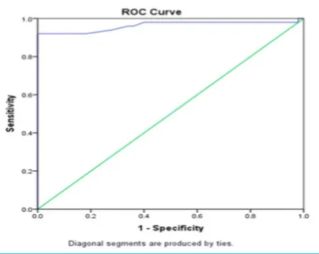
cut-off of 0.475 Absorbance unit had sensitivity and specificity of 92% and 82% respectively (area under curve- 0.96).
However, no significant differences in mean ischemia modified albumin values between three categories of acute
coronary syndrome were seen. Sensitivity of ischemia modified albumin assay (92%) was significantly higher compared
to electrocardiogram (72%), cardiac troponin I (18%), and creatine kinase-MB(42%).
Conclusions:
Ischemia modified albumin is elevated in acute coronary syndrome patients with better diagnostic
performance compared to electrocardiogram, cardiac troponin I, and creatine kinase-MB for early diagnosis, however,
with limited ability to discriminate between ST-elevation myocardial infarction, non-ST-elevation myocardial infarction
and unstable angina.
Keywords: Acute coronary syndrome; human serum albumin; ischemia modified albumin.
J Nepal Health Res Counc 2018 Jan-Mar;16(38):16-21
Utility of Ischemia Modified Albumin as an Early Marker
for Diagnosis of Acute Coronary Syndrome
Bijaya Mishra,1 Sunil Pandey,1 Surya Raj Niraula,2 Bijendra Kumar Rai,3 Prahlad Karki,4 Nirmal Baral,1 Madhab Lamsal1
1Department of Biochemistry, 2Department of School of Public Health and Community Medicine, 3Department of General Practice and Emergency Medicine, 4Department of Internal Medicine, BPKIHS, Dharan, Sunsari, Nepal.
METHODS
The present hospital based cross-sectional study was conducted in Department of Biochemistry in collaboration with Department of Internal Medicine- Cardiology Division and Department of General Practice and Emergency Medicine at B.P. Koirala Institute of Health Sciences, Dharan, for duration of one-year from 1st August 2014 to 30th July 2015. Ethical Clearance was obtained from Institutional Ethical Review Board (IERB), BPKIHS.
A total of 50 ACS diagnosed case and 50 healthy individual in control group were enrolled. Information of the study was provided, confidentiality of information was assured, and written consent was obtained from family member for the participation.
Applying the purposive sampling technique, patients presenting in Emergency Department with complain of chest pain and diagnosed as ACS by the attending physician were included in ACS group. Diagnosis was made on the basis of clinical history, ECG, qualitative cTnI and CK-MB activity that were performed immediately as a part of standard of care and patient management protocol of our Institute. Individuals with abnormal liver and renal function tests were excluded from the study.
Following initial assessment and diagnosis, the patients were divided into 3 categories of ACS as ST-elevation myocardial infarction(STEMI), non-STEMI and unstable angina (UA) by the physicians. No angiographic confirmation was considered due to lack of limited resource facility. The results of IMA were not made available to the attending physicians at the time of diagnosis and management. Samples sent for the investigations were collected and stored at -80oC for IMA assay until further analysis. Repeated freezing and thawing of samples was avoided.
Individuals without diagnosed medical conditions and not under any medication, who visited hospital for their routine check up were recruited in the control group after following up their investigation reports. Patients not willing to participate, and with hypoalbuminemia, jaundice, chronic kidney diseases, ischemic stroke, altered liver and renal functions test and pregnancy were excluded from the study.
The IMA assay was used to compare its value between the control group and the ACS group, in addition to evaluate its diagnostic performance when compared to cardiac biomarkers (cTnI and CK-MB) and ECG when used individually and in various combinations, in the ACS
group.
The IMA assay procedure was based on the principle that myocardial ischemia leads to structural change in amino terminal (NH2-Asp-Ala-His-Lys) of HSA determined by decrease dexogenous cobalt (Co(II)) binding, followed by the measurement of unbound Co(II) using dithiothreitol (DTT) as a coloring agent. IMA concentration is directly proportional to the color intensity formed.
IMA assay protocol involved addition of 50μL of 0.1% cobalt chloride (LobaChemie, CoCl2.6H2O) prepared in H2O to 200μL serum, followed by gently mixing, and incubation for 10 minutes. Fifty μL of DTT (Sigma, 1.5 mg/ml H2O) was added as colorizing reagent and incubated. The reaction was stopped two minutes later by addition of 1.0 mL of 0.9% NaCl. Using Sheerwood spectrophotometer, the development of color with DTT was compared to serum-cobalt blank, which was prepared similarly without the addition of DTT, and measured at 470 nm. IMA concentration was determined as difference in ABSU between test sample and sample blank.6
A rapid chromatographic immunoassay for qualitative detection of cTnI was performed using the one-step troponin I test device (ABONTMcTnI). Serum CK-MB activity was measured by the principle of optimized kinetic method in COBAS Roche c311 auto-analyzer. A 12-lead electrocardiogram was also performed on patients.
Data were entered in Microsoft Excel 2008 and transferred to IBM SPSS 11.5 version Descriptive statistics such as mean, standard deviation (SD), and percentage were calculated. Categorical data were present into tabular and graphical form and were tested using chi-square test. Independent Student’s t-test was applied to compare the means between two groups. For three categories, one-way ANOVA was performed. Receiver Operating Characteristics (ROC) curve was used to determinethe optimal cut-off values which provided the maximum sensitivity and specificity. For determination of diagnostic performance of the combined variables, McNemar test was applied.
RESULTS
Next, the ABSU data of IMA test value from ACS and control group was used to construct a ROC curve shown in Figure 2. The optimal diagnostic cut-off IMA value, with the maximum sensitivity (92%) and specificity (82%), was determined to be 0.475, with an AUC 0.961 (95% CI, 0.918-1.00). Thus, we considered 0.475 ABSU as an optimal diagnostic cut-off value for our study.
Figure 2. ROC curve comparing performance of IMA assay in ACS diagnosis.
The diagnostic performance of IMA, ECG, cTnI and CK-MB, all when used individually and in combination, for diagnosing ACS were analyzed for their clinical sensitivity, specificity, positive predictive value (PPV) and negative predictive value (NPV). The two, three and four test combination of IMA, ECG, cTnI and CK- MB were considered positive when any one of the test parameter was positive, and considered negative when all the later three tests were negative. The significance of diagnostic performance of the entire tests, alone and in various combinations were compared with IMA using McNemar test, as shown in Figure 3.
Further, the adjustment of IMA for serum albumin using the Lippi formula was also performed. The median albumin concentration in the control and the ACS groups were 4.00 (range 3.4 to 5.0) and 4.13 (range 3.1 to 5.1) respectively.
IMA Index = IMA x (individual serum albumin concentration/ median albumin of thestudy population) As shown in Table 3, there were, no significant differences between IMA and IMA index in both the groups.
Table 3. IMA and IMA index between the two groups.
Groups IMA IMA index *p value
ACS 0.823±0.19 0.809±0.177 0.257 Control 0.410±0.081 0.431±0.096 0.010 Data expressed as Mean±SD ; *Paired samples t-test
Table 1. Baseline characteristics among the control group and ACS group.
Variables Control
(n=50) ACS (n=50) value*p Age (years) 41.98±5.40 62.32±16.63 0.001
Male 72% 68% 0.660#
SBP (mmHg) 116.80±8.96 133.80±27.24 <0.001 DBP (mmHg) 75.00±11.56 83.20±14.34 0.002 Serum
albumin (g/ dl)
4.20±0.507 4.11±0.422 0.320
*Independent Student’s t-test; # chi square
Distribution of IMA assay: IMA assay performed in both groups revealed IMA value of 0.410 ABSU with SD 0.081 in control group, whereas in ACS group mean IMA of 0.823 ABSU with SD 0.191, the difference between the two groups being statistically significant (p<0.0001), as illustrated in Figure 1.
Figure 1. Comparison of mean IMA value between ACS and Control group.
The ACS group was categorized as STEMI 28%, NSTEMI 16% and unstable angina 56%. One-way ANOVA was applied to compare mean IMA values between the 3 categories, which revealed the differences in the mean within these categories to be statistically insignificant, as shown in Table 2.
Table 2. Comparison of the mean IMA value between the ACS categories.
Categories Mean±SD p value*
STEMI 0.843±0.146 1.0†
NSTEMI 0.925±0.094 1.0‡
DISCUSSION
Many patients presenting to ED may present with myocardial ischemia without the onset of myocardial necrosis. However, use of markers for diagnosing ACS is based on the presence of myocardial necrosis as a surrogate indicator for myocardial ischemia. Diagnostic tools including ECG, cTnI and CK-MB, although remains mainstay for the diagnosis of AMI, have limited role in predicting myocardial ischemia. Thus, a rapidly detectable and highly sensitive marker for myocardial ischemia would be appropriate to identify the patients in the early course of the disease, thus providing opportunity to intervene the progression of myocardial ischemia to myocardial infarction.6,7 IMA was developed, later approved by US FDA, and was found to be very promising for the early detection of myocardial ischemia.5
The mean age of ACS group (62.32 years) was significantly higher than control group (41.98 years) in our study. Similar difference of age between the two groups was also seen in study done by Bar–Or et al. and Abadie et al.3,8 Disparity between ages in our study groups was due difficulty in finding healthy elder individuals. However, study has shown no observed difference in IMA value between gender, and when studied in different age group.9
IMA results are instrument dependent, thus, it is suggested that each facility perform an independent
ROC analysis to define the optimal cut-off values of IMA assay results for their study.8 In our study, ROC curve revealed IMA cut-off of 0.475 ABSU optimal for distinction between the ACS and control group. This cut-off value used was approximately equivalent to 0.400 ABSU used by Bar–Or et al;3 0.500 ABSU by Bhagavan et al;10 and 0.0551 ABSU by Patil et al.11 AUC of 0.957 was also equivalent to that of the later two studies.
Exploring the feasibility of IMA values when used alone, and along with CK-MB, cTnI and ECG, the results of IMA was superior, and additive to, when used alone or in various combinations with the conventional diagnostic tools. This findings was in support to various other studies.11-14 The combination of IMA to CK-MB, cTnI and EGC significantly increased the sensitivities of these diagnostic tools, but the sensitivity of IMA itself did not increase significantly either when used alone (92%) or in total combination with other diagnostic tools (94%), which showed discordance to other studies.6,11,17Anwaruddin et al. in their study showed that the combination of IMA+Mb+CK-MB+cTn, increased the sensitivity for IMA from 80% to 97% for detecting ischemia.6 Takhshid et al. in their study showed a significant increase in the sensitivity of IMA from 84% to 96% when IMA was used in combination with cTnI and ECG17. Patil et al. 2013 also reported the sensitivity and specificity of IMA as 88% and 93 %, respectively, and when combined with cTnI the IMA sensitivity increased to 96%.11 Thus, all these studies suggested the use of IMA in conjunction with cardiac biomarkers and ECG despite the high performance of IMA. Compared to the fore mentioned studies, the disparity seen with our study might reflect the differences in baseline characteristics, the patient selection criteria, and the optimal cut-off value used. In addition, it is recommended that each laboratory should establish its own reference value and the optimal clinical cut-off of IMA, which may vary depending upon geographic, dietary and environmental factors.5
Similar to the studies done to by Bhagavan et al.10 and Wudkowska et al.,18 our study also exhibited the inability of IMA test value to discriminate between the STE-ACS (STEMI) from NSTE-ACS (NSTEMI and unstable angina). As no “gold standard” test for myocardial ischemia was available, the diagnostic performance of IMA assays was computed against the final diagnosis based on the interpretation of clinical findings and appropriate tests results. In addition, considering unstable angina and AMI together as myocardial ischemia might affect any final distinction between transient myocardial ischemia and myocardial necrosis.17
As the serum albumin concentration in the ACS and control individuals were within the reference interval,and, the exclusion of all the conditions that affects albumin concentration,a positive correlation existed between IMA and IMA index (albumin adjusted IMA value)19 in our study groups.
The study was however bounded by its limitations. Due to the lack of “gold standard” test for myocardial
ischemia, the diagnostic performance of IMA relied upon the diagnosis made by different attending physicians based on clinical findings and interpretation of the results of standard convention diagnostic tool for myocardial infarction. In addition,due to specificity issues, the study was limited by strict exclusion criteria; as a consequence, the applicability of the IMA assay in the patients presenting in the ED with ACS might be questioned.
CONCLUSIONS
Measurement of IMA as a marker of myocardial ischemia in the absence of myocardial necrosis and/or preceding myocardial necrosis, can be useful in diagnosis of ACS patients. We conclude IMA assay as a promising biochemical marker for the early diagnosis of ACS; however, the test is a poor discriminator between STE-ACS and NSTE-STE-ACS.
ACKNOWLEDGEMENTS
I express my gratitude for the contributions of Mr
Rajendra KC, Dr Prajwal Pandey and Aurik Pandey who
constantly supported and guided me throughout the
study period in all the aspects. Our thanks to all the
Faculty members, students and staffs of Department of
Biochemistry for all the help provided for the study. We
are grateful to the Nepal Health Research Council (NHRC) for providing us with the PG grant 2014 to conduct the study.