Structural Changes of the Testis and Changes in Semen
Quality Parameters Caused by Intraperitoneal and Peroral
Administration of Selenium in Rats
Michal Cabaj
1, Róbert Toman
1, Mária Adamkovi
č
ová
2, Peter Massányi
2, Svätoslav
Hluchý
1, Norbert Luká
č
2, Jozef Golian
21
Slovak University of Agriculture, Faculty of Agrobiology and Food Resources, 94976 Nitra, Tr. A. Hlinku 2, Slovakia
2
Slovak University of Agriculture Faculty of Biotechnology and Food Sciences, 94976 Nitra, Tr. A. Hlinku 2, Slovakia
Abstract
The aim of this study was to find the structural changes in the testis and semen quality parameters of rat after a single intraperitoneal and repeated peroral selenium administration. Rats were killed 36 hours following the intraperitoneal administration of selenium selenite (2 mg.kg-1 b.w.; 98% purity) and after 90 days of the peroral repeated
administration of selenium in drinking water (5 mg.l-1). Testis samples were evaluated by histological and
morphometrical methods in light microscopy. Evaluation of semen samples were examined with CASA method. 36 hours after the selenium i.p. administration, damage of cellular associations, release of necrotised epithelial cells to tubule lumen and fibrotisation and extension of interstitium were observed. Morphometry methods have shown the reduction of seminiferous epithelium volume (P<0.001), extension of interstitium (P<0.001) and increased area of intraepithelial spaces (P<0.01). In p.o. group similar but more intense changes were noted; in addition, occasionall degeneration of seminiferous tubuli and rarely total damage in histoarchitecture of seminiferous epithelium were observed. CASA analysis revealed significant decrease in all parameters except the concentration of spermatozoa. Additionally, we suppose that p.o. dose 5 mg.l-1 sodium selenite in drinking water is minimum lethal dose level for
young rats. Selenium after i.p. and p.o. administration causes damage of seminiferous epithelium and interstitium. It leads to changes in relative proportion of functional tissues of the testis. Reduced spermatogenesis and harmful effects in semen parameters are characteristic especially for peroral repeated (subchronic) administration. These changes are time- and dose-dependent. In both dosage methods subfertility or infertility can appear.
Keywords: CASA, histology, morphometry, rat, selenium, sperm, testis
1. Introduction
In recent years interest of scientific teams and then also public about essential microelement selenium greatly increased. Its effects are in the environment as well as in a living organism linked mostly to its biochemical form, concentration and periodicity of exposure. These parameters determine the beneficial or harmful effects of selenium. In endemic or contaminated areas
*Corresponding author: Michal Cabaj, +421 37 641
4503, [email protected]
(outside the optimal levels) of environment, selenium may act as limiting trace element or contaminant, both with harmful effects for living organisms of the ecosystem. The same effect for organisms has selenium in inappropriately diet or feed ration.
selenoenzymes and other biochemical forms participate in the antioxidant protection system of cells and their organelles against free radicals damage and the associated/linked diseases [2]. Some forms of selenium can act as prooxidant on various types of cancerous cells and this is considered as the way of their protective function in prevention of some types of cancers and the overall anticarcinogenic activity [3-7]. Selenium participates on deiodination of thyreoidal hormones and thereby indirectly affects energy metabolism [8-9]. Selenium significantly impacts sensitive metabolic regulations of male reproductive system. In the form of selenoenzymes and selenoproteins (PHGPx/GPx4, selenoprotein P and others) is essential for testicular development, spermatogenesis and sperm functions [10-14]. In idiopathic infertile men, positive correlations between selenium concentration and plasma testosterone were detected [13]. Many authors suggest the use of selenium alone or in combination with other antioxidants for fertility promotion and treat some forms of male sub- or infertility [13,15,16]. On the other hand, less known are effects of excess of selenium on the male reproductive system, complex mechanism of actions and relation between the results of different types of experiments. Kaushal and Bansal [17] reported prooxidant effects of selenium on germ cells after administration of an excess of sodium selenite to the mouse in their feed.
The aim of experiment was to describe and evaluate structural changes in the testis and sperm quality parameters of the rat after single intraperitoneal and repeated peroral administration of selenium.
2. Materials and methods
Experimental animals and design
30 Wistar rats were randomly divided to three groups: control group (C), group with single intraperitoneal administration of selenium (IP) and group with repeated peroral (subchronic) administration of selenium (PO). Age of rats at the beginning of the experiment was: 120 days for groups C and IP and 30 days for PO group. Rats of IP group were injected with selenium as sodium
selenite 2 mg.kg-1 b.w. (Na
2SeO3; 98% purity;
Reachem, Bratislava) in saline solution. PO rats recieved same sodium selenite in their drinking water (5 mg.l-1 for 90 days) with free access. At
the end of experiment rats of all group were anesthetized (ethyl ether) and humanely killed (IP group 36 hours after selenium administration and PO group after 90 days of Se administration). Anatomical dissection was performed and samples of testicles were fixed in modified Davidson’s solution [18]. Animals were housed individually in plastic cages with wood shavings bedding. In the experimental laboratory basic requirements of environmental conditions (temperature 20 to 22°C, air humidity 55±10%, 12 hours dark/light regime) and unlimited access to drinking water and feed (M3, Máchal, Czech republic) were maintained according to Government regulation no. 289/2003. Experiment was performed in an approved experimental laboratory SK PC 50004 SPU in Nitra, Slovak republic.
Histological and morphometrical evaluation
Samples of testis were stained with hematoxylin-eosin and were examined using the light microscopy (Nikon Eclipse E600, Japan). Morphometrical evaluation of photomicrographs was performed using computerized evaluation morphometric software M.I.S. Quick Photo with assistance of microscope Olympus AX 70 (Olympus, Japan) and further evaluated with quantitative morphometrical method modified by Uhrín and Kulíšek [19]. Testicular weight (g), mean seminiferous tubule diameter (μm), relative volume of testicular structures (seminiferous epithelium, intraepithelial empty spaces, tubule lumen, interstitial tissue, blood vessels) (%) were determined.
Evaluation of semen quality parameters
microscope Olympus BX 51 (Olympus, Japan). Following parameters were evaluated:
spermatozoa concentration % of motile spermatozoa
% of progresive motile spermatozoa DAP - distance average path (µm) DCL - distance curved line DSL - distance straight line
VAP - velocity average path (µm.s-1)
VCL - velocity curved line VSL - velocity straight line STR - straightness (VSL/VAP) LIN - linearity (VSL/VCL) WOB - wobble (VAP/VCL) ALH - amplitude of lateral head
displacement (µm.s-1)
BCF - beat cross frequency (Hz)
Statistical evaluation
Results were presented as mean±standard deviation (x±SD). For statistical evaluation of results one-way analysis of variance (one-way ANOVA) and Software Statgraphics Centurion XV with alpha level set as α = 0.05 was used.
3. Results and discussion
Photomicrographs of control group testis tissue have shown normal shape and histological structure. The space between seminiferous tubuli was filled by condensed interstitial tissue. The outer edge of the tubuli perimeter was closely lined with basement membrane and towards the lumen in irregular layers there were added germ cells in different stages of development, between which there were sketched Sertoli cells with characteristic nucleus. Depending on the stage of spermatogenic cycle at the top of apical Sertoli cells cytoplasm there were embedded spermatids. Lumens of seminiferous tubuli were filled with masses of released spermatozoa. In the interstitial tissue, blood vessels with fundamentally different diameters surrounded by numerous of Leydig cells and sometimes typical filaments structure of interstitial tissue were well seen. Weight of testis and diameter of semniniferous tubuli are shown in Table 1, the relative volume of the testis is
presented in table 2 and semen quality parameters are shown in table 3.
Table 1. Testis weight (g) and diameter of seminiferous tubuli (μm)
Group Testicular weight Seminiferous tubuli diameter
x±SD
Control 1.45±0.25 250.37±15.09 Selenium IP 1.44±0.08 255.52±15.94
Selenium PO 1.41±0.16 228.91±38.00
x-mean, SD-standard deviation
Excess of selenium in the body causes disruption of homeostasis which leads to acute stress reaction [20,21] with characteristic result – increased production of free radicals [20-22]. This overproduction of free radicals causes damage of cells and their structures [17,22-24], increased lipid peroxidation [22,25], damage of DNA [22,24-25] with the subsequent histo- and physiopathological manifestations [17,22,27]. Changes induced by excess of selenium are dose- (intensity of stress response) and time-dependent (duration of action of free radicals) [1]. These mechanisms seem to be a link between the toxic effects of many essential elements administered in excess.
After a single intraperitoneal administration of 2 mg.kg-1 sodium selenite, changes in weight of
after administration 4, 8 or 16 ppm of sodium selenite in drinking water to rats which indicates tissue damage and breakdown. Their findings correspond with our histopathological findings in seminiferous epithelium and interstitium. Reduction in volume of seminiferous epithelium
(
P<0.001
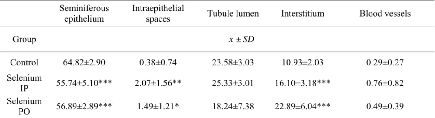
), free spaces in the epithelium (P<0.01), to the detriment of extension of interstitium (P<0.001) and insignificant extension of lumen of seminiferous tubuli and blood vessels were noted (Table 2).Table 2. Relative volume of testis structures (%)
x-mean, SD-standard deviation; * P<0.05; ** P<0.01;*** P<0.001
The most important change in the proportions of morphometrically evaluated components of testis was between damaged seminiferous epithelium and interstitial tissue. These changes might be potentially caused (in view of the short duration of treatment) by effects of free radicals (direct prooxidative effect of selenium and selenium-mediated stress reaction) in addition with direct effects of stress hormones (epinephrine, cortisol) to the structure and functions of testis. Evaluation of semen quality parameters has shown only insignificant changes. The most expressive change was increase in concentration (0.39±0.71 vs. 0.17±0.12), motility (39±23 vs. 29.66±15.88) and progressive motility (19.45±14.92 vs. 13.82±10.46), other parameters when compared with the control group (Table 3). In PO group after subchronic peroral administration of sodium selenite (5 mg.l-1/90 days) one of rats died. Palmer
and Olson [31] not recorded mortality following administration 2 and 3 ppm of sodium selenite to adolescent rats in drinking water. Increasing mortality was observed after administration of 6 and 8 ppm sodium selenite [31]. The dose used in our experiment (5mg.l-1 of drinking water/90
days) might be potentially the minimum lethal dose level for adolescent rats for the given route of administration. Weight of testis and diameter of seminiferous tubuli were not significantly decreased (Table 1). Similar but more intense histopathological changes than were noted in IP group were observed. Histoarchitecture of
semniniferous tubuli was damaged, in the epithelium less or more extensive free spaces in the areas of released necrotized germ cells released to the tubule lumen were visible. Additionally some parts of seminiferous epithelium were partly to completely degenerated - some stages of spermatogenic cells (from spermatogonia to late spermatids) was partly to totally undeveloped and in these places there was only Sertoli cells with their connections (Figure 3). In many places of seminiferous tubuli there was separating basement membrane with or without few germ cells. Morphometrically, the same statistically significant changes like in IP group was observed, except insignificantly decreased volume of interstitial tissue (Table 2). Selenium in excess causes oxidative stress in testicular germ cells, damage of RNA and DNA, reduction of the expression of genes required for synthesis of proteins essential for spermatogenesis [17,26]. Optimal production of spermatozoa in rats occurs first in the 45th postnatal days and
optimal production is achieved first in the 75th
days [31] consequently subchronic administration of selenium in our design of experiment potentionally influenced also testicular development. Evidence of this hypothesis might be partly or completely degenerated seminiferous tubuli. All evaluated parameters of semen quality in PO group were also significantly decreased, except insignificant decrease of spermatozoa concentration (Table 3). All parameters describing Seminiferous
epithelium
Intraepithelial
spaces Tubule lumen Interstitium Blood vessels
Group x ± SD
Control 64.82±2.90 0.38±0.74 23.58±3.03 10.93±2.03 0.29±0.27 Selenium
IP 55.74±5.10*** 2.07±1.56** 25.33±3.01 16.10±3.18*** 0.76±0.82 Selenium
motility are closely related to sperm mitochondrial section, site of energy production. Kaur and Parshad [25] reported morphological damage of mitochondrial section of spermatozoa after the peroral feeding of 4 ppm sodium selenite to the rats which supports our findings of reduced parameters of motility. These data suggest that the mitochondrial section might be directly or indirectly the target site of spermatozoon damage caused by selenium.
Table 3. Semen quality parameters
Group Control Selenium
IP Selenium PO Parameters x±SD
Concentration 0.17±0.12 0.39±0.71 0.12±0.10
%Motile 29.66±15.88 39±23 12.16±6.61**
%Progressive 13.82±10.46 19.45±14.92 3.87±3.91*
DAP 21.01±4.58 21.70±5.00 8.63±7.68**
DCL 33.78±8.22 33.71±8.11 13.54±10.75**
DSL 16.32±2.89 16.87±3.58 6.99±6.10**
VAP 53.39±12.65 54.46±12.30 21.16±18.59**
VCL 84.87±21.58 84.32±19.85 33.05±25.81**
VSL 41.42±7.85 42.31±8.67 17.29±14.95**
STR 0.79±0.05 0.78±0.05 0.40±0.30**
LIN 0.52±0.08 0.53±0.05 0.28±0.22*
WOB 0.65±0.08 0.66±0.03 0.33±0.27**
ALH 5.49±1.13 5.54±0.95 2.49±2.12*
BCF 17.90±1.75 17.92±3.08 7.66±6.27***
x – mean; SD – standard deviation; *P<0.05; **P<0.01;
***P<0.001
Figure 1. Control rat testis 1-seminiferous epithelium; 2-tubule lumen with spermatozoa; 3-interstitial tissue with Leydig cells; 4-blood vessels
Figure 2. Rat testis after i.p. selenium administration 1-seminiferous epithelium; 2-tubule lumen with released germ cells; 3-fibrotised interstitium with net of
reticular and collagen fibers; arrows-intraepithelial spaces – site of release germ cells
Figure 3. Rat testis after p.o. selenium administration 1-site of partly degenerated-undeveloped seminiferous epithelium only with spermatogonia; 2-tubule lumen with sperm; 3-interstitial tissue with Leydig cells;
4-blood vessel (longitudinal section)
4. Conclusions
Dose of sodium selenite 5 mg.kg-1 administered to
spaces of epithelium were increased. Changes in semen quality parameters (parameters of motility) have been significant only in PO group and appear to be time dependent. In both groups subfertility or infertility can appear. For complete explantation of the mechanism of selenium toxicity to male reproduction system further complex studies are needed.
Acknowledgements
This work has been supported by the Excellence Center for Agrobiodiversity Conservation and Benefit project implemented under the Operational Programme Research and Development financed by European Fund for Regional Development.
References
1. ATSDR - Agency for toxic substances and disease registry, 2003. Toxicological profile for selenium. U.S. Department of health and human services, 2003. pp. 457
2. Sun, Y., Mu, Y., Ma, S., Gong, P., Yan, G., Liu, J., Shen, J., Luo, G., The molecular mechanism of protecting cells against oxidative stress by 2-selenium-bridged beta-cyclodextrin with glutathione peroxidase activity, Biochim. Biophys. Acta, 2005, 17, 199-204 3. Drake, E. N., Cancer chemoprevention: selenium as a prooxidant, not an antioxidant, Med. Hypotheses., 2006, 67, 318-22
4. Ganther, H. E., Selenium metabolism, selenoproteins and mechanisms of cancer prevention: complexities with thioredoxin reductase, Carcinogenesis, 1999, 20, 1657-66
5. Plano, D., Baquedano, Y., Ibáñez, E., Jiménez, I., Palop, J. A., Spallholz, J. E., Sanmartín, C., Antioxidant-prooxidant properties of a new organoselenium compound library, Molecules, 2010, 15, 7292-312
6. Spallholz, J. E., Free radical generation by selenium compounds and their prooxidant toxicity, Biomeb. Environ. Sci., 1997, 10, 260-70
7. Xiang, N., Zhao, R., Zhong, W., Sodium selenite induces apoptosis by generation of superoxide via the mitochondrial-dependent pathway in human prostate cancer cells, Cancer Chemoter. Pharmacol., 2009, 63, 351-62
8. Köhrle, J., The trace element selenium and the thyroid gland. In Biochimie, 81, 1999, 5, 527-33. 9. Köhrle. J., Gärtner, R.,. Selenium and thyroid, Best Pract. Res. Clin. Endocrinol. Metab., 23, 815-27 10. Akinloye, O., Arowojolu, A. O., Shittu, O. B., Adejuwon, C. A., Osotimehin, B., Selenium status of idiopathic infertile Nigerian males, Biol. Trace Elem. Res., 2005, 104, 9-18
11. Boitani, C., Puglisi, R.,. Selenium, a key element in spermatogenesis and male fertility, Adv. Exp. Med. Biol., 2008, 63, 65-73
12. Flohé, L., Selenium in mammalian spermiogenesis, Biol. Chem., 2007, 388, 987-95
13. Keskes-Ammar, L., Feki-Chakroun, N., Rebai, T., Sahnoun, Z., Ghozzi, H., Hammami, S., Zghal, K., Fki, H., Damak, J., Bahloul, A.,. Sperm oxidative stress and the effect of an oral vitamin E and selenium supplement on semen quality in infertile men, Arch. Androl., 2003, 49, 83-94
14. Kaushal, N., Bansal, M. P.,. Role of selenium in regulation of spermatogenesis: involvement of activator protein 1. Biofactors, 2005, 23, 151-162
15. Safarinejad, M. R., Safarinejad, S.,. Efficacy of selenium and/or N-acetyl-cysteine for improving semen parameters in infertile men: a double-blind, placebo controlled, randomized study, J. Urol., 2009, 181, 741-751
16. Moslemi, M. K., Tavanbakhsh, S.,. Selenium-vitamin E supplementation in infertile semen parameters and pregnancy rate, J. Gen. Med., 2011, 23, 99-104
17. Kaushal, N., Bansal, M. P., Selenium variation induced oxidative stress regulates p53 dependent germ cell apoptosis: plausible involvement of HSP70-2, Eur. J. Nutr., 2009, 48, 221-227
18. Latendresse, J. R., Warbrittion, A. R., Jonassen, H., Creasy, D. M., Fixation of testes and eyes using a modified Davidson's fluid: comparison with Bouin's fluid and conventional Davidson's fluid, Toxicol. Pathol., 2002, 30, 524-533
19. Uhrín, V., Kulíšek, V., Využitie morfometrických metód pre stanovenie hrúbky svalových vlákien, Živočíšna výroba, 1980, 53, 935-942
20. Miller, L. L., Rasmussen, J. B., Palace, V. P., Hontela, A.,. The physiological stress responce and oxidative stress biomarkers in rainbow trout and brook trout selenium-impacted streams in a coal mining region, Journal of applied toxicology. 2009, 29, 681-688
21. Potmis, R. A., Novinakere, V. K., Rasekh, H. R., Early, J. L. 2nd,. Effect of selenium (Se) on plasma ACTH, beta-endorphin, corticosterone and glucose in rat: influence of adrenal enucleation and metyrapone pretreatment, Toxicology, 1993, 79, 1-9
22. Kaushal, N., Bansal, M. P.,. Disminished reproductive potential of male mice in responce to selenium-induced oxidative stress: involvement of HSP70, HSP70-2, and MSJ-1, J. Biochem. Mol. Toxicol. 2009, 23, 125-136
23. Shalini S, Bansal M. P., Role of selenium in regulation of spermatogenesis: Involvement of activator protein 1. Biofactors 2005, 23, 151-162
25. Kaur, R., Parshad, V. R., Effects of dietary selenium on differentiation, morphology and functions of spermatozoa of the house rat, Rattus rattus L., Mutat. Res. 1994, 309, 29-35
26. Ranawat, P., Bansal, M. P., Apoptosis induced by modulation in selenium status involves p38 MAPK and ROS: implications in spermatogenesis, Mol. Cell. Biochem., 2009, 330, 83-95
27. Kaur, R., Kaur, K., Effects of dietary selenium on morphology of testis and cauda epididymis in rats, Indian J. Physiol. Pharmacol., 2000, 44, 265-272 28. Kaur, R., Shama, S., Rampal, S., Effect of sub-chronic selenium toxicosis on lipid peroxidation, glutathione redox cycle and antioxidant enzymes in calves, Vet. Hum. Toxicol., 2003, 45, 190-192
29. Nebbia, C., Brando, C., Burdino, E., Rasero, R., Valenza, F., Arisio, R., Ugazio, G.,. Effects of the chronic administration of sodium selenite on the rat testes, Res. Commun. Chem. Pathol. Pharmacol., 1987, 58, 183-97
30. Palmer, I. S.; Olson, O. E., Relative toxicities of selenite and selenate in the drinking water of rats. J. Nutr. 1974, 104, 306-314