R E S E A R C H
Open Access
Alpha-defensins 1-3 release by dendritic cells is
reduced by estrogen
Maria M Escribese
1,2,3*†, Marta Rodríguez-García
4,5†, Rhoda Sperling
6, Stephanie M Engel
7, Teresa Gallart
4and
Thomas M Moran
1,2Abstract
Background:During pregnancy the immune system of the mother must protect any activation that may negatively affect the fetus. Changes in susceptibility to infection as well as resolution of some autoimmune disorders represent empirical evidence for pregnancy related alterations in immunity. Sex hormones reach extremely high levels during pregnancy and have been shown to have direct effects on many immune functions including the antiviral response of dendritic cells. Among the immunologically active proteins secreted by monocyte derived DCs (MDDC) are the alpha-defensins 1-3. This family of cationic antimicrobial peptides has a broad spectrum of microbicidal activity and has also been shown to link innate to adaptive immunity by attracting T cells and immature DCs, which are essential for initiating and polarizing the immune response.
Methods:We compare culture-generated monocyte derived DCs (MDDCs) with directly isolated myeloid dendritic cells (mDCs) and plasmacytoid dendritic cells (pDCs) and measure their alpha-defensins 1-3 secretion by ELISA both, in basal situations and after hormone (E2 or PG) treatments. Moreover, using a cohort of pregnant women we isolated mDCs from blood and also measure the levels of these anti-microbial peptides along pregnancy. Results:We show that mDCs and pDCs constitutively produce alpha-defensins 1-3 and at much higher levels than MDDCs. Alpha-defensins 1-3 production from mDCs and MDDCs but not pDCs is inhibited by E2. PG does not affect alpha-defensins 1-3 in any of the populations. Moreover, alpha-defensins 1-3 production by mDCs was reduced in the later stages of pregnancy in 40% of the patients.
Conclusions:Here, we demonstrate that mDCs and pDCs secrete alpha-defensins 1-3 and present a novel effect of E2 on the secretion of alpha-defensins 1-3 by dendritic cells.
Background
Defensins are a family of cationic antimicrobial peptides [1] that demonstrate a broad-spectrum of microbicidal activity against bacteria, fungi and viruses [2]. Moreover, recent evidence suggest that defensins also interact with host immune cells playing an important role in both the innate and adaptive immune response [3]. Two groups of human defensins have been described based on their structural characteristics:a- andb-defensins [1]. Both types have antimicrobial activity against bacteria and fungi as well as against enveloped virus such as influ-enza, herpes virus, cytomegalovirus and HIV [2].
a-defensins 1-3 specifically, are secreted by monocytes, monocyte-derived DCs [4], NK cells and gδT cells in addition to neutrophils [2]. Moreover, a-defensins 1-3 have been detected throughout the female reproductive tract (FRT) in non-pregnant women [5,6] as well as in the amnion, chorion, placenta, amniotic fluids and cervi-cal mucus plug from pregnant women [7-9]. Changes in these peptides have also been reported to be involved in several pregnancy complications such as preterm deliv-ery and preeclampsia [7,10].
Dendritic cells (DCs), as immunological sentinels, are crucial intermediates linking innate immunity to adap-tive immunity. Upon exposure to microbial invaders, they undergo a maturational change characterized by the up-regulation of surface molecules involved in the interaction with T cells and the release of numerous cytokines. This culminates in their migration to lymph * Correspondence: [email protected]
†Contributed equally
1
Department of Microbiology, Mount Sinai School of Medicine, New York, NY, USA
Full list of author information is available at the end of the article
nodes, where microbe-derived peptides are presented to specific T cells and initiation of the adaptive immune response occurs [11,12]. Most studies utilize DCs gener-ated from CD14+ monocyte precursors, monocyte derived dendritic cells (MDDCs) cultured in cytokine-supplemented media. However, fully functional DCs can be directly isolated from human blood based upon the surface expression of CD1c+ (mDCs) without the need for cytokine differentiation. Other DC populations such as the plasmacytoid DCs (pDCs) have been proposed as important mediators of innate immunity because of their ability to produce large amounts of type I inter-feron (IFN) following viral infection.
Steroid sex hormones exert a powerful effect on var-ious immune system functions [13,14]. The monthly hormonal changes associated with the female menstrual cycle also correlate with deviations in immunity [15]. However, the most drastic changes in immunity occur during pregnancy when the levels of estrogen (E2) and progesterone (PG) rise to extremely high concentrations in the third trimester [16,17]. Some defensins, such as HD-5 and HBD1-4 undergo variations at mucosal sur-faces during the menstrual cycle [6,18], suggesting that a-defensins 1-3 may be regulated by changes in hormo-nal levels [19].
Numerous interactions between the endocrine and the immune systems have been described [20-22]. Several groups have shown that estrogen administration leads to clinical improvement in experimental autoimmune ence-phalomyelitis due to changes in DCs function and the promotion of a Th2 immune response [23]. In contrast, disease exacerbation has been reported to occur during pregnancy in systemic lupus erythematosus (SLE) patients [24]. Progesterone (PG) also causes a shift to a Th2 dominated response by blocking the Th1 pathway, inducing IL-10 and blocking IL12 [13,14].
E2 has a wide range of physiological activities, and it is thought to act through two receptors ERa and ERbby direct and indirect binding to promoter and/or enhancer regions within target genes [25]. Receptors for PG and E2 have been detected on some leukocytes [26,27] and in particular on DCs [28]. At least three distinct classes of anti-estrogens have been described. In this study we have used a type I antagonists represented by ICI 182,780 that inhibits estrogen activity bothin vivo and in vitro. In contrast, type II and III, represented by Tamoxifen and Raloxifene mimic the biological effects of E2 in some specific tissues but oppose E2 action in other [29].
In this report we demonstrate that directly isolated DCs secrete a-defensins 1-3, and that E2 acts to inhibit this secretion. This same trend is observed in mDCs iso-lated from pregnant women in their first trimester (low
E2) and third trimester (high E2). Together these data suggest a direct effect of E2 over the secretion of a-defensins 1-3 by dendritic cells.
Methods
Isolation and culture of human DCs
PBMCs were isolated as previously described [30]. Briefly, Ficoll density gradient centrifugation (Histopa-que; Sigma Aldrich) was performed using buffy coats from healthy human donors (Mount Sinai Blood Donor Center and New York Blood Center). CD14+ cells were immunomagnetically purified using anti-human CD14+ antibody-labeled magnetic beads and iron-based Midi-macs LS columns (Miltenyi Biotec). After elution from the columns, these cells were plated (0.7 × 10⁶ cells/ml) in DC medium (RPMI [Invitrogen], 1% Human Serum, 100 units/ml of penicillin, and 100μg/ml streptomycin [Invitrogen]) supplemented with 500 U/ml human gran-ulocyte-macrophage colony-stimulating factor (GM-CSF; Peprotech) and 1,000 U/ml human interleukin-4 (IL-4; Peprotech) and incubated for 5 to 6 days at 37°C. mDC and pDCs were immunologicaly purified using human CD1c (BDCA-1) and CD303 (BDCA-4) anti-body-labeled magnetic beads (Miltenyi Biotec) and iron based MiniMacs LS columns. After elution from col-umns, cells were plated (1 × 10⁶cells/ml) and treated or not with E2 or PG.
Hormone treatment and viral infection
After 5 to 6 days in culture at a 1 × 10⁶cell/ml concen-tration, MDDCs were treated either with 17b-estradiol (E2; Sigma) or with progesterone (PG; Sigma) at concen-trations of 10μg/ml, 1μg/ml, 0.1μg/ml and mock (0 ug/ ml). In some cases the MDDCs treated for 24 h with 10 ug/ml of E2 or PG were infected with New Castle Disease Virus NDV-B1 for 10 h at and MOI(multiplicity of infec-tion) of 0.5. mDCs and pDCs were treated with each spe-cific condition right after their isolation. After mock or E2/PG treatment supernatants were collected and a-defensins 1-3 secretion was measured by ELISA.
ICI 182,780 treatment
ELISAs
Capture enzyme-linked immunosorbent assays (ELISAs) for HNP1-3 (Hycult Biotechnology) was used according to the manufacturers’ instructions to quantify the a-defensins 1-3 in the DC supernatants from pregnant women. Plates were read in an ELISA reader from Bio-tek Instruments assay following the manufacturer’s pro-tocol and data were analyzed using software from Applied Cytometry Systems. Levels of serum estradiol and progesterone were measured by Lenetix Medical Screening Laboratory, Inc., Mineola, New York.
Flow Cytometry
Cells were stained with CD14 FITC/CD11cPE (from BD Bioscience), to verify the differentiation process from monocytes to MDDCs. The staining was performed according to the manufacturer’s instructions (Beckman Coulter or BD-Pharmingen). The expression level of each marker was determined by flow cytometry after gating on the lineage-specific marker, using a FC 500 flow cytometer from Beckman Coulter. Data were ana-lyzed using CXP software (Beckman Coulter).
RNA extraction and Quantitative real-time PCR from human DCs
RNA extraction and RT-PCR was performed as described before [30]. Briefly, samples of 1 × 10⁶ DCs differentially treated according to the experimental pro-tocol were pelleted, and RNA was isolated and treated with DNase using Absolutely RNA RT-PCR miniprep kit (Stratagene, La Jolla, CA, USA). RNA was quantified using a Nanodrop spectrophotometer (Nanodrop Tech-nologies). The yields of RNA were approximately 20-50 ug/sample. qRT-PCR was performed by using a pre-viously published SYBR green protocol with an ABI7900 HT thermal cycler [30]. Each transcript in each sample was assayed two times, and the mean cycle threshold was used to calculate the fold change for each gene. Four housekeeping genes were used for global normali-zation in each experiment (actin, Rps11, glyceraldehyde-3-phosphate dehydrogenase, and tubulin genes) [31]. Data validity by modeling of reaction efficiencies and analysis of measurement precision was determined as described previously [30].
Patient samples
Maternal blood and serum samples utilized in these experiments were obtained from theViral Immunity in Pregnancy (VIP) study, an ongoing NIH-funded project which enrolls pregnant women during the first 20 weeks of gestation and follows them through 6 months post delivery in order to assess the interplay between mater-nal systemic immunologic responses and enhanced sus-ceptibility to viral pathogens. The study is IRB approved
by the Program for the Protection of Human Subjects at the Mount Sinai School of Medicine. Peripheral blood samples are collected from women at five specific time points: study entry (< 20 weeks’gestation), 25-27 weeks’ gestation, 34-36 weeks’gestation, 6 weeks post-delivery and 6 months post-delivery. Each woman’s perinatal or 6 weeks postpartum blood draw can be compared against her own 6 month post-partum “normal” blood draw, thus allowing each woman to serve as her own control, implicitly accounting for interindividual varia-tion in a number of factors. Women are recruited from the general obstetrics practice at the Mount Sinai Hos-pital in NYC. Exclusion criteria include: (1) Individuals with known or suspected HIV infection; (2) Individuals with a history of splenectomy, known auto-immune dis-orders (systemic lupus erythematosus or rheumatoid arthritis), or require chronic systemic immunosuppres-sive therapy; (3) Individuals with a history of previous bone marrow or organ transplant; (4) Individuals who have received any immune globulin (including Rhogam) or blood derived products within 3 months of study enrollment; (5) Individuals who became pregnant using a donor egg for this pregnancy; (6) Individuals with a history of malignancy; and, (7) Individuals that will be managed in the third trimester of pregnancy with epo-gen. Individuals with asthma were eligible to participate as long as they did not receive chronic oral steroid treat-ment. Individuals with anemia, defined by a Hemoglobin < 10 g/dl were excluded by the local IRB because of safety concerns re: volume of blood donation required by the study.
Statistical Analysis
Data were transformed with natural-logarithms. Mixed models were used to examine changes in defensins dur-ing pregnancy accountdur-ing for repeated measures taken from the same women over time. The analyses were performed using PROC MIXED of SAS, Version 9.2 (SAS Inc., Cary, NC). All P values were two-tailed, with P < 0.05 considered as statistically significant. Statistical analysis for changes in a-defensins 1-3 induce by hor-mones treatments was performed by analysis of variance, and a P value less than .05 was considered significant.
Results
Myeloid DCs (mDCs) and plasmacytoid (pDCs) secrete a-defensins 1-3
pDCs (BCDA-4+) were directly isolated from buffy coats as well as MMDCs. After 6 h in culture the levels of a-defensins 1-3 secreted into the supernatants were mea-sured. As a control we used monocytes (negative con-trol) and PBMC (positive concon-trol), that has been
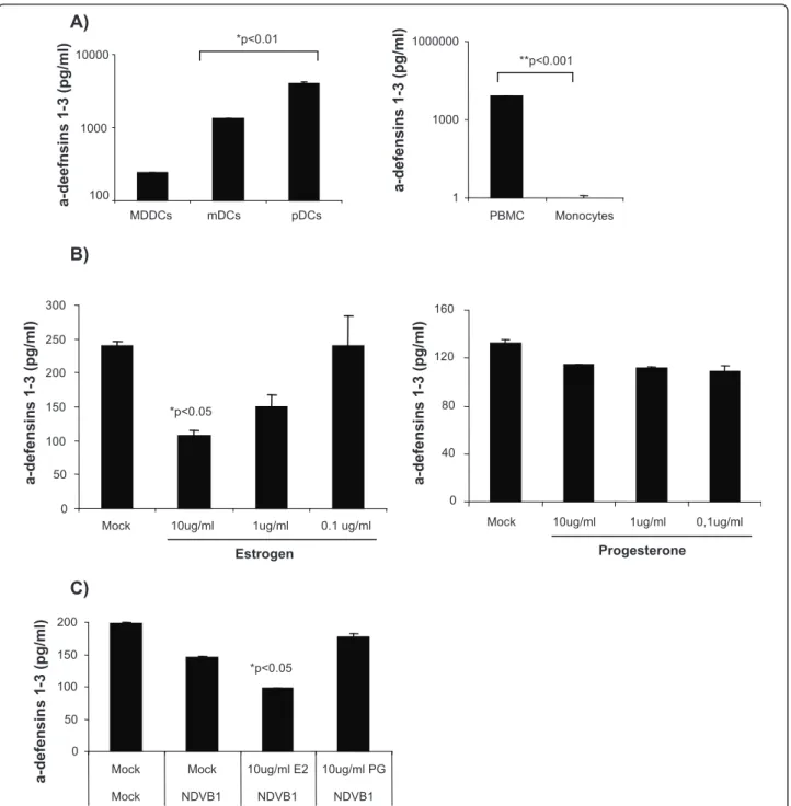
described to secrete a-defensins 1-3 constitutively [32] (Figure 1A, right panel). A comparison of the constitu-tive secretion ofa-defensins 1-3 from MDDCs, mDCs and pDCs by ELISA show that the levels ofa-defensins 1-3 detected in the supernatants from mDCs and pDCs
B)
A)
100 1000 10000
MDDCs mDCs pDCs
a-d
eefn
sin
s
1-3 (pg/ml)
*p<0.01
C)
Estrogen
*p<0.05
0 50 100 150 200 250 300
Mock 10ug/ml 1ug/ml 0.1 ug/ml
a-d
efen
sin
s 1-3
(pg/ml)
Progesterone
0 40 80 120 160
Mock 10ug/ml 1ug/ml 0,1ug/ml
a-d
efen
sin
s 1-3
(pg/ml)
**p<0.001
1 1000 1000000
PBMC Monocytes
a-d
efen
sin
s 1-3
(pg/ml)
*p<0.05
0 50 100 150 200
Mock Mock 10ug/ml E2 10ug/ml PG
Mock NDVB1 NDVB1 NDVB1
a-d
efen
sin
s 1-3
(pg/ml)
Figure 1MDDCs secretea-defensins 1-3: E2 and PG effect. A) ELISA quantification ofa-defensins 1-3 secreted by MDDCs, mDCs and pDCs (left panel) or PBMCs and monocytes (right panel) in the supernatant after 6 h of culture. Data are presented as mean plus or minus SD from 3
independent experiments.*P< 0.01 and *P < 0.001 versus MDDCs levels. Data are presented in a log scale. Levels ofa-defensins 1-3 measured
by ELISA in: (B) MDDCs treated for 24 h with 10, 1 and 0.1 ug/ml of E2 (left panel) or PG (right panel). (C) MDDCs pretreated for 24 h with E2 or
PG (10 ug/ml) before NDV-B1 infection for 10 h. Data are presented as mean plus or minus SD from 3 independent experiments.*P< 0.05versus
were 10 and 15-fold higher, respectively, than those detected in the supernatants from MDDCs (Figure 1A, left panel) (p < 0.01).
Estrogen but not Progesterone down-regulates a-defensins 1-3 secretion by DCs
Estrogen exerts an inhibitory effect on mDC and MDDC secretion of type I interferon (IFN) and some inflamma-tory cytokines [28]. Therefore we measured the effect of estrogen (E2) and progesterone (PG) ona-defensins 1-3 secretion by MDDCs, mDCs and pDCs. Treatment of MDDCs with E2 resulted in a dose-dependent reduction in a-defensin 1-3 secretion. Conversely, PG treatment had no effect on the secretion of a-defensins 1-3 in MDDCs (Figure 1B). Moreover, we also show that E2 but not PG still have a significant (*p < 0.05 vs mock treated DCs) inhibitory effect in the secretion of a-defensins 1-3 in viral infected MDDCs (Figure 1C), that correlates with our previous report describing the immunosuppressive effect of E2 during viral infection [28].
We also analyzed the effect of E2-treatment on the secretion of a-defensins 1-3 by mDCs and in accor-dance with the results observed for MDDCs, a signifi-cant reduction in the levels of a-defensins 1-3 was detected in cells treated with high concentrations of E2 but PG treatment did not have any significant effect (Figure 2A). In contrast, the a-defensins 1-3 secretion by pDCs was not affected by estrogen (Figure 2B). These results show that E2 suppressesa-defensins 1-3 secretion by mDCs and MDDCs but not in pDCs.
Effect of ICI 182,780 in the secretion ofa-defensins 1-3 by DCs
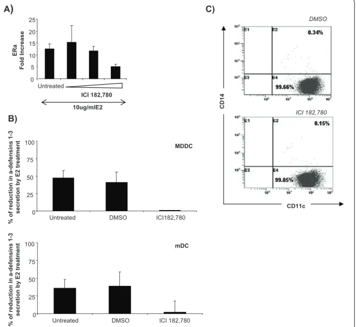
In order to determine if the reduction ofa-defensins 1-3 secretion was specific and mediated through the estro-gen receptor, DCs were treated with the estroestro-gen recep-tor (ER) antagonist ICI 182,780 at a concentration of 1000 nM, 100 nM and 10 nM for 3 days prior to the E2 treatment. Since E2 upregulates ER expression, ERa transcription in the presence of E2 is a useful measure of the effectiveness of ICI 182,780. At the highest con-centration of ICI 182,780 in the presence of 10 ug/ml E2 a significant reduction of 2.5 -folds in ERa expres-sion was observed (Figure 3A). This concentration of the drug was able to reverse the inhibition of E2 on a-defensins 1-3 secretion by both types of DCs (Figure 3B). However, low concentrations for Faslodex (100 and 10 nM) do not significantly change (1.24 and 0.92-folds, respectively) ERaexpression in relation with the levels detected in untreated MDDCs. Moreover, 1000 nM ICI 182,780 did not affect the differentiation process of the MDDCs measured by CD11c and CD14 expres-sion at day 5 of culture (Figure 3C). These results
confirmed that the effect of E2 on a-defensins 1-3 secretions was mediated through estrogen receptor sig-naling and that a novel E2 regulated mechanism pre-ventsa-defensins 1-3 secretion in DCs.
Secretion of a-defensins 1-3 by DCs during pregnancy
Since estrogen levels rise significantly in women from the first to the third trimester of pregnancy we speculated that if estrogen was exerting regulatory control over a-defensins 1-3 secretion, then the mDCs collected late in pregnancy should produce less of these anti-microbial peptides. A cohort of women from an ongoing study of immunity during pregnancy being performed at Mount Sinai School of Medicine (VIP study) were chosen based upon their significant difference in serum estrogen levels between the first and third trimester (Figure 4A). mDCs were isolated from these subjects and the secretion of a-defensins 1-3 was measured by capture ELISA after 12 h in culture. However, patient variation was great and no statistical difference between the groups could be demon-strated.a-defensins 1-3 release by DCs isolated from 40% of the patients showed at least a two-fold reduction between the 1stand 3rdtrimester as estrogen levels rose. However, many (50%) showed no significant change and two patients showed a more than a two fold rise in secre-tion (10%) (Figure 4B).
Discussion
During normal pregnancy, the maternal immune system remains systemically immunocompetent [33,34]. How-ever, it is faced with a dilemma that it must continue to protect the mother against infection while inhibiting immune activation against the semiallogeneic fetus. There is clear evidence that in the later stages of preg-nancy women are uniquely susceptible to microbial infection. The prevailing view, though not shared by all, is that the predominant immune response is shifted from a gamma interferon based Th1 response to a less inflammatory Th2 response. The period of enhanced sensitivity to infection corresponds to the extremely high concentrations of E2 and PG that are reached in the later stages of pregnancy [16,17]. Both E2 and PG have been shown to exert a powerful effect on various aspects of the innate and adaptive immune response [13,14]. In a previous publication we investigated the effect of these hormones on the activation of DCs cul-tured from monocytes as well as those isolated directly from the blood. PG did not exert any effect on DCs in either the steady state or after activation by viruses. In contrast, E2 was a potent inhibitor of DC activation trig-gered by a number of human viruses but not by toll-like receptors ligands (TLRs) [28].
range of microorganisms and by participating in adap-tive immunity by attracting monocytes, T cells, or immature DCs [35,36]. Moreover,a-defensins 1-3 have been detected throughout the female reproductive tract (FRT) in non-pregnant women [5,6] as well as in the amnion, chorion, placenta, amniotic fluids and cervical mucus plug from pregnant women [7-9]. Previous reports by us and others demonstrate the expression of a-defensins 1-3 by immune cells populations including MDDCs [4], NK cell, B cells, gδTcells, mast cells, neu-trophils [2]. Most human DC studies are performed using MDDCs produced by culturing these cells in the presence of the cytokines GM-CSF and IL4. It is not known whether cells produced this way simulate in vivo DC populations. However, mDCs as well as pDCs can be isolated directly from human blood by the expression of the CD1c marker (mDC) or BCDA4 (pDC) on their surface [37]. In this report we analyzed directly isolated mDCs and pDCs and show that they both secrete signif-icantly more a-defensins 1-3 than MDDCs. These
results suggest that culturing of DCs may reduce their a-defensin 1-3 secretion and that previous work may have underestimated the contribution of DCs to in vivo a-defensin 1-3 production [38].
17-b-estradiol (E2), one of the most important forms of estrogen, acts through binding to two known hor-mone receptors, estrogen receptors ERaand ERb [25], which are expressed in specific tissues as well as in immune cells, including macrophages, CD8+ T cells, CD4+ T cells, B cells, monocytes, and DCs [39]. Cells pre-treated with ICI 182, 780, a specific ER antagonist, became insensitive to the inhibitory effect of E2, demon-strating a direct and specific effect of E2 on the release ofa-defensins 1-3 by DCs. However, intrinsic or subse-quently acquired resistance to the therapy remains a major obstacle in treatment with this compound as has been describe by Zhao et al., using a cell line of breast cancer [40]. These open new questions that should be addressed in further investigations with dendritic cells. In our system, ICI 182,780 reduces the expression of the
B)
0 1000 2000 3000 4000 5000
Mock 10ug/ml
a-d
efen
sin
s 1-3
(pg/ml))
Estrogen
A)
Estrogen *p<0.05
0 500 1000 1500 2000
Mock 10ug/ml 1ug/ml 0.1 ug/ml
a-d
efen
sin
s 1-3
(pg/ml)
Progesterone 0
500 1000 1500 2000
Mock 10ug/ml 1ug/ml 0.1 ug/ml
a-d
efen
sin
s 1-3
(pg/ml)
Figure 2mDCs and pDCs secretea-defensins 1-3: E2 and PG effect. Levels ofa-defensins 1-3 measured by ELISA in: (A) mDCs treated for 24 h with 10, 1 and 0.1 ug/ml of E2 (left panel) or PG (right panel). (B) pDCs treated with 10 ug/ml of E2 for 8 h. Data are presented as mean
ER, as a result of the induction of a proteasome-depen-dent degradation induced by this compound [41]. How-ever, ICI 182,780 treatment does not affect the differentiation of monocytes into MDDCs as measured by percentage of CD11c+CD14-cells.
E2 but not PG suppressed thea-defensins 1-3 pro-duction from the mDCs at high concentration but had no effect on pDCa-defensins 1-3 secretion. This result
is consistent with our earlier observation that E2 did not inhibit activation or cytokine secretion from pDCs. A possible explanation for these observations comes from the work of Hartman et al who demonstrated that murine pDCs derive exclusively from estrogen-resistant myeloid progenitors [42] which may explain the failure of E2 to altera-defensin 1-3 production by these cells.
0 5 10 15 20 25
Untreated
ICI 182,780
10ug/mlE2
ERa
F
o
ld Inc
rease
A
)
B)
CD1
4
CD11c DMSO
ICI 182,780
0 25 50 75 100
Untreated DMSO ICI182,780
%
o
f
re
d
u
ct
io
n
in
a
-de
fe
ns
ins
1
-3
s
e
cr
eti
on b
y E
2
tre
at
m
ent MDDC
0 25 50 75 100
Untreated DMSO ICI 182,780
%
o
f
re
d
u
ct
io
n
in
a
-de
fe
ns
ins
1
-3
s
e
cr
eti
on b
y E
2
tre
at
m
ent mDC
C)
Based upon our in vitro studies we expected that mDCs collected from the blood in the later part of preg-nancy would show a reduction ina-defensins 1-3 pro-duction. Moreover, evidence suggests that elevated levels of a-defensins 1-3 in serum correlated with adverse pregnancy outcomes [43,44]. While we observed a trend towards less a-defensins 1-3 production from DCs collected in the 3rd trimester relative to those col-lected from the same patient in the 1sttrimester, the dif-ference was not statistically significant. Almost 40% of the patients did show a very substantial drop in a-defensins 1-3 release from their mDCs at third
trimester, while the remaining patients secreted a-defensins 1-3 at nearly identical levels to what was observed in the 1st trimester. It is worth noting, how-ever, that the levels of estrogen detected in serum were lower than the dose that most effectively inhibited a-defensin 1-3 secretion in vitro.
Among the 50% failed to show a reduction, the consis-tency of the mDC a-defensin 1-3 secretion within patients and variation between patients (ranging from 10 ng/ml to 60 ng/ml) was significant. This high inter-donor variability has been well described and likely cor-responds to variations in the copy number of the genes
A)
B)
0 10000 20000 30000 40000 50000 60000 70000 80000
10004100 31
10248100 20
101 94
100 58
100 12
100 10
100 05
100 27
10013100 14
100 19
10030101 99
100 29
102 07
102 47
a-d
ef
en
si
n
s 1
-3
(pg
/ml
)
First trimester Third trimester
that encode for a-defensins 1-3 between individuals [45].
The sum of thea-defensin 1-3 production by mDCs suggested a net loss in the later stages of pregnancy. If other cells that produce a-defensins 1-3 were likewise affected, seruma-defensins1-3 during pregnancy would be expected to drop.
In this report we document that myeloid and plasma-cytoid DCs directly isolated from the blood produce substantial amounts of a-defensins 1-3 constitutively. pDCs are not affected, but both mDCs and MDDCs produce less a-defensins 1-3 in the presence of high levels of E2. Alpha-defensins 1-3 release from mDCs collected in the third trimester of pregnancy either fell or stayed the same in the majority of the patients.
Conclusions
Our results demonstrate a specific effect for E2 on the secretion of a-defensins 1-3 by dendritic cells. Moreover, we also observed this effect in dendritic cells isolated from pregnant women at third trimester, when the levels of E2 reach extremely high concentrations. Together, this data provide new insights that contribute to explain immune changes during pregnancy.
List of abbreviations
DC: Dendritic cells; FRT: Female reproductive track; NK: Natural killer; E2:
Estrogen (17-β-estradiol); PG: Progesterone; HD5: alpha-defensin HD5;
HBD1-4: Human beta defensins 1-4; MDDC: Monocyte derived dendritic cells; mDCs: myeloid dendritic cells; pDCs: plasmacytoid dendritic cells; IFN:
Interferon; ERα: Estrogen receptor alpha; ERβ: Estrogen receptor beta; PBMC:
Peripheral blood mononuclear cells.
Acknowledgements
MME, RS, SE, SW are supported by NIH contract NO1 - AI 50028. TG is mainly supported mainly by Research Grants FIS2003-1200 and FIS2006-1259 from the Spanish Ministry of Health and MRG is the recipient of a research
award‘’Emili Letang’’(Hospital Clinic, Barcelona).
Author details
1
Department of Microbiology, Mount Sinai School of Medicine, New York,
NY, USA.2Immunology Institute, Mount Sinai School of Medicine, New York,
NY, USA.3Department of Molecular Microbiology and Infections Biology, CIB,
CSIC, Madrid, Spain.4Service of Immunology, Hospital Clinic Universitari de
Barcelona, Barcelona, Spain.5Department of Physiology, Dartmouth Medical
School, Lebanon, New Hampshire, USA.6Department of Obstetrics,
Gynecology and Reproductive Sciences, Mount Sinai School, of Medicine,
New York, NY,USA.7Department of Preventive Medicine, Mount Sinai School
of Medicine, New York, NY, USA.
Authors’contributions
MME designed and performed research, analyzed data, and wrote the manuscript; MGR performed research and wrote the manuscript; SE performed statistical analysis; RS wrote the manuscript; TG designed research and wrote the manuscript; and TMM designed research and wrote the manuscript. All authors read and approved the final manuscript
Competing interests
The authors declare that they have no competing interests.
Received: 16 May 2011 Accepted: 23 August 2011 Published: 23 August 2011
References
1. Yang D, Biragyn A, Hoover DM, Lubkowski J, Oppenheim JJ:Multiple roles
of antimicrobial defensins, cathelicidins, and eosinophil-derived neurotoxin in host defense.Annu Rev Immunol2004,22:181-215.
2. Klotman ME, Chang TL:Defensins in innate antiviral immunity.Nat Rev
Immunol2006,6(6):447-456.
3. Shi J, Aono S, Lu W, Ouellette AJ, Hu X, Ji Y, Wang L, Lenz S, van Ginkel FW,
Liles M, Dykstra C, Morrison EE, Elson CO:A novel role for defensins in
intestinal homeostasis: regulation of IL-1beta secretion.J Immunol2007,
179(2):1245-1253.
4. Rodriguez-Garcia M, Oliva H, Climent N, Garcia F, Gatell JM, Gallart T:
Human immature monocyte-derived dendritic cells produce and secrete alpha-defensins 1-3.J Leukoc Biol2007,82(5):1143-1146.
5. King AE, Critchley HO, Kelly RW:Innate immune defences in the human
endometrium.Reprod Biol Endocrinol2003,1:116.
6. King AE, Fleming DC, Critchley HO, Kelly RW:Differential expression of the
natural antimicrobials, beta-defensins 3 and 4, in human endometrium.
J Reprod Immunol2003,59(1):1-16.
7. King AE, Kelly RW, Sallenave JM, Bocking AD, Challis JR:Innate immune
defences in the human uterus during pregnancy.Placenta2007,
28(11-12):1099-1106.
8. Cole AM:Innate host defense of human vaginal and cervical mucosae.
Curr Top Microbiol Immunol2006,306:199-230.
9. Quayle AJ:The innate and early immune response to pathogen
challenge in the female genital tract and the pivotal role of epithelial cells.J Reprod Immunol2002,57(1-2):61-79.
10. King AE, Paltoo A, Kelly RW, Sallenave JM, Bocking AD, Challis JR:
Expression of natural antimicrobials by human placenta and fetal membranes.Placenta2007,28(2-3):161-169.
11. van Duivenvoorde LM, van Mierlo GJ, Boonman ZF, Toes RE:Dendritic
cells: vehicles for tolerance induction and prevention of autoimmune diseases.Immunobiology2006,211(6-8):627-632.
12. Granucci F, Zanoni I, Feau S, Capuano G, Ricciardi-Castagnoli P:The
regulatory role of dendritic cells in the immune response.Int Arch Allergy Immunol2004,134(3):179-185.
13. Orsi NM, Tribe RM:Cytokine networks and the regulation of uterine
function in pregnancy and parturition.J Neuroendocrinol2008,
20(4):462-469.
14. Beagley KW, Gockel CM:Regulation of innate and adaptive immunity by
the female sex hormones oestradiol and progesterone.FEMS Immunol Med Microbiol2003,38(1):13-22.
15. Arruvito L, Sanz M, Banham AH, Fainboim L:Expansion of CD4+CD25+and
FOXP3+ regulatory T cells during the follicular phase of the menstrual cycle: implications for human reproduction.J Immunol2007,
178(4):2572-2578.
16. Bukovsky A, Cekanova M, Caudle MR, Wimalasena J, Foster JS, Henley DC,
Elder RF:Expression and localization of estrogen receptor-alpha protein
in normal and abnormal term placentae and stimulation of trophoblast differentiation by estradiol.Reprod Biol Endocrinol2003,1:13.
17. Edelstam G, Karlsson C, Westgren M, Lowbeer C, Swahn ML:Human
chorionic gonadatropin (hCG) during third trimester pregnancy.Scand J Clin Lab Invest2007,67(5):519-525.
18. Quayle AJ, Porter EM, Nussbaum AA, Wang YM, Brabec C, Yip KP, Mok SC:
Gene expression, immunolocalization, and secretion of human defensin-5 in human female reproductive tract.Am J Pathol1998,
152(5):1247-1258.
19. Schaefer TM, Wright JA, Pioli PA, Wira CR:IL-1beta-mediated
proinflammatory responses are inhibited by estradiol via down-regulation of IL-1 receptor type I in uterine epithelial cells.J Immunol
2005,175(10):6509-6516.
20. Daynes RA, Araneo BA, Hennebold J, Enioutina E, Mu HH:Steroids as
regulators of the mammalian immune response.J Invest Dermatol1995,
105(1 Suppl):14S-19S.
21. Khardori R, Adamski A, Khardori N:Infection, immunity, and hormones/
endocrine interactions.Infect Dis Clin North Am2007,21(3):601-615, vii.
22. Lang TJ:Estrogen as an immunomodulator.Clin Immunol2004,
113(3):224-230.
23. Pettersson A, Ciumas C, Chirsky V, Link H, Huang YM, Xiao BG:Dendritic
cells exposed to estrogen in vitro exhibit therapeutic effects in ongoing experimental allergic encephalomyelitis.J Neuroimmunol2004,
24. Carmona F, Font J, Cervera R, Munoz F, Cararach V, Balasch J:Obstetrical outcome of pregnancy in patients with systemic Lupus erythematosus. A study of 60 cases.Eur J Obstet Gynecol Reprod Biol1999,83(2):137-142.
25. Nicholson RI, Johnston SR:Endocrine therapy–current benefits and
limitations.Breast Cancer Res Treat2005,93(Suppl 1):S3-10.
26. Olsen NJ, Kovacs WJ:Gonadal steroids and immunity.Endocr Rev1996,
17(4):369-384.
27. Cutolo M, Sulli A, Seriolo B, Accardo S, Masi AT:Estrogens, the immune
response and autoimmunity.Clin Exp Rheumatol1995,13(2):217-226.
28. Escribese MM, Kraus T, Rhee E, Fernandez-Sesma A, Lopez CB, Moran TM:
Estrogen inhibits dendritic cell maturation to RNA viruses.Blood2008,
112(12):4574-84.
29. Hall JM, Couse JF, Korach KS:The multifaceted mechanisms of estradiol
and estrogen receptor signaling.J Biol Chem2001,276(40):36869-36872.
30. Fernandez-Sesma A, Marukian S, Ebersole BJ, Kaminski D, Park MS, Yuen T,
Sealfon SC, Garcia-Sastre A, Moran TM:Influenza virus evades innate and
adaptive immunity via the NS1 protein.J Virol2006,80(13):6295-6304.
31. Gonzalez-Maeso J, Yuen T, Ebersole BJ, Wurmbach E, Lira A, Zhou M,
Weisstaub N, Hen R, Gingrich JA, Sealfon SC:Transcriptome fingerprints
distinguish hallucinogenic and nonhallucinogenic 5-hydroxytryptamine 2A receptor agonist effects in mouse somatosensory cortex.J Neurosci
2003,23(26):8836-8843.
32. Yang D, Chen Q, Chertov O, Oppenheim JJ:Human neutrophil defensins
selectively chemoattract naive T and immature dendritic cells.J Leukoc
Biol2000,68(1):9-14.
33. Jilani N, Das BC, Husain SA, Baweja UK, Chattopadhya D, Gupta RK,
Sardana S, Kar P:Hepatitis E virus infection and fulminant hepatic failure
during pregnancy.Journal of gastroenterology and hepatology2007,
22(5):676-682.
34. Mullooly JP, Barker WH, Nolan TF Jr:Risk of acute respiratory disease
among pregnant women during influenza A epidemics.Public Health Rep
1986,101(2):205-211.
35. Rodríguez-García MOH, Climent N, Escribese MM, García F, Moran TM,
Gatell JM, Gallart T:Impact of alpha-defensins1-3 on the maturation and
differentiation of human monocyte-derived DCs. Concentration-dependent opposite dual effects.Clin Immunol2009,131(3):374-384.
36. Chang TL, Vargas J, DelPortillo A, Klotman ME:Dual role of
alpha-defensin-1 in anti-HIV-alpha-defensin-1 innate immunity.J Clin Invest2005,115(3):765-773.
37. Osugi Y, Vuckovic S, Hart DN:Myeloid blood CD11c(+) dendritic cells and
monocyte-derived dendritic cells differ in their ability to stimulate T lymphocytes.Blood2002,100(8):2858-2866.
38. Rodriguez-Garcia M, Climent N, Oliva H, Casanova V, Franco R, Leon A,
Gatell JM, Garcia F, Gallart T:Increased alpha-defensins 1-3 production by
dendritic cells in HIV-infected individuals is associated with slower disease progression.PloS one5(2):e9436.
39. Cvoro A, Tatomer D, Tee MK, Zogovic T, Harris HA, Leitman DC:Selective
estrogen receptor-beta agonists repress transcription of proinflammatory genes.J Immunol2008,180(1):630-636.
40. Zhao H, Lo YH, Yu L, Wang SC:Overcoming resistance to fulvestrant
(ICI182,780) by downregulating the c-ABL proto-oncogene in breast cancer.Molecular carcinogenesis2011,50(5):383-389.
41. Lanvin O, Bianco S, Kersual N, Chalbos D, Vanacker JM:Potentiation of
ICI182,780 (Fulvestrant)-induced estrogen receptor-alpha degradation by the estrogen receptor-related receptor-alpha inverse agonist XCT790.
J Biol Chem2007,282(39):28328-28334.
42. Harman BC, Miller JP, Nikbakht N, Gerstein R, Allman D:Mouse
plasmacytoid dendritic cells derive exclusively from estrogen-resistant myeloid progenitors.Blood2006,108(3):878-885.
43. Okazaki T, Ota Y, Yuki N, Hayashida A, Shoda A, Nishikawa M, Oshima K,
Fukasawa I, Watanabe H, Inaba N:Plasma levels of alpha-defensins 1-3
are an indicator of neutrophil activation in pregnant and post-partum women.J Obstet Gynaecol Res2007,33(5):645-650.
44. Prieto JA, Panyutich AV, Heine RP:Neutrophil activation in preeclampsia.
Are defensins and lactoferrin elevated in preeclamptic patients?J Reprod
Med1997,42(1):29-32.
45. Aldred PM, Hollox EJ, Armour JA:Copy number polymorphism and
expression level variation of the human alpha-defensin genes DEFA1 and DEFA3.Human molecular genetics2005,14(14):2045-2052.
doi:10.1186/1477-7827-9-118
Cite this article as:Escribeseet al.:Alpha-defensins 1-3 release by dendritic cells is reduced by estrogen.Reproductive Biology and Endocrinology20119:118.
Submit your next manuscript to BioMed Central and take full advantage of:
• Convenient online submission
• Thorough peer review
• No space constraints or color figure charges
• Immediate publication on acceptance
• Inclusion in PubMed, CAS, Scopus and Google Scholar
• Research which is freely available for redistribution