Abstract
Blood clots affect over half a million Americans each year and have a high mortality rate. Current anticoagulants have been historically successful, but can
dangerously increase bleeding risk. Using chromogenic substrate assays, I tested almost 1,000 compounds from the Tidwell library of cationic compounds and analyzed their
ability to inhibit five specific coagulation proteases that are involved in blood clot formation. A small number of promising compounds inhibited only one coagulation enzyme. Another member of my lab performed Prothrombin Time (PT) and Activated
Partial Thromboplastin Time (APTT) assays for each of these compounds. Further analysis showed that the compounds that were deemed successful in the PT and APTT
had very little overlap with those that successfully inhibited only one coagulation enzyme. These results suggest that current clinical assay methods may not be ideal indicators of thrombotic state. These studies provide useful insight into how to assess
Introduction
Venous thromboembolisms (VTE), specifically deep vein thromboses (DVT) and pulmonary embolisms (PE), pose a significant health risk in America. According to the
American Heart Association, over 250,000 Americans are hospitalized annually due to thrombotic events and some studies estimate up to 600,000 Americans suffer a VTE each year [1, 2]. A thrombosis occurs when the coagulation system is inappropriately activated
inside a blood vessel, forming a blood clot that obstructs blood flow. Thrombotic events have high morbidity and mortality rates. About twenty percent of DVTs and pulmonary
embolisms are immediately fatal and approximately thirty percent of people diagnosed will die within thirty days [1].
Even though there is such a high rate of incidence and death, there are very few
anticoagulant drugs currently available to treat vascular thrombosis. The two most common anticoagulant drugs, heparin and warfarin, have significant drawbacks. Heparin
has been used clinically for over 70 years and is still considered a high-risk
pharmaceutical [3]. It is useful due to its immediate effects, but it requires parenteral administration and can cause osteoporosis with long-term use [4]. Warfarin can be
administered orally and is given long-term in low doses to reduce clotting risk. Unfortunately, warfarin has a narrow therapeutic window and overdosing can lead to
dangerous hemorrhage. Fatal bleeds are seen in 1% to 3% of patients [5]. Still, heparin and warfarin are often prescribed together in a dual system to break up a clot and reduce immediate clot risk. Recently, the FDA has approved several new, direct-acting
need in the population. Developing small, targeted molecules that can interact with coagulation proteases could offer better specificity and selectivity than seen in warfarin
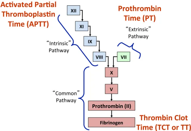
or heparin and reduce negative side effects. Below is a schematic showing the intricacies of coagulation, which adds to the difficulty of creating new anticoagulants (Figure 1.)
Figure 1. The details of coagulation, showing the intrinsic, extrinsic, and common pathways.
In 1964, the process of coagulation was first described as a “cascade of
proenzyme-enzyme transformations” until the final product of fibrin mesh was reached [6].
Figure 2. Division of coagulation into the cascade model. Each Roman numeral represents a serine protease or cofactor that is activated during coagulation and in turn
activates the following protease until fibrin is produced.
Two of the most common clinical assays used to assess thrombotic state are Prothrombin Time (PT) and activated Partial Thromboplastin Time (aPTT). PT measures
the amount of time it takes for the “extrinsic” pathway to be activated and produce fibrin. aPTT measures the time for “intrinsic” pathway activation. Unfractionated heparin
extends aPTT, but each laboratory must determine the therapeutic time range based on their own aPTT system [7]. Warfarin extends Prothrombin Time. Some new
anticoagulants, specifically rivaroxaban and dabigatran, do not require routine
or necessary. aPTT gives an approximation of dabigatran activity, but is not linear over clinically used concentrations. Instead, diluted thrombin time (dTT) is a more sensitive
assay to the effects dabigatran [10]. In short, most major anticoagulants are sensitive to different assays, which make assessing thrombotic state difficult in a clinical setting.
This research was conducted as part of an American Heart Association grant originally designed to identify and evaluate novel, small molecules as therapeutic anticoagulants to decrease harm and death due to thrombotic events. The grant focused
on analyzing the Tidwell Library of compounds, synthesized by Dr. Richard Tidwell, a medicinal chemist in the Department of Pathology and Laboratory Medicine. Previously,
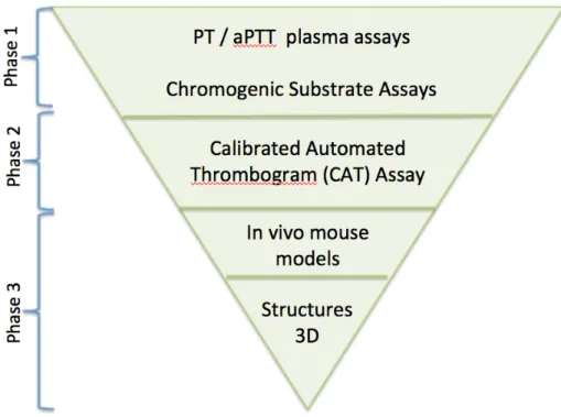
the compounds were screened as anti-viral agents and anti-protozoan agents, among other things. Approximately 1500 compounds were made available to our lab to be screened as anticoagulant agents. We developed a tiered system to assess antithrombotic properties,
Figure 3. Our tiered plan to analyze the Tidwell library of cationic compounds. While we are moving into Phase 2 and still analyzing the Tidwell compounds, our current data has provided insight into our methods and the current clinical assays used to assess thrombotic state.
Materials and Methods
Tidwell Library
The Tidwell lab generously provided us with 5 mg samples of their library of
cationic compounds; approximately 50 compounds were received weekly for the duration of the project. To date we have received 1400 compounds. Each compound was taken up
Compounds that did not dissolve completely 500 µL of DMSO were incubated at 37°C for 15 minutes. If still insoluble, the compounds were incubated at 60°C for 15
minutes. If incubation at 60°C was ineffective, an additional 500 µL of DMSO was added to lower the concentration to 5 mg/mL. Incubation was repeated if necessary. If still not
dissolved, 1000 µL of DMSO were added to lower the concentration to 2.5 mg/mL and the compound was incubated as necessary. If still insoluble, the compound was declared insoluble for our purposes and stored. To date, there are 138 insoluble compounds.
Prothrombin Time (PT)
Prothrombin Time assays were run by Jasmine Dennis.
The Neoplastine-Cl Plus reagent was pre-warmed at 37oC for 30 minutes. Plasma (VisuCon-F pooled plasma, Affinity Biologicals) and Tidwell compounds were warmed
in a 37oC dry bath for 10-15 minutes until thawed. Cuvette strips with one magnetic steel ball in each (Diagnostica Stago) were warmed for 3 minutes. Tidwell compounds were
diluted in plasma to 100 µg/mL. Controls of plasma and plasma with 1% DMSO were made. This assay was run in duplicate on a Diagnostica Stago STart 4 Hemostasis Analyzer, which measured the amount of time it took for the plasma to clot after the
reagent was added.
Each cuvette strip (containing 50 µL of plasma-diluted inhibitor) was placed in an
incubation well for 60 seconds. After that, the cuvette was moved to the recording well. Simultaneously, 100 µL of Neoplastine-Cl Plus reagent was added and recording began. For values that exceeded 20 seconds, compounds were diluted 1:2 with plasma and tested
Activated Partial Thromboplastin Time (APTT)
Activated Partial Thromboplastin Time assays were run by Jasmine Dennis.
This assay was run in duplicate on a Diagnostica Stago STart 4 Hemostasis Analyzer, which measured the amount of time it took for the plasma to clot. The PTT-A
reagent was pre-warmed at 25oC and CaCl2 (25 mM, pH 7.4) at 37oC for 20 minutes.
Plasma (VisuCon-F pooled plasma) and Tidwell compounds were warmed in a 37oC dry bath for 10-15 minutes until thawed. Cuvette strips with one magnetic steel ball in each
(Diagnostica Stago) were warmed for 3 minutes. Tidwell compounds were diluted in plasma to 100 µg/mL. Controls of plasma and plasma with 1% DMSO were made.
Each cuvette strip (containing 50 µL of plasma-diluted inhibitor and 50 µL of PTT-A reagent) was placed in an incubation well for 180 seconds. Then, the cuvette was moved to the recording well where 50 µL of CaCl2 was added and recording began
simultaneously. For compounds that exceeded 120 seconds, compounds were diluted 1:2 with plasma and the assay was repeated until the value was below 50 seconds.
Chromogenic Substrate Assay
The assay was performed in 96-well U-bottomed assay plates. The buffer used
was HEPES 20 mM, NaCl 150 mM, polyethylene glycol 0.1%, and CaCl2 5 mM pH 7.4.
The buffer base was prepared in advance and calcium was added daily to the needed
amount. Purified, activated coagulation enzymes (thrombin, FIXa, FXIa, FXa, and FVIIa) were stored at their stock solutions in glycerol in the -20°C freezer to protect from
degradation. They were added to buffer at appropriate individual concentrations. Due to
were used (Appendix 2). All substrates had a final concentration of 200 µM. The Tidwell inhibitors were added to the buffer at a final concentration of 25 µg/mL.
The substrate’s cleavage (reflecting enzyme activity and therefore inhibitor strength) was measured by reading the absorbance at 405 nm for 20 minutes using a
ThermoMax Microplate Reader (Molecular Devices). Inhibitor potency was expressed as a percentage by comparing each inhibitor well to a control well.
Results and Discussion
PT and APTT
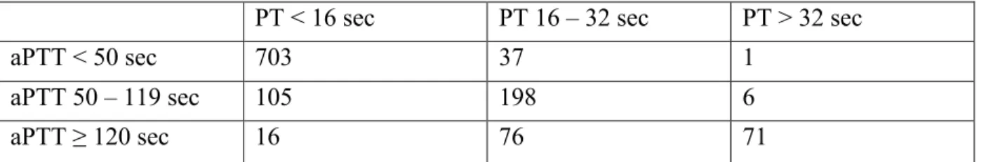
In total, 1213 Tidwell compounds were assayed using PT and aPTT. The
compounds were categorized according assay times (Table 1). These times were chosen to reflect standard, elevated, and dangerously prolonged values of PT and aPTT. The
majority of the compounds did not prolong PT or aPTT significantly.
PT < 16 sec PT 16 – 32 sec PT > 32 sec
aPTT < 50 sec 703 37 1
aPTT 50 – 119 sec 105 198 6
aPTT ≥ 120 sec 16 76 71
Table 1. Tidwell Compound results based on PT and APTT data. 71 compounds had very prolonged PT and aPTT times. The majority of the compounds (703) did not prolong
either assay.
Chromogenic Substrate Assays
Originally, our lab was going to use PT and aPTT data to choose a select number
of compounds to further analyze using chromogenic substrate assays. Because our PT and aPTT results fell in the non-prolonged range for both assays, we decided to analyze
been analyzed using Chromogenic Substrate Assays. A compound was labeled an inhibitor if it decreased enzyme activity by 70% compared to control. Based on those
criteria, 566 compounds showed no significant inhibition of any coagulation protease. We did find 90 compounds that inhibited three or more coagulation proteases and dubbed
them “pan-inhibitors”. All of these compounds inhibited thrombin, FXa, and at least one other enzyme. Sixty-six compounds were dual enzyme inhibitors. As shown in Table 2, all but three of the dual-enzyme inhibitors inhibit thrombin or factor Xa. We are unsure
of an explanation for this trend involving thrombin and FXa, but it may be a future area of inquiry.
FIXa FXIa FXa FVIIa
Thrombin 2 7 39 2
FIXa 0 2 3
FXIa 10 0
FXa 1
Table 2. Numbers of dual-enzyme inhibitors. It is interesting to note that there are 66 dual-enzyme inhibitors. All but three inhibited either thrombin or FXa. The chromogenic substrate assays identified 76 single-enzyme inhibitors (Table
3) .The majority of these inhibitors were thrombin inhibitors. These inhibitors are of particular interest to our lab due to their targeted nature, especially FXIa. Factor XI deficiency causes Hemophilia C, a form of hemophilia that has a lower and milder
Thrombin FIXa FXIa FXa FVIIa
39 8 10 19 8
Table 3. Numbers of single-enzyme inhibitors. These inhibitors are of particular interest to our lab. Other studies have shown that single enzyme inhibitors can reduce thrombotic
risk without increasing bleeding tendency.
Comparison of PT/aPTT results and Chromogenic Substrate Assays
We have analyzed fewer compounds using chromogenic substrate assays than
PT/aPTT, but the sample size is large enough that we expect the current data will reflect the overall trends of the Tidwell compounds. Figure 4 compares the PT and aPTT data
Figure 4. A graphic representation of PT and aPTT data combined with chromogenic substrate assay data. The blue circles represent single enzyme inhibitors, scaled by size. The numbers to the left of the circle note how many single enzyme inhibitors fall in each PT/aPTT time category. The purple circles represent multi-enzyme inhibitors (3 or more proteases inhibited). The number to the right of the circle notes how many are in each
category.
We expected the compounds that fell in the highly prolonged range (PT > 32 sec, aPTT ≥ 120 sec) to be very potent single enzyme inhibitors. Instead, our data show the 71
compounds in that range are primarily multi-enzyme inhibitors. Most of the single enzyme inhibitors, regardless of potency, fell in the non-prolonged range (PT >16
inhibition of coagulation proteases. In a previous study done in this lab by Chantelle Rein-Smith, multi-enzyme inhibitors were successful anticoagulants, but also
dramatically and fatally decrease blood pressure in mouse injury models. Therefore, prolonged PT and aPTT are not good indications of anticoagulant potential based on our
standards.
Analysis of Current Anticoagulants
As acknowledged before, there are serious drawbacks to historic anticoagulants, namely heparin and warfarin. There has been a shift in anticoagulant research to very
targeted compounds that inhibit specific serine proteases or cofactors; targeted compounds have had success in reducing thrombotic risk without increasing bleeding tendency. Unfortunately, many of these compounds currently on the market cannot be
assayed using PT or aPTT.
Dabigatran etexilate mesylate (marketed as Pradaxa) is a relatively new, oral,
direct thrombin inhibitor. Compared to warfarin, it has a lower risk of clot-related strokes, brain bleeds, and death [12]. And, unlike warfarin, dabigatran does not require frequent monitoring [13]. Dabigatran has very little effect on PT at clinical
concentrations. aPTT is only suitable to provide a qualitative analysis of dabigatran in patient plasma; the aPTT curve flattens out at high clinical concentrations, so aPTT is not
appropriate to assess the quantitative effect of dabigatran [14].
A study of another targeted anticoagulant, apixaban, showed that only one PT reagent was sensitive enough to detect apixaban levels across clinical concentrations.
It should be noted that not all new, targeted anticoagulants fail to prolong PT or aPTT assays. For example, rivaroxaban is a targeted factor Xa inhibitor and prolongs PT
and aPTT [16].
Our results agree with current research that shows PT and aPTT are not enough to
assess thrombotic state in patients using novel targeted anticoagulants. These common clinical assays are not obsolete, but they can no longer fully evaluate patient status with such a wide range of anticoagulants available.
Conclusion
Our lab is continuing with analysis of Tidwell compounds to identify future
anticoagulants. Many compounds have shown potential in the initial assays. Our next step is to analyze the effects of the inhibitors on overall thrombin generation using a
Calibrated Automated Thrombogram (CAT) assay. With the most successful compounds from our in vitro experiments, we hope to move into mouse models. Our strategy is to use a mouse model of a saphenous vein ferric chloride injury to test the in vivo anticoagulant
properties of our compounds.
Overall, the initial assays were very telling about the way we approach
identification of anticoagulants. It has previously been shown that currently marketed novel targeted anticoagulants can have altered results in clotting assays, including PT and aPTT [17]. Our data are consistent with these observations and provide one explanation
as to why this may occur. Individual factor inhibition demonstrated prolongation of neither PT nor aPTT. The use of these assays is better suited for analysis of multifactor
substrate assays were much more useful to identify successful targeted compounds, but currently these assays are not common in clinical settings. Based on our data,
Acknowledgements
I would like to thank all the people who made my research experience possible
and helped me along the path to writing an honor thesis: the Office of Undergraduate Research; the UNC Biology department; my Biology sponsor, Dr. William Marzluff; and, above all, my PI and advisor, Dr. Frank Church.
Frank, thank you for your support in all that I do. You encourage me daily and
help me realize that I can take on the world.
Mac, thank you for your guidance and your sarcasm. You pushed me to think
outside the box and I cannot thank you enough for answering all my questions along the way.
Jasmine, thank you for all the hard work you invested in this project. This thesis
would only be half of what it is without you.
Savannah, Liz, Matt, Mark, Peter, Laura, Grace, thank you for making the lab
a place I loved, a place I felt supported, and a place where work didn’t feel like work.
My family and friends, thank you for helping me maintain my sanity through the
Literature Cited
1. Beckman MG, Hooper WC, Critchley SE, Ortel TL. Venous thromboembolism: A public health concern. Am J Prev Med 2010 4; 38 (4, Supplement):S495-501.
2. Jaff MR, McMurtry MS, Archer SL, Cushman M, Goldenberg N, Goldhaber SZ, Jenkins JS, Kline JA, Michaels AD, Thistlethwaite P, et al. Management of massive and submassive pulmonary embolism, iliofemoral deep vein thrombosis, and chronic thromboembolic pulmonary hypertension: A scientific statement from the american heart association. Circulation 2011 April 26; 123 (16):1788-830.
3. Medication Errors [Internet]: US Department of Health and Human Services; c2015 [cited 2015 March/18]. Available
from: http://psnet.ahrq.gov/primer.aspx?primerID=23.
4. Mureebe, L. Direct thrombin inhibitors: Alternatives to heparin. Vascular 2007 -11-01;15(6):372; 372,375; 375.
5. Zareh M, Davis A, Henderson S. Reversal of warfarin-induced hemorrhage in the emergency department. Western Journal of Emergency Medicine 2011;12(4):386-92.
6. MacFarlane RG. An enzyme cascade in the blood clotting mechanism, and its function as a biochemical amplifier. Nature 1964; 202:498-9.
7. Olson JD, Arkin CF, Brandt JT, Cunningham MT, et al. College of american
pathologists conference XXXI on laboratory monitoring of anticoagulant therapy: Laboratory monitoring of unfractionated heparin therapy. Arch Pathol Lab Med 1998 Sep 1998;122(9):782.
8. Thomas TF, Ganetsky V, Spinler SA. Rivaroxaban: An oral factor xa inhibitor. Clin Ther 2013 Jan 2013;35(1):4-27.
9. Blommel ML, Blommel AL. Dabigatran etexilate: A novel oral direct thrombin inhibitor.. Am J Health Syst Pharm 2011;68(16):1506-19.
10. van Ryn J, Grottke O, Spronk H. Measurement of dabigatran in standardly used clinical assays, whole blood viscoelastic coagulation, and thrombin generation assays. Clinics in Laboratory Medicine 2014; 34 (3):497-501.
12. FDA Drug Safety Communication: FDA study of Medicare patients finds risks lower for stroke and death but higher for gastrointestinal bleeding with Pradaxa
(dabigatran) compared to warfarin [Internet]: FDA; c2014 [cited 2015 April 5]. Available from:
http://www.fda.gov.libproxy.lib.unc.edu/Drugs/DrugSafety/ucm396470.htm.
13. Connolly SJ, Ezekowitz MD, Yusuf S, Eikelboom J, Oldgren J, Parekh A, Pogue J, Reilly PA, Themeles E, Varrone J, et al. Dabigatran versus warfarin in patients with atrial fibrillation. N Engl J Med 2009 09/17; 2015/04;361(12):1139-51. 14. van Ryn J, Stangier J, Haertter S, Liesenfeld K, Wienen W, Feuring M, Clemens A.
Dabigatran etexilate – a novel, reversible, oral direct thrombin inhibitor:
Interpretation of coagulation assays and reversal of anticoagulant activity. Clinical Focus 2010;103(6):1116.
15. Dale BJ, Ginsberg JS, Johnston M, Hirsh J, Weitz JI, Eikelboom JW. Comparison of the effects of apixaban and rivaroxaban on prothrombin and activated partial thromboplastin times using various reagents. Journal of Thrombosis and Haemostasis 2014;12(11):1810-5.
16. Samama M, Contant G, Spiro T, Perzborn E, Flem L, Guinet C, Gourmelin Y, Rohde G, Martinoli J. Laboratory assessment of rivaroxaban: A review. Thrombosis Journal 2013;11(11).
Appendix
Enzyme Stock concentration Final concentration
Thrombin 2 µM 4 nM
Factor IXa 40 µM 200 nM
Factor XIa 1 µM 2 nM
Factor Xa 2.5 µM 5 nM
Factor VIIa 40 µM 100 nM
Appendix 1. Enzyme concentrations of the chromogenic substrate assay.
Enzyme Final Concentration Substrate Final Substrate Concentration
Thrombin 4 nM Pefachrome FIXa 200 µM
Factor IXa 200 nm Pefachrome FIXa 200 µM
Factor XIa 2 nM Pefachrome FIXa 200 µM
Factor Xa 5 nM Pefachrome FVIIa 200 µM
Factor VIIa 100 nM Pefachrome FVIIa 200 µM