Strong tolerance to hepatitis B virus (HBV) surface antigens limits the therapeutic effect of the conventional hepatitis B surface antigen (HBsAg) vaccination in both preclinical animal models and patients with chronic hepatitis B (CHB) infec-tion. In contrast, we observed that clinical CHB patients presented less immune tolerance to the preS1 domain of HBV large surface antigen. To study whether targeting the weak tolerance of the preS1 region could improve therapy gain, we explored vaccination with the long peptide of preS1 domain for HBV virions clearance. Our study showed that this preS1-polypeptide rather than HBsAg vaccination induced robust immune responses in HBV carrier mice. The anti-preS1 rapidly cleared HBV virions in vivo and blocked HBV infection to hepatocytes in vitro. Intriguingly, vaccination of preS1-polypeptide even reduced the tolerized status of HBsAg, opening a therapeutic window for the host to respond to the HBsAg vaccine. A sequential administration of antigenically distinct preS1-polypeptide and HBsAg vaccines in HBV carrier mice could finally induce HBsAg/hepatitis B surface antibody serological conversion and clear chronic HBV infec-tion in carrier mice. Conclusion: These results suggest that preS1 can function as a therapeutic vaccine for the control of CHB. (HEPATOLOGY2017;66:1067-1082)
P
ersistent HBV infection still represents asub-stantial threat to public health, despite the exis-tence of effective prophylactic vaccines. More than 2 billion people are infected with hepatitis B virus (HBV), and 350 million become chronic HBV carriers worldwide. Nearly 1 million people die from hepatitis
B–related diseases every year.(1)Thus, there remains an
urgent need for effective treatment strategies to limit the enormous burden of viral hepatitis on global health. The HBV genome encodes three overlapping
viral surface antigens, named small (S), middle (M), and large (L) proteins, respectively. “S protein,” known as hepatitis B surface antigen (HBsAg), is the common C-terminal domain of these three proteins, which is the most abundant surface antigen. M protein is derived from a transcript initiating at the upstream start codon of HBsAg. L protein is composed of the N-terminal preS1 domain and the adjacent M
pro-tein.(2) HBsAg is the most active component of
con-ventional HBV vaccines. Although it elicits strong
Abbreviations: AAV, adeno-associated virus; AHB, acute hepatitis B; ALT, alanine aminotransferase; ANOVA, analysis of variance; APC, allophyco-cyanin; AST, aspartate aminotransferase; cDNA, complimentary DNA; CHB, chronic hepatitis B; CTL, cytotoxic T lymphocyte; ELISA, enzyme-linked immunosorbent assay; ELISPOT, Enzyme-Linked ImmunoSpot; gDNA, genomic DNA; HBcAg, hepatitis B core antigen; HBeAg, hepatitis B e anti-gen; anti-HBs, antibody to hepatitis B surface antianti-gen; HBsAb, hepatitis B surface antibody; HBsAg, hepatitis B surface antianti-gen; L-HBsAg, large HBsAg; rHBsAg, recombinant HBsAg; HBV, hepatitis B virus; HC, healthy controls; HRP, horseradish peroxidase; IFA, incomplete Freund adjuvant; IFNc, interferon-gamma; IgG, immunoglobulin G; IHC, immunohistochemical; kb, kilobase; LNs, lymph nodes; mAb, monoclonal antibody; MACS, magnetic cell separation system; PBMCs, peripheral blood mononuclear cells; PMM, primary hepatocytes maintenance medium; SUMO, Small Ubiqui-tin Modifier; WT, wild type.
Received November 23, 2016; accepted April 20, 2017.
Additional Supporting Information may be found atonlinelibrary.wiley.com/doi/10.1002/hep.29239/suppinfo.
Supported by the National Key Basic Research Program of China (nos. 2012CB910203 and 2012CB519000) and the National Grand Program on Key Infectious Diseases (no. 2012ZX10002006) to Y.-X.F. and H.P.; National Nature and Science Foundation of China (nos. 81471579 and 81641063) to H.P.; and NIH funding (R01AI095097) to L.S. and Y.-X.F.
Vaccines
Targeting
PreS1
Domain
Overcome
Immune
Tolerance
in
Hepatitis
B
Virus
Carrier
Mice
YingjieBian ,1,2ZhengZhang,3ZhichenSun,1,2JuanjuanZhao,3DanmingZhu,4YangWang,5SherryFu,5JingyaGuo,1
release of HBV virions,(17)making it a potential target for HBV therapy.
Though the B-cell and T-cell epitopes of the preS1
sequence are well characterized(18-20) and short preS1
peptides have been shown to protect from HBV
infec-tion to the chimpanzee,(21)the tolerized state of preS1
and its unique contribution for vaccination in chronic HBV infection has not been well defined. Given that the preS1 domain contains HBV-binding epitopes for cell entry and exists at much lower levels than HBsAg, we sought to determine whether it presented a much weaker tolerized status than that of HBsAg in CHB hosts, and thus may be feasible to efficiently induce immune responses to the preS1 domain for protective HBV immunity. Indeed, we observed both anti-body and specific T-cell responses to the preS1 region other than HBsAg in clinical CHB patients. The anti-preS1 antibody correlated well with the reduction of preS1 and HBV DNA. In a further study, a murine CHB model with persistent viremia and immune
tol-erance to the viral antigens was used.(22,23) We
observed that the preS1-polypeptide was a potential immunogen in HBV carrier mice and its vaccination could clear HBV virions. Unexpectedly, we observed that preS1-polypeptide vaccination even reduced the tolerized status of HBsAg, providing a potential thera-peutic strategy for treating CHB patients.
Materials and Methods
PATIENTS
In this study, patient clinical information is summa-rized in Table 1. Sera and peripheral blood
ARTICLE INFORMATION:
From the1IBP-UT Group for Immunotherapy, CAS Key Laboratory for Infection and Immunity, Institute of Biophysics, Chinese
Acad-emy of Sciences, Beijing, China;2University of Chinese Academy of Sciences, Beijing, China; 3Research Center for Biological Therapy,
Beijing 302 Hospital, Beijing, China;4Alphamab Co. Ltd., Suzhou, China;5Department of Pathology, UT Southwestern Medical Center,
Dallas, TX;6Lineberger Comprehensive Cancer Center, University of North Carolina at Chapel Hill, Chapel Hill, NC; and7Treatment
and Research Center for Infectious Diseases, 302 Hospital of Chinese PLA, Beijing, China.
ADDRESS CORRESPONDENCE AND REPRINT REQUESTS TO:
Yang-Xin Fu, M.D., Ph.D.
Department of Pathology, UT Southwestern Medical Center ND6.200A, UTSW
6000 Harry Hines Boulevard Dallas, TX 75235-9072
E-mail: [email protected]
Tel:11-216-648-6537
or
Hua Peng, Ph.D.
Key Laboratory of Infection and Immunity Institute of Biophysics Chinese Academy of Sciences 15 Datun Road
Chaoyang District Beijing 100101, China
Tel:186-10-64881152
E-mail: [email protected]
immunogenicityasapreventivevaccineinhealthy
peo-ple, the current HBsAg vaccines cannot induce
anti-body to hepatitis B surface antigen (anti-HBs) for
viral-clearance in chronic hepatitis B (CHB)
pa-tients.(3) High levels of viral antigens in circulation
have been shown to induce host immune tolerance
with impaired dendritic cell, natural killer, or T-cell
andB-cellfunctionsandthuscontributetoHBV
per-sistence.(4-7)Howtobreakorbypassimmunetolerance
and induce anti-HBV immune responses is still a
majorchallengeinthedevelopmentofHBV
therapeu-ticvaccines.
The correlationbetweenhighviralantigenloadand
dysfunction of the immune system in chronic
infec-tionshas beendocumented.(7,8)Arecentstudyfurther
showed thatthethresholdofantigenexpressionisthe
dominant factor in determining the fate of T cells in
theliver.(9)However,incertainmousemodels,studies
also demonstrated that some antigens of HBV could
induce effective immune responses because of trace
expression quantity or high immunogenicity, like the
HBV nonstructure protein polymerase(10) and the
“nontolerogen,”hepatitisB coreantigen(HBcAg).
(11-13) These implied the potential alternative HBV
vac-cine candidates other than the tolerized HBsAg and
hepatitis B e antigen (HBeAg) antigens. In HBV
infection, the defective viral particles containing
HBsAg generally outnumber infectious HBV virions
by up to 1,000:1.(2) Unlike HBsAg, the preS1 region
exists primarily inmature infectious HBVvirions and
thus its level is much lower than that of HBsAg.(14)
Besides, the preS1 domain mediates the viral
inter-action with the cellular receptor for hepatocyte
mononuclear cells (PBMCs) isolated from blood
sam-ples of healthy controls (HC; n58), acute hepatitis B
(AHB) patients (n510), and CHB patients (n525)
were collected and stored by the Beijing 302 Hospital (Beijing, China). Serum levels of HBs and anti-preS1 were measured by enzyme-linked immunosor-bent assay (ELISA). HBsAg, HBeAg, and preS1 anti-gen levels were measured by ELISA. HBV DNA in serum was measured by qPCR. Consent for collection of serum and PBMC samples was given by each patient in writing and authorized by the hospital ethics review committee.
MICE AND REAGENTS
C57BL/6 mice were purchased from Vital River
Laboratories Animal Technology Co. (Beijing,
China). HLA-A2.1 transgenic mice (C57BL/6-Tg(HLA-A2.1)1Enge/JNju) were purchased from the Nanjing Biomedical Research Institute of Nanjing University (Nanjing, China). Mice were maintained under specific pathogen-free condition in the
BSL-21animal facility, and animal experiments were
fol-lowed with protocol no. DWSWAQ (ABSL-2) 2012205 at the Institute of Biophysics, Chinese Acad-emy of Sciences (Beijing, China). Six- to 8-week-old male mice were used in all experiments. All animal experiments were performed in compliance with the Guidelines for the Care and Use of Laboratory Ani-mals and were approved by the Biomedical Research Ethics Committee of the Institute of Biophysics of the Chinese Academy of Sciences.
Recombinant proteins used in this study include PreS1 standard-polypeptide (PrimeGene Biotechnol-ogy Co., Ltd, Shanghai, China), recombinant HBsAg (rHBsAg; Key-Biotechnology Co., Ltd, Beijing, China) for vaccination, and HBsAg (ayw serotype; Pri-meGene Biotechnologya) for Enzyme-Linked Immu-noSpot (ELISPOT) testing. The large-HBsAg (L-HBsAg)-containing protein for vaccination was a
commercial product that was purified from patient sera (Key-Biotechnology). Both the commercial standard preS1 and the lab-produced preS1 polypeptides are the proteins of 12 Kdalton and contain only the sequence of the preS1 domain of HBV large-HBsAg, which is comprised of preS1, preS2, and S regions. ELISA kits were used for testing HBsAg, HBeAg (Shanghai Kehua Bio-engineering Co., Ltd, Shanghai, China), and preS1 (Shanghai Alpha Biotechnology Co., Ltd, Shanghai, China). For the ELISA to test anti-preS1, plates were coated with the commercial standard preS1
protein at 2 mg/mL in phosphate-buffered saline and
samples were added at 1:10, 1;100, and 1:1,000 dilu-tions. Then, a secondary antibody of goat-anti-mouse immunoglobulin G (IgG; horseradish peroxidase [HRP] conjugated; Cwbiotech, Beijing, China) was added for chromogenic reaction. The purchased anti-preS1 antibody was an IgG2a monoclonal antibody, which was raised against HBV preS1 antigen (Santa Cruz Biotechnology, Santa Cruz, CA). The anti-preS1 XY007 monoclonal antibody (mAb) was screened and produced in our lab by yeast display tech-niques. The peptides for anti-HBs (ayw) testing 111-140 amino acids (PGSSTTSTGPCRTCMTTAQ GTSMYPSCCCT) were synthesized by China Pepti-des Co., Ltd (Shanghai, China).
VIRUS AND ADENO-ASSOCIATED
VIRUS/HBV1.3 INFECTION
The HepG2-hNTCP cell line and HBV-D
(sub-type, ayw) virus were kindly provided by Professor Wenhui Li (National Institute of Biological Sciences, Beijing, China). The AAV-HBV1.3 virus was pur-chased from the Beijing FivePlus Molecular Medicine Institute (Beijing, China). This recombinant virus car-ries 1.3 copies of the HBV genome (genotype D, sero-type ayw) and is packaged in adeno-associated virus (AAV) serotype 8 capsids. Adult C57BL/6 and C57BL/6-Tg (HLA-A2.1) mice were injected with TABLE 1. Clinical Characteristics of Study Cohort
Patients
Age
(years6SD)
Sex
(n5Male,
%)
Viremia (IU/mL,
6SD)
HBeAg
(n5Positive,
%)
Anti-HBeAg
(n5Positive,
%)
ALT
(IU/mL6SD)
AST
(IU/mL6SD)
HC (n58) 44.43 613.66 n54, 50 —* n50, 0 —* —* —*
AHB (n510) 34.78 611.44 n56, 60 1.46E1066
2.99E106
n52, 20 n58, 80 757.006
437.67
337.786
302.92
CHB (n525) 25.268.89 n519, 76 1.55E1086
1.5E108
n525, 100 n50, 0 225.286
337.88
110.246
126.34
Liver HBV DNA was extracted from 50 mg of liver tissue using a gDNA kit (Tiagen Biotech, Beijing, China). Total RNA was extracted from livers of AAV-HBV1.3-infected mice with TRIzol reagent (Invitrogen, Carlsbad, CA, USA). RNA was reverse-transcribed (RT-PCR) using the RevertAid First Strand cDNA synthesis Kit (Thermo Scientific, Wal-tham, MA, USA). Samples were analyzed by using qPCR with the following HBV-specific primers:
HBV-HBsAg–real–F:50-CACATCAGGATTCCTA
GGACC-30; HBV-HBsAg-real-R: 50-GGTGAGT
GATTGGAGGTTG-30. HBV-3.5kb-real-F:50-GA
GTGTGGATTCGCACTCC- 30; HBV-3.5kb-real–
R:50-GAGGCGAGGGAGTTCTTCT-30.
HBV-total-real-F:50-TCACCAGCACCATGCAAC-30;
and HBV-total-real-R:50-AAGCCACCCAAGGCA
CAG-30. Real- time PCR was performed using the
SYBR Premix Ex Taq kit (Takara, Japan) with an ABI Fast 7500 Real-Time PCR System.
HBV INFECTION AND
INHIBITION ASSAYS
IN VITRO
In vitro HBV infection and inhibition assays were
performed as reported.(16) Briefly, 13107 copies of
genome-equivalent HBV were inoculated into culture
medium of 13105 HepG2-hNTCP cells in 48-well
plates in the presence of the preS1 antibody or anti-preS1 serum and incubated for 24 hours. Cells were then washed with medium three times and maintained in primary hepatocytes maintenance medium (PMM) medium (William’s E medium [Gibco, Waltham, MA,
USA] with 5lg/mL of transferrin, 5 ng/mL of sodium
selenite, 3lg/mL of insulin [insulin/transferrin/sodium
selenite; Corning, NY, USA], 2 mM of L-glutamine,
10 ng/mL of epidermal growth factor [Sigma-Aldrich, St Louis, MO, USA], 2% dimethyl sulfoxide, 100 U/
mL of penicillin, and 100lg/mL of streptomycin).
Medium was changed every 2 days. Viral infection at different time points was analyzed by measuring viral antigen and viral RNAs in culture medium.
ELISPOT ASSAY
Lymph nodes (LNs), spleen, and liver were har-vested at the indicated time points after immunization with rHBsAg or preS1-polypeptide vaccines. Intrahe-patic lymphocytes were isolated by enzymatic diges-tion. Briefly, liver tissues were digested by collagenase
IV (Roche, Basel, Switzerland) at 378C for 15 minutes.
the experimentally indicated amounts of recombinant
virus(dilutedto 200mLwith saline)bytail vein
injec-tion. After4 weeks ormore,stable HBV carrier mice
were usedforvaccination.(22) Micewere bled through
theophthalmicveinattheindicatedtimepointsinthe
respective experiments to monitor HBsAg, HBV
preS1antigen(preS1),HBsantibody,preS1antibody,
andHBVgenomicDNA(gDNA)inserum.
PreS1-POLYPEPTIDE
VACCINE
PREPARATION
ThecodingsequenceofpreS1-polypeptidewas
syn-thesized by PCR fromcomplimentary DNA(cDNA)
extracted fromlivers of AAV-HBV1.3-infected mice.
We used the following primers: 50-CGGGATCCat
ggggcagaatctttccacca and 30-CCGCTCGAGctaggcct
gaggatgagtgtttct.(24)Then,thesequencewassubcloned
into plasmidSUMO-pET-28a toyieldtheexpression
plasmid.TheplasmidwastransformedintoEscherichia
coliBL21(DE3)cells.Expressionoffusionproteinwas
inducedby1mmol/Lofisopropyl-beta-D
-thiogalacto-pyranoside at 378C and left overnight. Then, the
inducedcellswereharvestedandsonicated.The
super-natantofthecelllysateswaspurifiedbyNi-NTA(GE
Healthcare, Wilkes-Barre, PA) column
chromatogra-phy. Finally, the 63His-SUMO (Small Ubiquitin
MOdifier)tagwascutbySUMO-protease,andpreS1
was separated by molecular size exclusion and
ion-exchange chromatography (Superdex-75, and HiTrap
QFF;GEHealthcare).
VACCINATION
Alum adjuvant (Alhydrogel 2%) and MPLA
Syn-thetic VacciGrade were purchased from InvivoGen
(Toulouse, France). CpG-1826 (TCCATGACGTT
CCTGACGTT) was synthesized by Life
Technolo-gies Corporation (Carlsbad, CA, USA). Incomplete
Freund’s adjuvant (IFA) was purchased from
Sigma-Aldrich (St.Louis, MO,USA) (SupportingFig. S1).
The adjuvants were mixed with 5mg of rHBsAg or
10mgofpreS1foruseinvaccination.Allvaccineswere
injectedsubcutaneously.
HBV-DNA
AND
RNA
DETECTION
Serum HBV DNA was extracted from 200mL of
serum and measured following the manufacturer’s
correlation test. A value of P<0.05 was considered a statistically significant correlation.
Results
PreS1 DOMAIN PRESENTS MORE
IMMUNOGENICITY THAN HBsAg
IN CLINICAL CHB PATIENTS
To dissect the tolerized status toward preS1 domain and HBsAg in HBV infection, we compared the levels of antigens and antibodies in clinical HBV patients. We observed that preS1 antigen presented in much
less quantity (10-fold) than HBsAg in serum (Fig.
1A). Anti-preS1, but not anti-HBs, was detectable in both AHB and CHB patients (Fig. 1B), correspond-ing to the specific T-cell response to preS1 domain, but not HBsAg, in CHB patients (Fig. 1C). All these suggest that CHB hosts have less tolerance toward the preS1 domain than HBsAg.
In the clinic, the appearance of anti-preS1 in patients
implies a better recovery from AHB.(25-27)So, we
inves-tigated whether there were relationships between anti-preS1 and reduction of HBV infection. We observed that anti-preS1 was negatively correlated with preS1 antigen (Fig. 1D), as well as HBV DNA (Fig. 1E). The results implied that the immune responses to the preS1 domain might be associated with a potential recovery from CHB infection. All these raise the possibility that preS1, presenting in less quantity, together with its essential role in formation of HBV virion, might serve as an immunogenic antigen and thus be an effective thera-peutic vaccine to clear HBV virions in CHB infection.
THE LEVEL OF PreS1 ANTIGEN
IS SIGNIFICANT LOWER THAN
THAT OF HBsAg IN THE MURINE
HBV MODEL
We have reported a murine model with intravenous infection of AAV-HBV1.3, which partially mimics immunological characteristics of chronic HBV
infec-tion.(22,23)Tolerance to HBsAg was induced by its level
in serum and could be reversed by reducing its titer with a neutralizing mAb, leading to immune responses to the
conventional HBsAg vaccine.(28) We compared the
antigen levels of preS1 and HBsAg in the same model. PreS1 antigen was 10-fold lower than HBsAg in the peripheral (Fig. 2A), regardless of the HBV infection doses (Fig. 2B), similar to that in clinical patients. We
Thesuspensionwascentrifugedat30gfor1minuteto
remove hepatocytes. Lymphocytes were then pelleted
by centrifugation at 400g for 10 minutes and further
purifiedwith40%and70%Percollsolutionsby
centri-fugation at 800g for20minutes at roomtemperature.
Cells were collected from the interface, and red
blood cells were removed with
ammonium-chloride-potassiumbuffertomakeasingle-cellsuspension.
CD4-deleted and CD8-deleted lymphocytes were
collected through magnetic separation with anti-APC
(allophycocyanin) Microbeads (Miltenyi Biotec Inc.,
BergischGladbach, Germany),followingthe
manufac-turer’s protocol. Briefly, splenocytes were first stained
with the APC-conjugated primary antibody, and cells
werethenmagneticallylabeledwithanti-APC
microbe-ads.Then, the cellsuspensionwasloaded ontoa
mag-netic cell separation system (MACS) column, which
wasplacedinthe magneticfieldofaMACSseparator.
The magnetically labeled CD41 or CD81 cells were
retained within the column. The unlabeled cells were
collectedasCD4-deletedorCD8-deletedcells.
PBMCs from HBV patients were stored in liquid
nitrogen.Andafterbeingthawedat378C,lymphocytes
werepelletedbycentrifugationat400gfor5minutesin
10mLof complete medium.Then, cells wereputinto
mediumtomakeasingle-cellsuspension.Fordetecting
antigen-specific immune response, lymphocytes were
incubated for 48 hours at 378C in complete medium
containing full-preS1 protein orHBsAg peptide pools
that covers all identified HBsAg T-cell epitopes in an
interferon-gamma (IFNc) ELISPOT plate (Merck
Millipore,Billerica,MA). Afterincubation, the IFN-c
secretion wasanalyzed using a biotinylatedanti-IFN-c
antibody and streptavidin-HRP (BD Biosciences,
Franklin Lakes, NJ). Finally, the spots were visualized
with30-amino-9-ethylcarbazolesubstrateandquantified
withanautoanalyzingsystem.
STATISTICAL
ANALYSIS
Error bars in data represent mean6SEM. Data
were analyzed using an unpaired two-tailed t test or
two-wayanalysisofvariance(ANOVA)bythe
Graph-Pad Prism statistical software (GraphPad Software
Inc.,San Diego,CA,USA).A value of P<0.05was
considered statistically significant (*P<0.05;
**P<0.01;***P<0.001;and****P<0.0001).The
cor-relation between HBsAg and preS1, anti-preS1 and
preS1, and anti-preS1 and HBV DNA in clinical
also observed that preS1 antigen presented in much less quantity than HBsAg in liver (Fig. 2C).
PreS1 domain has been reported to be contained
mainly on HBV virions in clinical samples.(14)To
con-firm this in the murine model, HBV carrier mice were intraperitoneally injected with the mAb, XY007, spe-cific to the preS1 region. We observed that XY007 efficiently cleared both serum preS1 antigen and HBV DNA to an undetectable level, but there was no signif-icant change for level of HBsAg (Fig. 2D). Thus, the preS1 antigen presents in low quantity in the HBV murine model, similar to that in CHB patients. All these raises the possibility that preS1 might function as a potential breakthrough point for breaking HBV immune tolerance.
PreS1 DOMAIN IS NOT
TOLERIZED IN HBV
CARRIER MICE
We first determined the immunogenicity of the preS1 domain by vaccinating C57BL/6 mice with preS1-polypeptide formulated in the indicated
adju-vants (Supporting Fig. S1). Then, the immune
responses of preS1-polypeptide vaccine and the con-ventional HBsAg vaccine were compared in HBV car-rier mice. As mentioned, though HBsAg vaccine elicits strong immune response in wild-type (WT) mice, it did not induce HBsAb seroconversion in car-rier mice (Fig. 3A). And there was no induction of specific T-cell response to HBsAg in contrast to its
FIG.1. PreS1domainislessimmunetolerizedthanHBsAginCHBpatients.(A)HBsAgandpreS1antigenlevelsinCHBpatients
(n 5 25) were tested by ELISA. The correlation between HBsAg and preS1 was analyzed using a two-tailed correlation test. (B)
Anti-HBs and anti-preS1 in HC (n 5 8), AHB (n 5 10), and CHB (n 5 25) were tested by ELISA (dilution 1:10). (C) PBMCs
from patients (AHB, n 5 2; CHB, n 5 14) were collected and then stimulated with 5 mg/mL of HBsAg or preS1. Specific T-cell
response to HBsAg and preS1 were tested byIFN-c secreting ELISPOT. (D,E) In CHB patients, the correlation between
anti-preS1 (OD450-630) and preS1 (log10ng/mL) and the correlation between anti-preS1 (OD450-630) and HBV DNA (log10IU/mL)
were analyzed. Error bars in data represent mean6SEM. *P<0.05; **P<0.01; ****P<0.0001 by a two-tailed correlation test
(A,D,E) or unpaired t test (B). Abbrevistion: OD, optical density.
robust immune response observed in WT mice (Fig. 3B). Thus, inducing HBsAb is a challenging clinical goal for the conventional HBsAg vaccine to achieve because of the immune tolerance induced by the circu-lating HBsAg.
However, unlike the tolerized HBsAg antigen, HBV carriers that were vaccinated with preS1-polypeptide generated specific antibodies to the preS1 domain, the level of which is comparable to that in WT mice (Fig. 3C). We also determined the specific T-cell responses to the preS1 region in draining LN, spleen, and liver after preS1-polypeptide vaccination, all of which were similar to
the responses developed in WT mice (Fig. 3D). These results suggest that, contrary to HBsAg, the preS1 domain is not a viral tolerogen in the HBV carrier model and could be used as a therapeutic vac-cine for CHB infection.
PreS1-POLYPEPTIDE
VACCINATION EFFECTIVELY
PREVENTS HBV INFECTION
In the clinical setting, the preS1 domain alone had never been used as a preventive vaccine for HBV
infec-tion. To determine whether preS1-polypeptide
FIG.2. PreS1antigenismuchlowerthanHBsAginHBVcarriermice.(A)C57BL/6mice(n54/group,6-8weeksold,male)were
intravenously infected with 1 3 1010 viral genome equivalents(vg) of AAV-HBV1.3 viruses in 200 mL of saline. Blood samples were
collected every week after infection. Antigen levels of HBsAg and preS1 were detected by ELISA. (B) Three doses of virus ranging
from 2 3 109to 1 3 1010vg were delivered to C57BL/6 mice (n 5 3/group, 6-8 weeks old, male). Four weeks later, the antigen level
in the serum was measured by ELISA. (C) C57BL/6 mice were infected with 1 3 1010vg of viruses. Mice were sacrificed to collect
liver tissue 8 weeks later. Protein levels of preS1 antigen and HBsAg in grinded tissue were detected by ELISA. (D) Carrier mice
were intraperitoneally injected with 200 mg of mAb XY007 specific to the preS1 domain, and 1 day later, preS1 and HBsAg antigen
levelsinserum weretested byELISA;HBVDNAinserumwasextractedandwastested byqPCRas themanufacturermentioned.
One representative result of three independent experiments for panel A (N 5 12/group), or four for panel B (N 5 16/group), or three
for panel D (N 5 15/group) is shown. Error bars in data represent mean 6 SEM. “ns” means “no significant difference.” *P< 0.05;
vaccination could be used for HBV prevention, mice
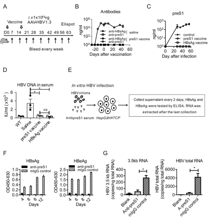
were inoculated subcutaneously with
preS1-polypeptide as the indicated time schedule in Fig. 4A. This vaccination induced preS1-specific antibody (Fig. 4B) and cleared the preS1 antigens in serum after AAV-HBV infection, similar to the clinical HBsAg vaccination (Fig. 4C). Most important, the HBV DNA in serum was also undetectable at the end of the experiment (Fig. 4D), indicating the viral clearance by endogenous antibody to the preS1 domain. These results demonstrate that preS1-polypeptide vaccination
can effectively prevent HBV infectionin vivo.
As HBV is a hepadnavirus that infects only humans
and a few primates,(29) mouse models cannot be used
to test the direct HBV infection/reinfection to the host
liverin vivo. To investigate whether the preS1 antisera
could block HBV entry/re-entry to hepatocytes, we
infected HepG2-hNTCP cells with HBV viruses in
the presence of anti-preS1 serum and incubated for 24
hours (Fig. 4E), as reported.(16) The production of
both HBsAg and HBeAg in cell culture media was significantly reduced when anti-preS1 serum was added, but not the control antibody (Fig. 4F). It indi-cated that the HBV infection to hepatocytes was
FIG.3. Unlike HBsAg,thepreS1domainisnottolerizedinHBVcarriermice.C57BL/6(n54/group,6-8weeksold,male)WT,
and stable HBV carrier mice were vaccinated with preS1-polypeptide or HBsAg formulated in IFA adjuvant, respectively. (A)
Anti-HBs in serum after HBsAg vaccination in WT and carrier mice were determined at the indicated time points by ELISA. (B) In
HBsAg-vaccinatedWT andcarriermice,23105lymphocytes fromLNandspleen werecollected28days afterthesecond
vaccina-tion. Then, cells were stimulated with 5 mg of full-length HBsAg or BSA, and the specific T-cell responses to HBsAg were tested by
IFN-c ELISPOT. (C) Anti-preS1 in serum after preS1-polypeptide vaccination in WT and carrier mice were determined at the
indi-catedtimepointsbyELISA.(D)InpreS1-polypeptide-vaccinatedWT andcarriermice,23105lymphocytesfromLN,spleen,and
liver were collected 28 days after the second vaccination. Then, cells were stimulated with 5 mg of preS1 polypeptide or BSA. Specific
T-cellresponsestothepreS1domainweretestedbyIFN-cELISPOT.Onerepresentativeresultofthree(WTmice,N514)orfour
(carriermice,N514)independentexperimentsisshown.Errorbarsindatarepresentmean6SEM.“ns”means“nosignificant
differ-ence.” ****P< 0.0001 by two-way ANOVA (A,C) or unpaired ttest (B,D). Abbreviations: BSA, bovine serum albumin; OD, optical
FIG.4. PreS1-polypeptidefunctionsas aneffectivepreventivevaccineforHBVinfection.(A)Time scheduleforpreS1-polypeptide
vaccinationtopreventHBVinfection.(B)Onlyanti-preS1,butnoanti-HBs,wasdetectedafterpreS1vaccinationandHBVinfection
inpreS1-polypeptide-vaccinatedmice.(C)PreS1 antigenlevelinserum afterHBVinfectionwastestedbyELISA.(D)HBVDNA
inserum wasextractedperthemanufacturer’sinstructionsandtestedbyqPCR.(E)SchematicdiagramofblockingHBVinfectionto
hepatocytes invitro. Briefly, HepG2-hNTCPcells were inoculated with 1 3 107genome equivalents of HBV in the presence of
anti-preS1serumorcontrolmouseIgGandincubatedfor24hours.Then,cellswerewashedwithmediumthreetimesandmaintainedin
PMM medium. The supernatant of the culture wascollected and medium was changed every 2 days. Levels of HBsAg (F) and
HBeAg (G) inthe supernatant weremeasured byELISA. HBVviral RNAs ininfected cellswere extracted at the indicated time
points;HBV-specific3.5-kbRNA(H)andHBVtotalRNA(I)werequantifiedbyqPCR.Onerepresentativeresultofthree
indepen-dent experiments is shown for panels A, B, C, and D (N 5 12/group) and for panels E, F, G, H, and I (N518 wells/group). Error
bars in data represent mean 6 SEM. “ns” means “no significant difference.” *P< 0.05 by unpaired ttest (D,H,I). Abbreviations: IgG,
ADMINISTRATION OF
PreS1-POLYPEPTIDE PARTIALLY
RESTORES HOST IMMUNE
RESPONSES TO HBsAg
In the study of applying preS1-polypeptide as pre-ventive vaccination for HBV infection, we unexpect-edly observed that the level of HBsAg significantly decreased (Fig. 6A). However, we could not detect sig-nificant anti-HBsAg in serum (Fig. 4B). The decrease, but not clearance, of HBsAg may result from the weak anti-HBsAg response induced by preS1-polypeptide vaccination, which could not completely neutralize the large-scale HBsAg circulating in serum. To test this hypothesis, we detected the specific B-cell response to HBsAg by ELISPOT and observed that preS1-polypeptide vaccination indeed induced HBsAg-specific antibody response (Fig. 6B). Given that the preS1 domain is always coexisting with HBsAg in HBV L-HBsAg during infection, the nontolerized preS1-specific T cells could cross-reactivate the
toler-ized B cells to HBsAg,(20) which thus might partially
restore host immune response toward HBsAg. To test the hypothesis, we detected the serum level of HBsAg in carrier mice after preS1 vaccination. Intriguingly, the preS1-polypeptide vaccination definitely reduced HBsAg (Fig. 6C) and induced the specific B-cell response to HBsAg, even in HBV carrier mice (Fig. 6D).
Thus, we proposed that preS1 vaccination may open a therapeutic window for the hosts who are deeply tolerized to the HBsAg vaccine. We designed a sequential vaccine of preS1-polypeptide before HBsAg in HBV carrier mice to test this hypothesis. The vacci-nation schedule was as shown in Fig. 7A. Compared to HBsAg vaccination alone, the priming with preS1-polypeptide before HBsAg vaccine cleared both preS1 and HBsAg antigens in HBV carrier mice (Fig. 7B). We determined the antibody in serum and observed that such a combination induced specific B-cell response to HBsAg and made anti-HBs seroconver-sion (Fig. 7C), a marker for clinical cure to CHB infection. And, most important, specific T-cell res-ponse to HBsAg could also be detected both in spleen and liver (Fig. 7D). The specific T-cell response induced the reduction both of the HBV RNAs and DNA in hepatocytes (Fig. 7E-G). This response to HBsAg was also accompanied by the diminished immunohistochemical (IHC) staining of HBV core antigen in the liver (Fig. 7H). Thus, the strategy of priming with preS1 before HBsAg vaccination might
blockedbyantiseratothepreS1domain.Thiswas
fur-therdemonstratedbythereductionofHBVviral
repli-cative intermediates, including the 3.5-kilobase (kb)
HBV RNA and the total HBV RNA (Fig. 4G).
To further confirm that anti-preS1 alone could
inhibit the infection and subsequent replication of
HBV in hepatocytes, we repeated the experiment by
mAb XY007 (Supporting Fig. S2). Thus, using the
murine HBV model and HepG2-hNTCP infection
system, we demonstrated that preS1-polypeptide was
potentially an effective vaccine for preventing HBV
infection.
PreS1-POLYPEPTIDE
SERVES
AS
A
THERAPEUTIC
VACCINE
IN
HBV
CARRIER
MICE
Todetermine thetherapeuticeffects ofpreS1
poly-peptidevaccine,wechallengedHBVcarrier micewith
preS1vaccine,andboostedwiththesamedose14days
later. Antigen and antibody responses were tested at
the indicated time points (Fig. 5A). As mentioned,
HBsAgvaccinecould notinduceimmuneresponse in
HBV carrier mice. Even on 35 days postvaccination
withHBsAg,noneofthecarriermiceshoweda
signif-icantreduction inpreS1 antigen(Fig. 5B) and
HBV-DNA in serum (Fig. 5C). On the contrary,
preS1-polypeptide vaccination induced anti-preS1 immune
response, and then preS1 antigen decreased sharply
afterinductionofanti-preS1.Itwasclearedcompletely
onday14postvaccineboost(Fig.5B).Moreover,
anti-preS1resultedinHBV-DNAclearanceinserum(Fig.
5C). The in vitro HBV inhibition assay showed that
anti-preS1 in HBV carrier mice resulted in a
signifi-cantreductioninthesecretionofHBsAgandHBeAg
in theHepG2-hNTCPcell in vitrosystem (Fig. 5D).
RNAlevelsofHBVinHepG2-hNTCPalsoindicated
that anti-preS1 induced in carrier mice blocked
HBV infection/reinfection to hepatocytes efficiently
(Fig.5E).
We repeated the experiment in the HLA-A2.1/
transgenicHBVcarriermicemodel.(23)Thesameasin
the C57BL/6 carrier model, vaccination with preS1
polypeptide, rather than HBsAg, induced clearance
of the preS1 antigen. ELISPOT results for T-cell
responsecorrespondedwelltoantibodyresponse(
Sup-portingFig.S3).Together,theseresultsindicatedthat
preS1 antigen is not tolerized in HBV carrier mice.
The viral-specific immune response to the preS1
domain can clear HBV viral particles and potentially
serve as an effective treatment for clinical CHB virus infection in the future.
Discussion
In this study, we have explored whether preS1-polypeptide vaccination is a potential treatment for CHB infection. We first analyzed the levels of preS1 antigen and HBsAg in clinical HBV patients and compared the immunogenicity of these two viral anti-gens in HBV carrier mice. The preS1 domain of L-HBsAg presents strong immunogenicity for both B-cell and T-B-cell responses in contrast to HBsAg, the major toleragen in CHB patients. Actually, the appear-ance of anti-preS1 indicated a potential recovery from
HBV infection. By using the HBV carrier model, we confirmed the immunogenicity of preS1 polypeptide.
The anti-preS1 induced by preS1-polypeptide
cleared HBV DNA in carrier mice and blocked HBV infection/reinfection to hepatocytes. Furthermore, preS1-polypeptide vaccination even weakened HBsAg tolerized status and the subsequent vaccination with HBsAg could induce anti-HBs seroconversion in HBV carrier mice.
Unlike preS1 polypeptide, HBsAg has been widely used as a prophylactic vaccine against HBV, but it can-not induce immune responses to clear HBsAg in CHB
patients and even in some healthy recipients.(4,30-32)
To further enhance vaccine immunogenicity and immune protection from HBV infection, the L-HBsAg containing preS1 domain has been included in
FIG.5. PreS1-polypeptidefunctionsastherapeuticvaccineinHBVincarriermice.(A)TimeschedulefortestingpreS1antigen
tol-erance in HBV carrier mice. C57BL/6 (n 5 4/group, 6-8 weeks old, male) WT, and stable HBV carrier mice were vaccinated with
preS1-polypeptideorHBsAg formulatedinIFAadjuvant,respectively. (B)After preS1-polypeptideorHBsAg vaccinationincarrier
mice,preS1antigenlevelsinserumweredeterminedbyELISAattheindicatedtimepoints.(C)HBVDNAinserumofeachgroup
wasextractedanddeterminedbyqPCR.(D)Anti-serainducedinHBVcarriermicewith preS1vaccinationblocked HBVinfection
toHepG2-hNTCPinvitro.LevelsofHBsAgandHBeAginsupernatantsweremeasuredbyELISAattheindicatedtimepoints.(E)
Total RNAs of infected cells were extracted onday 10 of HBV infection in vitro, and HBV-specific RNAs were measured with
qPCR. One representative result of four independent experiments is shown for panels A and B (N 5 21/group), three for panel C
(N 5 12/group), and three for panels D and E (N 5 12 wells/group). Error bars in data represent mean 6 SEM. “ns” means “no sig-nificant difference.” *P< 0.05; **P< 0.01; ***P< 0.001; ****P< 0.0001 by two-way ANOVA (B) or unpaired t test (C,D,E).
the third-generation HBV vaccines.(33-35)However, by using the HBV carrier mouse model, we observed that, same as the conventional HBsAg vaccine, the L-HBsAg-containing protein vaccination showed no therapeutic effects in the tolerized model either (Supporting Fig. S4). Failing in induction of immune response to the preS1 domain by the L-HBsAg-containing protein vaccination might be attributed to an overwhelming immune tolerance to HBsAg, a
physical link of preS1 domain to HBsAg, or too little amount of preS1 region containing in the
vac-cine.(36,37) It is also possible that alum, the currently
clinically used adjuvant, might not be potent enough to induce detectable responses. Whether the preS1 region itself alone can be applied as a vaccine has not been directly tested. Here, we found that the antige-nicity of preS1 was weaker than that of HBsAg; it can only induce robust responses with stronger adjuvants,
FIG.6.PreS1-polypeptidevaccinationinducetheB-cellresponsetoHBsAginHBVmice.(A)AfterpreS1-polypeptidevaccination,mice
wereinfectedwithHBVandthelevelofHBsAginserumwastestedbyELISA,whichindicatedthatpreS1prevaccinationdiminished
HBsAglevelinserum.(B)InpreS1-prevaccinatedmice,23105splenocyteswerecollected28daysafterHBVinfection.SpecificB-cell
responsetoHBsAgwastestedbyB-cellELISPOTassay.(C)InHBVcarriermice,preS1-polypeptidewasvaccinatedatdays28and42
afterAAV-HBVinoculation.HBsAgvariationsincarriermiceweretestedbyELISA.(D)InpreS1-polypeptide-vaccinatedHBVcarrier
mice,23105splenocyteswerecollected.SpecificB-cellresponsetoHBsAgwastestedbyB-cellELISPOTassay.One(A,B,C)ortwo
(D) representative results of three (N 5 11/group) independent experiments are shown. Error bars in data represent mean 6 SEM. *P<
FIG.7. HBsAg boost immunization amplifies preS1-polypeptide-induced anti-HBV response for viral clearance. (A) Time schedule
for preS1-polypeptide combination with HBsAg as vaccine to treat HBV carrier mice. (B) After carrier mice were vaccinated with
preS1-polypeptide1HBsAg, the preS1 and HBsAg antigens in serum were tested by ELISA. (C)B-cell response to HBsAg was
testedbyB-cellELISPOT,andanti-HBsinserumwastestedbyELISA.(D)Miceweresacrificedonthelastday,and23105
lym-phocytes from LN, spleen, and liver were collected. Then, cells were stimulated with 5 mg of preS1 polypeptide, HBsAg, or BSA, and
specific T-cell response was measured by IFN-csecretion T-cell ELISPOT assay. (E) HBV total RNAs and (F) intermediate
prod-ucts 3.5-kb RNA in liver tissue were determined with qPCR. (G) HBV DNA in liver was determined by qPCR. (H) IHC staining
forHBcAginhepatocytes(Magnification:200X).Onerepresentativeresultofthree(N511/group;B,C,D)ortwo(N57;E,F,G,H)
independent experiments are shown. Error bars in data represent mean 6 SEM. *P< 0.05; **P< 0.01; ***P< 0.001; ****P< 0.0001 by
two-way ANOVA (B,C) or unpaired ttest (D,E,F,G). Abbreviations: Ad, adenovirus; BSA, bovine serum albumin; i.v, intravenous;
HBV reinfection to healthy hepatocytes in patients, and thus dilute and even diminish the HBV-infected hepatocytes gradually. Unexpectedly, we observed that preS1-polypeptide vaccination partially restored im-mune response to HBsAg during HBV infection and thus opened a therapeutic window for the host to fully respond to the HBsAg vaccine. To amplify the response to HBsAg induced by preS1-polypeptide vac-cination and induce stronger and multispecific T-cell responses for complete viral clearance in HBV carrier mice, the sequential combination of preS1-polypeptide before the HBsAg vaccination was administered. We showed that preS1-polypeptide priming before vacci-nation with HBsAg restored both the B-cell and T-cell response to HBsAg in the periphery and liver, ulti-mate resulting in the HBsAb seroconversion and grad-ual clearance of HBV in the liver. IHC staining also showed a dramatic decrease in the number of core-positive hepatocytes in livers of HBV carrier mice. This strategy indeed makes HBsAg, the preventive vaccine, achieve an unexpected therapeutic effect in HBV carrier mice. However, it is not yet clear how preS1 region vaccination helped to restore immune response to HBsAg. In HBV infection, the preS1 domain is always fused with HBsAg within L-HBsAg; thus, T cells to the preS1 region may help in the
immune response to HBsAg.(20,43) Moreover, the
immune complex of endogenous anti-preS1 with HBV virions may further enhance anti-HBV immune response. The function of the B-cell and T-cell responses to the preS1 domain in the restoration of anti-HBV immunity is under investigation. In sum-mary, preS1-polypeptide vaccine can reverse HBV tolerance in HBV carrier mice and may serve as a potentially effective therapeutic strategy for treating CHB infection.
Acknowledgments:Y.B., Y.-X.F., and H.P. designed the experiments. We thank Dr. Wenhui Li for pro-viding the HepG2-hNTCP cell line and HBV virus
(ayw subtype) for HBV infection experiment in vitro.
We thank Daryl Harmon for editorial assistance.
REFERENCES
1) World Health Organization. Hepatitis B. Fact sheet. Geneva, Switzerland: World Health Organization; 2015.
2) Ganem D, Prince AM. Hepatitis B virus infection—natural his-tory and clinical consequences. N Engl J Med 2004;350:1118-1129.
like IFA. However, this adjuvant limits
preS1-polypeptide vaccination usage in the clinical setting.
We are exploring several other formulation for preS1
polypeptide vaccination, including a nanovaccine that
containspreS1regiontoenhanceitsimmunogenicity.
In theclinic, anti-preS1appears early inthe course
of AHB.(38) Even the occasional appearance of
anti-preS1inafewpatientswithchronicaggressive
hepati-tis or treated with antiviral agents indeed correlates
wellwith betterhealthimprovement.(39) However,the
random clinical analyses of the appearance of
anti-preS1inCHBhavenotbeenconclusiveforthe
corre-lationbetweenchangesinimmunestatusandviral
per-sistence.InCHBinfection,solublecirculatingHBsAg
induceslymphocytesanergythatfailtoreceivethe
sec-ondary danger signals for sustaining their activation.
However,thepreS1domainiscontainedonHBV
viri-ons, which viral DNA can significantly enhance the
immunogenicity of preS1.By comparing theimmune
status between preS1 and HBsAg in clinicalpatients,
we observed that preS1 has much less immune
toler-ance. And the appearance of antibody and T-cell
responsestothepreS1domaininclinicalpatients
cor-related with a better prognosis of the significant
decrease of serum HBV DNA. Recently, an entry
inhibitor peptide derived from the preS1 region,
Myrcludex-B,has beeninvestigatedasatreatmentfor
HBV infection. This peptide has been shown to
inhibitHBVinfectioninvivo andhinderthe
amplifi-cation of the covalently closed circular DNA pool in
initially infected hepatocytes.(40) Here, we
demon-strated that preS1-polypeptide vaccination can induce
astrong antibody response inHBV carrier mice, thus
makingitauniquecandidatevaccinefortreatingHBV
infection.ComparedwithMyrcludex-B,such
vaccina-tionwould bemuchmorepotentin inducingintrinsic
anti-preS1 and would provide long-term protection
fromHBV infection. Further clinicaltrials for
target-ingpreS1regionarewarranted.
In fact, CD41, but not CD81, T-cell responses
were induced by preS1-polypeptide vaccination (
Sup-porting Fig. S5A,B), with no liver damage indicated
by normalalanineaminotransferase (ALT)and
aspar-tateaminotransferase(AST) levelsinserum(
Support-ing Fig. S5C,D). Consistently, the only cytotoxic T
lymphocyte(CTL)epitopeinthepreS1region
identi-fied is forhuman HLA-A11-restricted.(41,42) Lack of
CTLs after preS1-polypeptide vaccination may have
liver escape from unexpected injuries, meanwhile the
preS1-vaccine-induced anti-preS1 antibodies in the
3) Dienstag JL, Stevens CE, Bhan AK, Szmuness W. Hepatitis B vaccine administered to chronic carriers of hepatitis b surface antigen. Ann Intern Med 1982;96:575-579.
4) Shi B, Ren G, Hu Y, Wang S, Zhang Z, Yuan Z. HBsAg inhibits IFN-a production in plasmacytoid dendritic cells through TNF-a and IL-10 INDUCTION IN MOnocytes. PLoS One 2012;7:e44900.
5) Duan XZ, Wang M, Li HW, Zhuang H, Xu D, Wang FS. Decreased frequency and function of circulating plasmocytoid dendritic cells (pDC) in hepatitis B virus infected humans. J Clin Immunol 2004;24:637-646.
6) Han Q, Lan P, Zhang J, Zhang C, Tian Z. Reversal of hepatitis B virus-induced systemic immune tolerance by intrinsic innate immune stimulation. J Gastroenterol Hepatol 2013;28:132-137. 7) Mueller SN, Ahmed R. High antigen levels are the cause of T
cell exhaustion during chronic viral infection. Proc Natl Acad Sci U S A 2009;106:8623-8628.
8) Edwards BH, Bansal A, Sabbaj S, Bakari J, Mulligan MJ, Goepfert PA. Magnitude of functional CD81
T-cell responses to the gag protein of human immunodeficiency virus type 1 cor-relates inversely with viral load in plasma. J Virol 2002;76:2298-2305.
9) Tay SS, Wong YC, McDonald DM, Wood NA, Roediger B, Sierro F, et al. Antigen expression level threshold tunes the fate of CD8 T cells during primary hepatic immune responses. Proc Natl Acad Sci U S A 2014;111:E2540-E2549.
10) Kakimi K, Isogawa M, Chung J, Sette A, Chisari FV. Immuno-genicity and toleroImmuno-genicity of hepatitis B virus structural and nonstructural proteins: implications for immunotherapy of persis-tent viral infections. J Virol 2002;76:8609-8620.
11) Chen M, Sallberg M, Hughes J, Jones J, Guidotti LG, Chisari FV, et al. Immune tolerance split between hepatitis B virus pre-core and pre-core proteins. J Virol 2005;79:3016-3027.
12) DiMattia MA, Watts NR, Stahl SJ, Grimes JM, Steven AC, Stuart DI, Wingfield PT. Antigenic switching of hepatitis B virus by alternative dimerization of the capsid protein. Structure 2013;21:133-142.
13) Milich DR, Schodel F, Hughes JL, Jones JE, Peterson DL. The hepatitis B virus core and e antigens elicit different Th cell sub-sets: antigen structure can affect Th cell phenotype. J Virol 1997; 71:2192-2201.
14) Petit MA, Zoulim F, Caipel F, Dubanchet S, Dauguet C, Trepo C. Variable expression of preS1 antigen in serum during chronic hepatitis B virus infection: an accurate marker for the level of hepatitis B virus replication. HEPATOLOGY1990;11:809-814.
15) Klingmuller U, Schaller H. Hepadnavirus infection requires interaction between the viral pre-S domain and a specific hepato-cellular receptor. J Virol 1993;67:7414-7422.
16) Yan H, Zhong G, Xu G, He W, Jing Z, Gao Z, et al. Sodium taurocholate cotransporting polypeptide is a functional receptor for human hepatitis B and D virus. Elife 2012;1:e00049. 17) Bruss V, Ganem D. The role of envelope proteins in hepatitis B
virus assembly. Proc Natl Acad Sci U S A 1991;88:1059-1063. 18) Ferrari C, Penna A, Bertoletti A, Cavalli A, Valli A, Schianchi
C, Fiaccadori F. The preS1 antigen of hepatitis B virus is highly immunogenic at the T cell level in man. J Clin Invest 1989;84: 1314-1319.
19) Hu WG, Wei J, Xia HC, Yang XX, Li F, Li GD, et al. Iden-tification of the immunogenic domains in HBsAg preS1 region using overlapping preS1 fragment fusion proteins. World J Gas-troenterol 2005;11:2088-2094.
20) Milich DR, McLachlan A, Moriarty A, Thornton GB. A single 10-residue pre-S(1) peptide can prime T cell help for antibody
production to multiple epitopes within the pre-S(1), pre-S(2), and S regions of HBsAg. J Immunol 1987;138:4457-4465. 21) Neurath AR, Seto B, Strick N. Antibodies to synthetic peptides
from the preS1 region of the hepatitis B virus (HBV) envelope (env) protein are virus-neutralizing and protective. Vaccine 1989; 7:234-236.
22) Yang D, Liu L, Zhu D, Peng H, Su L, Fu YX, Zhang L. A mouse model for HBV immunotolerance and immunotherapy. Cell Mol Immunol 2014;11:71-78.
23) Dion S, Bourgine M, Godon O, Levillayer F, Michel ML. Adeno-associated virus-mediated gene transfer leads to persistent hepatitis B virus replication in mice expressing HLA-A2 and HLA-DR1 molecules. J Virol 2013;87:5554-5563.
24) Lu YY, Li K, Cheng J, Wang L, Liu Y, Zhang LX. Cloning and expression of the preS1 gene of hepatitis B virus in yeast cells. Hepatobiliary Pancreat Dis Int 2002;1:238-242.
25) Wei J, Wang YQ, Lu ZM, Li GD, Wang Y, Zhang ZC. Detection of anti-preS1 antibodies for recovery of hepatitis B patients by immunoassay. World J Gastroenterol 2002;8:276-281. 26) van Ditzhuijsen TJ, Kuijpers LP, Koens MJ, Rijntjes PJ, van
Loon AM, Yap SH. Hepatitis B pre-S1 and pre-S2 proteins: clinical significance and relation to hepatitis B virus DNA. J Med Virol 1990;32:87-91.
27) Whalley SA, Murray JM, Brown D, Webster GJ, Emery VC, Dusheiko GM, Perelson AS. Kinetics of acute hepatitis B virus infection in humans. J Exp Med 2001;193:847-854.
28) Zhu D, Liu L, Yang D, Fu S, Bian Y, Sun Z, et al. Clearing persistent extracellular antigen of hepatitis B virus: an immuno-modulatory strategy to reverse tolerance for an effective therapeu-tic vaccination. J Immunol 2016;196:3079-3087.
29) Ganem D, Varmus HE. The molecular biology of the hepatitis B viruses. Annu Rev Biochem 1987;56:651-693.
30) Op den Brouw ML, Binda RS, van Roosmalen MH, Protzer U, Janssen HL, van der Molen RG, Woltman AM. Hepatitis B virus surface antigen impairs myeloid dendritic cell function: a possible immune escape mechanism of hepatitis B virus. Immu-nology 2009;126:280-289.
31) Xu Y, Hu Y, Shi B, Zhang X, Wang J, Zhang Z, et al. HBsAg inhibits TLR9-mediated activation and IFN-alpha production in plasmacytoid dendritic cells. Mol Immunol 2009;46:2640-2646. 32) Woltman AM, Op den Brouw ML, Biesta PJ, Shi CC, Janssen
HL. Hepatitis B virus lacks immune activating capacity, but actively inhibits plasmacytoid dendritic cell function. PLoS One 2011;6:e15324.
33) McDermott AB, Cohen SB, Zuckerman JN, Madrigal JA. Hep-atitis B third-generation vaccines: improved response and con-ventional vaccine non-response—evidence for genetic basis in humans. J Viral Hepat 1998;5(Suppl 2):9-11.
34) Yap I, Chan SH. A new pre-S containing recombinant hepatitis B vaccine and its effect on non-responders: a preliminary obser-vation. Ann Acad Med Singapore 1996;25:120-122.
35) Shouval D, Roggendorf H, Roggendorf M. Enhanced immune response to hepatitis B vaccination through immunization with a Pre-S1/Pre-S2/S vaccine. Med Microbiol Immunol 2015;204:57-68. 36) Prange R, Clemen A, Streeck RE. Myristylation is involved in intracellular retention of hepatitis B virus envelope proteins. J Virol 1991;65:3919-3923.
37) Gerlich WH. Prophylactic vaccination against hepatitis B: achievements, challenges and perspectives. Med Microbiol Immunol 2015;204:39-55.
39) Hellstrom U, Lindh M, Krogsgaard K, Sylvan S. Demonstration of an association between detection of IgG antibody reactivity towards the C-terminal region of the preS1 protein of hepati-tis B virus and the capacity to respond to interferon therapy in chronic hepatitis B. J Gastroenterol Hepatol 2008;23:804-810.
40) Volz T, Allweiss L, Ben MM, Warlich M, Lohse AW, Pollok JM, et al. The entry inhibitor Myrcludex-B efficiently blocks intrahepatic virus spreading in humanized mice previously infected with hepatitis B virus. J Hepatol 2013;58:861-867. 41) Jin Y, Shih WK, Berkower I. Human T cell response to the
sur-face antigen of hepatitis B virus (HBsAg). Endosomal and non-endosomal processing pathways are accessible to both endogenous and exogenous antigen. J Exp Med 1988;168:293-306.
42) Desmond CP, Bartholomeusz A, Gaudieri S, Revill PA, Lewin SR. A systematic review of T-cell epitopes in hepatitis B virus: identification, genotypic variation and relevance to antiviral thera-peutics. Antivir Ther 2008;13:161-175.
43) Milich DR. Immune response to hepatitis B virus proteins: rele-vance of the murine model. Semin Liver Dis 1991;11:93-112.
Author names in bold designate shared co-first authorship.
Supporting Information
Additional Supporting Information may be found at