GENETIC AND EPIGENETIC MECHANISMS REGULATING SMOOTH MUSCLE CELL DIFFERENTIATION
Kevin Dale Mangum
A dissertation submitted to the faculty at the University of North Carolina at Chapel Hill in partial fulfillment of the requirements for the degree of Doctor of Philosophy in
Pathology in the School of Medicine.
Chapel Hill 2017
Approved by:
Christopher P. Mack Victoria L. Bautch Jiandong Liu
© 2017
ABSTRACT
Kevin Dale Mangum: Genetic and Epigenetic Mechanisms Regulating Smooth Muscle Cell Differentiation
(Under the direction of Christopher P. Mack)
Smooth muscle differentiation is a complex process, involving numerous
molecular, genetic, and epigenetic mechanisms. Notably, smooth muscle cells (SMCs) retain marked plasticity in their ability to convert between synthetic and more
differentiated contractile gene programs. In vascular diseases, including hypertension, atherosclerosis, and restenosis, SMCs dedifferentiate from their healthy, mature state to a more immature “phenotypically modulated” cell type capable of migrating,
proliferating, and producing extracellular matrix, all of which contribute to disease. Additionally, genetic alterations in various components of the smooth muscle
transcriptional machinery result in cardiovascular disease and even death. Thus, a more complete understanding of the exact mechanisms regulating SMC differentiation is crucial for the development of novel targets in the diagnosis and treatment of vascular disease.
The work herein interrogates several points along the RhoA axis and defines their roles in SMC differentiation. First, the genetic and epigenetic mechanisms regulating expression of a smooth muscle-specific gene, GRAF3, are uncovered.
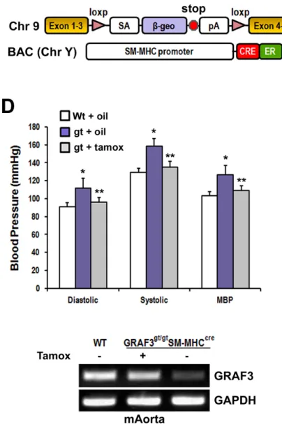
GRAF3, also referred to as ARHGAP42, was first described by my collaborators in Joan Taylor’s Lab as a smooth muscle selective Rho-GAP essential for blood pressure
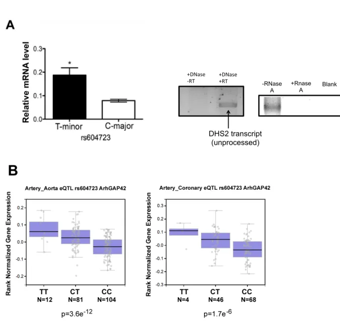
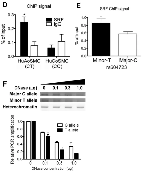
with changes in blood pressure, and the rs604723 T-allele variant located in a highly conserved DHS increased GRAF3 expression by promoting SRF binding to this region. In addition to SRF, we show that the transcription activity of this region as well as GRAF3 expression are controlled by the transcription factors, RBPJ and TEAD1.
In subsequent chapters, we describe novel mechanisms regulating function of MRTF-A. Given that MRTF-A is essential for full activation of smooth muscle-specific gene expression, we hypothesize that these newly identified mechanisms regulate SMC differentiation. We describe our approach for identifying post-translational modifications and binding partners that regulate MRTF-A function. In our search for novel MRTF-A binding partners, we identified the putative histone lysine methyltransferase, PRDM6, and demonstrated that it was required for SMC differentiation. In overexpression experiments in COS-7 cells, we detected significant methylation on MRTF-A.
ACKNOWLEDGMENTS
It goes without saying that the past four and a half years have been quite the journey and influenced by so many individuals. I have to start off by acknowledging my scientific mentor and role model, Chris Mack. Your enthusiasm, curiosity, and, yes, even skepticism were highly inspiring to me as a budding scientist, and, for that matter, are what I consider the qualities of a true scientist to be. What I’m grateful for the most is your unwavering approachability, which made it so easy to test drive experimental ideas and hypotheses. No matter if it was first thing in the morning and I hadn’t even stepped into my office, you always welcomed new ideas, no matter the practicality. As a result, science became even more addicting, and my passion for it flourished under your mentorship. Thank you. I’ll forever be indebted to you for that.
I must also thank Joan Taylor, who helped me find protocols, design primers, etc. on countless occasions. Dr. Taylor, you’ve been such a crucial member of my
committee and a tremendous asset to the development of my research goals. To the rest of my committee members, Vicki Bautch, Jiandong Liu, and Praveen Sethupathy, thank you for your willingness to serve on my committee. Your input over the past few years has been helpful.
Next, it’s only fitting that I acknowledge the UNC MD/PhD program. During my second year of medical school, I decided that I wanted to pursue my PhD. The first talk I had about joining the program was with Dr. Orringer, who never hesitated and
helped my transition into the Program are Alison Regan, Carol Herion, and Mohanish Deshmuk. Thank you all for taking a chance on me. I can’t tell begin to explain how much saying yes in that moment impacted me.
It’s hard to imagine four years without good company. I really hit the jackpot in the Mack-Taylor Lab by being surrounded be so many kind, dedicated, and intelligent individuals. Laura-Weise Cross, even though you’ve already graduated, I was so lucky to work with you for the majority of my grad school experience. Kaitlin Lenhart, a previous graduate student in the Taylor Lab, always provided me with encouragement and helpful advice at the start of my graduate school career. Thank you, Rachel Dee for always smiling and being so wonderful to work with. I’m also fortunate to have worked with my other lab members: Xue Bai, Qiang Zhu, and Zack Opheim.
To my family, thank you for supporting me during my endeavors, especially my Mom and Dad. I know it’s been a long road and you’ve been there every step of the way. To Tanner Mangum, you made it so easy for me to take a step back from science and laugh a little, which has been so invaluable. Thank you Trish for being there as well. Your support means everything to me. I have to acknowledge my grandparents, and roots in general, for helping me stay humble. Meme, especially, thank you for teaching me that “there’s more than one way to skin a cat.” I truly believe that this contributed to my scientific creativity from a very early age.
To my friend Negeen Hamedani, thank you for your encouragement over the last five years. I’m so grateful to have you as a friend.
TABLE OF CONTENTS
LIST OF FIGURES ... x
LIST OF TABLES ... xiii
LIST OF ABBREVIATIONS ... xiv
CHAPTER 1: BACKGROUND AND SIGNIFICANCE ... 1
Blood vessel development, structure, and function ... 1
Developmental diversity of SMCs ... 3
Role of SMC phenotypic switching in cardiovascular disease ... 3
Transcriptional regulation of SMC differentiation ... 8
Epigenetic regulation of SMC differentiation ... 23
ENCODE Consortium, UCSC Genome Browser, and GTEx Database ... 30
Genetic and molecular basis of blood pressure regulation ... 31
Objectives of this dissertation research ... 39
CHAPTER 2: BLOOD PRESSURE-ASSOCIATED POLYMORPHISM CONTROLS ARHGAP42 EXPRESSION VIA SERUM RESPONSE FACTOR DNA BINDING ... 40
Overview ... 40
Introduction ... 41
Materials and Methods ... 42
Results ... 51
Allele-specific differences of ARHGAP42 expression in SMCs ... 51
ARHGAP42 genotype and human hypertension ... 53
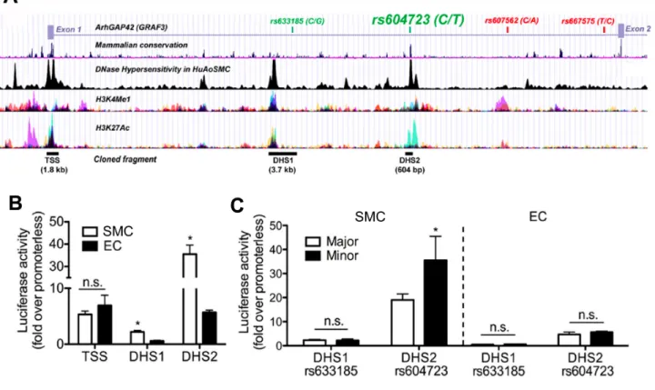
Identification of regulatory elements within the ARHGAP42 gene ... 55
The minor allele sequence at rs604723 binds SRF ... 59
SRF is required for ARHGAP42 expression and for the effects of the rs604723 variation ... 62
ARHGAP42 expression is upregulated by RhoA signaling ... 62
Activation of ARHGAP42 expression attenuates the development of hypertension ... 65
Discussion ... 76
CHAPTER 3: REGULATION OF ARHGAP42 EXPRESSION BY RBPJ and TEAD1..80
Overview ... 80
Introduction ... 81
Materials and Methods ... 84
Results ... 88
Engineering a novel dCas-SRF fusion protein to target endogenous loci ... 88
Identification of the core regulatory region required for GRAF3 transcription ... 89
RBPJ and TEAD1 bind to a conserved sequence within the DHS ... 89
Activity of the GRAF3 DHS is required by Notch/RBPJ and TEAD1 ... 94
Endogenous GRAF3 expression in SMC is required by RBPJ and TEAD1 ... 98
Cooperativity between RBPJ and TEAD1 ... 100
The long non-coding RNA, AK124326, inhibits GRAF3 expression ... 102
Discussion ... 107
CHAPTER 4: IDENTIFICATION OF MRTF-A POST-TRANSLATIONAL MODIFICATIONS AND BINDING PARTNERS ... 111
Overview ... 111
Introduction ... 112
Materials and Methods ... 115
Results ... 118
Identification of SMC-specific MRTF-A binding partners ... 118
Validation of PRDM6 as an MRTF-A binding partner ... 118
PRDM6 is required for SMC differentiation ... 125
Identification of post-translational modifications on MRTF-A ... 125
Identification of lysine methyltransferases that methylate MRTF-A ... 129
K27 is required for nuclear import of MRTF-A ... 131
SMYD2 interacts with MRTF-A ... 135
SMYD2 inhibits MRTF-A nuclear localization ... 135
SMYD2 inhibits MRTF-A-dependent smooth muscle transcription ... 139
Actin dynamics regulate MRTF-A band shifs ... 139
Discussion ... 143
CHAPTER 5: CONCLUSIONS, PERSPECTIVES, AND FUTURE DIRECTIONS ... 147
Pharmacologic regulation of RhoA- and Rho-dependent pathways ... 147
Regulation of MRTF-A-dependent transcription as a way to direct SMC differentiation ... 151
LIST OF FIGURES
Figure 1.1. SMC vessel structure and phenotypic switching ... 4
Figure 1.2. RhoA signaling regulates MRTF nuclear localization in SMC ... 13
Figure 1.3. Domain structure and conservation of myocardin and the MRTFs ... 15
Figure 1.4. Contrasting the transcription mechanisms that regulate SMC differentiation in the healthy versus phenotypically modulated SMC ... 24
Figure 1.5. Signaling mechanisms regulating SMC contraction ... 33
Figure 1.6. Pharmacologic and genetic regulation of the RhoA signaling axis ….…….36
Figure 2.1. ARHGAP42 expression in SMC is regulated by allele-specific mechanisms and controls blood pressure ……….………52
Figure 2.1 (continued). ARHGAP42 expression in SMC is regulated by allele-specific mechanisms and controls blood pressure……….………54
Figure 2.2. An enhancer within the ARHGAP42 first intron displays strong SMC-specific and allele-specific activity and is required for endogenous ARHGAP42 expression ... 58
Figure 2.2 (continued). An enhancer within the ARHGAP42 first intron displays strong SMC-specific and allele-specific activity and is required for endogenous ARHGAP42 expression ... 60
Figure 2.3. The minor T allele at rs604723 promotes SRF binding ………...……..61
Figure 2.3 (continued). The minor T allele at rs604723 promotes SRF binding ... 63
Figure 2.4. The allele-specific activity of the DHS2 enhancer is SRF-dependent ... 64
Figure 2.5. ARHGAP42 expression is activated by RhoA signaling and cell stretch .... 67
Figure 2.6. ARHGAP42 expression limits the development of hypertension ... 68
Figure 2.6 (continued). ARHGAP42 expression limits the development of hypertension ... 69
Supplemental Figure II. Tamoxifen treatment of Arhgap42gt/gtSM-MHCCreERT2 mice restored blood pressure homeostasis ... 70
Supplemental Figure IV. Tamoxifen treatment of DOCA-salt-treated Arhgap42gt/gt SM-MHCCreERT2 mice restored arhgap42 expression in
mesenteric arteries ... 72
Supplemental Figure V. TMEM133 is an extension of the ARHGAP42 3’ UTR ... 73
Figure 3.1. SRF targeted to the conserved DHS increases endogenous GRAF3 expression ... 91
Figure 3.2. Preliminary mapping of the core regulatory region within the GRAF3 DHS ... 92
Figure 3.3. A core DNase I Hypersensitive regulatory region drives GRAF3 transcription ... 93
Figure 3.4. The core DHS regulatory region binds RBPJ and TEAD1 ... 96
Figure 3.5. RBPJ and TEAD1 binding sites are required for GRAF3 transcription ... 97
Figure 3.6. GRAF3 transcription is regulated by Notch signaling ... 99
Figure 3.7. RBPJ/Notch is required for GRAF3 expression ... 101
Figure 3.8. TEAD1 is required for GRAF3 expression ... 103
Figure 3.9. The LNC RNA AK124326 negatively regulates GRAF3 expression ... 105
Figure 3.10. mir-505-3p suppresses GRAF3 expression ... 106
Figure 4.1. The N-terminal RPEL domains and basic/Q-rich/SAP region of MRTF-A mediate its interaction with PRDM6 ... 123
Figure 4.2. Actin bridges MRTF-A and PRDM6 through MRTF’s N-terminal RPEL domains ... 124
Figure 4.3. PRDM6 interacts directly with the SRF-binding reigon of MRTF-A ... 126
Figure 4.4. PRDM6 is required for SMC differentiation ... 127
Figure 4.5. MRTF-A is methylated in vivo ... 130
Figure 4.6. Additional predicted methylation sites within MRTF-A ... 132
Figure 4.7. SMYD2 and SET7/9 methylate MRTF-A in vitro and in vivo ... 133
Figure 4.9. K27 is essential for MRTF-A nuclear import ... 136
Figure 4.10. SMYD2 interacts with MRTF-A ... 137
Figure 4.11. SMYD2 inhibits MRTF-A nuclear localization ... 140
Figure 4.12. SMYD2 inhibits MRTF-A-dependent promoter activity ... 141
LIST OF TABLES
Table 2.1 Analysis of ARHGAP42 genotype and blood pressure in
human populations ... 56
Supplemental Table I. Characteristics of clinical cohort ... 74
Supplemental Table II. Analysis of Arhgap42 genotype in human population ... 75
Table 4.1. MRTF-A binding partners in mouse SMC ... 119
Table 4.1 (continued). MRTF-A binding partners in mouse SMC ... 120
LIST OF ABBREVIATIONS
ACE: Angiotensin Converting Enzyme AII: Angiotensin II
AJ: Adherens Junction ATP: Adenosine Triphosphate BP: Blood Pressure
BSA: Bovine Serum Albumin CAD: Coronary Artery Disease
CADASIL: Cerebral Autosomal-Dominant Arteriopathy with Subcortical Infarcts and Leukoencephalopathy
CNN: Calponin
COS: CV-1 in Origin with SV40 genes
CRISPR: Clustered Regularly Interspaced Short Palindromic Repeats CRM-1: Chromosomal Mainenance-1
CT: Threshold Cycle
DAPI: 4',6-Diamidino-2-Phenylindole, Dihydrochloride
DAPT: N-[(3,5-Difluorophenyl)acetyl]-L-alanyl-2-phenyl]glycine-1,1-dimethylethyl ester
DHS: Dnase-I Hypersensitive Site
DMEM: Dulbecco's Modified Eagle Medium DNA: Deoxyribonucleic Acid
DOCA: Deoxycorticosterone acetate DTT: Dithiothreitol
EDTA: Ethylenediaminetetraacetic Acid EEL: External Elastic Lamina
EMSA: Electrophoretic Mobility Shift Assay ENCODE: Encyclopedia of DNA Elements ERK: Extracellular signal-related kinase ES: Embryonic Stem
EV: Empty Vector
FAK: Focal Adhesion Kinase FBS: Fetal Bovine Serum FCS: Fetal Calf Serum
GAP: GTPase Activating Protein
GAPDH: Glyceraldehyde 3-phosphate dehydrogenase GDP: Guanosine-5'-diphosphate
GEF: Guanine nucleotide exchange factor GFP: Green Fluorescent Protein
GO: Gene Ontology
GPCR: G Protein Coupled Receptor
GRAF: GTPase Regulator Associated with Focal Adhesion Kinase GST: Glutathione-S-transferase
GTP: Guanosine-5'-triphosphate
GWAS: Genome Wide Association Study
HEPES: 4-(2-hydroxyethyl)-1-piperazineethanesulfonic acid HTN: Hypertension
ID: Identify/Identification IEL: Internal Elastic Lamina IP: Immunoprecipitation IRB: Institutional Review Board IV: Intravenous
KCL: Potassium Chloride
LARG: Leukemia-associated Rho GEF
LD: Linkage Disequilibrium LNC: Long Non-coding LZ: Leucine Zipper
MADS: MCM1, Agamous, Deficiencs, SRF MAF: Mean Allele Frequency
MAML: Mastermind-like MCAT: Muscle-CAT MEK: MAP/ERK kinase MHC: Myosin Heavy Chain MLC: Myosin Light Chain
MLCK: Myosin Light Chain Kinase
MRTF: Myocardin-Related Transcription Factor
MYOSLID: MYOcardin-induced Smooth muscle LncRNA, Inducer of Differentiation MYPT: Myosin Phosphatase Targeting Protein
NLS: Nuclear Localization Sequence/Signal NTC: Non-Targeting Control
PAGE: Polyacrylamide Gel Electrophoresis PBS: Phosphate Buffered Saline
PCR: Polymerase Chain Reaction PDGF: Platelet Derived Growth Factor
PDZ: Psd-95 (Post Synaptic Density Protein), DlgA (Drosophila Disc Large Tumor Suppressor) and ZO1 (Zonula Occludens-1 Protein)
PH: Plekstrin Homology PKA: Protein Kinase A PKC: Protein Kinase C
PKMT: Protein Lysine Methyltransferase PMSF: Phenylmethylsulfonyl Fluoride
PTM: Post Translational Modification
RBPJ: Recombination Signal Binding Protein For Immunoglobulin Kappa J Region
RIPA: Radioimmunoprecipitation assay buffer
RNA: Ribonucleic acid
ROCK: Rho-associatd protein kinase RT: Real Time
SAM: S-adenosylmethione SAP: SAF-A/B, Acinus and PIAS
SDS: Sodium Dodecyl Sulfate SEM: Standard Error of the Mean
SFM: Serum Free Media SM: Smooth Muscle
SMA: Smooth Muscle Alpha-Actin SMC: Smoth Muscle Cell
SMMHC: Smooth Muscle Myosin Heavy Chain SMYD: SET And MYND Domain Containing
SNF: SWItch/Sucrose Non-Fermentable SNP: Single Nucleotide Polymoerphism SRF: Serum Response Factor
STAT: Signal transducer and activator of transcription SUMO: Small Ubiquitin-like Modifier
SWI: SWItch/Sucrose Non-Fermentable TAD: Transcriptional Activation Domain TAGLN: Transgelin
TEAD: TEA domain family member 1 TEF: Transcriptional Enhancer Factor TET: Ten-eleven Translocation
TGF: Transforming Growth Factor TK: Tyrosine Kinase
TSS: Transcription Start Site
UCSC: University of California, Santa Cruz UNC: University of North Carolina
UTR: Untranslated Region
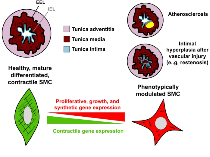
CHAPTER 1: BACKGROUND AND SIGNIFICANCE Blood vessel development, structure, and function
Blood vessels supply the developing embryo and adult with sufficient oxygen and nutrients required to support organ growth, function, and life. The mature artery is
comprised of three main layers: the tunica intima, tunica media, and tunica adventitia (Figure 1). The tunica intima, which is the innermost layer, consists of a single lining of endothelial cells and is separated from the tunica media by the internal elastic lamina. The tunica media is composed of multiple sheets of vascular smooth muscle cells (SMCs), which contract to regulate vessel diameter, tone, and, consequently,
downstream tissue perfusion. The external elastic lamina separates the tunica media and the tunica adventitia, which is formed from connective tissue. The connective tissue of the adventitia houses peripheral nerves, immune cells, fibroblasts, and resident vascular progenitor cells that are thought to play a role in vascular disease and repair (1, 2).
alone are unable to maintain and stabilize the developing vascular system (7, 8). Maturation and stabilization of the developing vasculature by SMCs is essential for a functional vascular network. After extensive remodeling of the endothelial plexus, the first step in vascular maturation is investment of the plexus by mural cells (smooth muscle cells and pericytes), which relies heavily on PDGF, TGFβ, and Notch signaling (8-10). Intact PDGF-B/PDGF-Rβ signaling is required for SMC investment during blood vessel development, as global homozygous deletion of the PDGF-B ligand or its
receptor results in hemorrhage and embryonic lethality from diminished SMC coverage in capillary beds (12). Another important regulator of SMC migration during vessel development is Notch signaling, loss of which leads to significantly reduced SMC coverage in developing vessels (13). This will be discussed in greater detail below.
other locations beginning at E12.5, and then in all smooth muscle tissues by E17.5 (16). Finally, calponin is expressed in the heart as early as E8.5 but persists until E13.5, at which time calponin transcripts are detected in smooth muscle-containing tissues (17). Developmental diversity of SMCs
SMCs have a profound developmental diversity, since they are not derived from a single embryonic origin (18). Rather, various primordial embryonic tissues give rise to the different SMC lineages that are found in the adult, and each lineage is regionally specified according to which tissue it arose from. Consequently, SMCs throughout the adult arterial tree form a patchwork of the different embryonic origins from which they derive. Genetic fate mapping approaches have been particularly helpful in delineating these different sources of SMCs, which include the neural crest, second heart field, proepicardium, somites, splanchnic mesoderm, mesothelium, and various
mesangioblasts and stem cells. Neural crest cells migrate then differentiate into SMCs within the ascending and arch portions of the aorta as well as the carotid arteries. Second heart field cells migrate to the cardiac outflow track where they form SMCs at the base of the aortic root and pulmonary trunk. SMCs of the coronary arteries are derived from the proepicardium, while those in the descending aorta arise from somites. Role of SMC phenotypic switching in cardiovascular disease
Figure 1.1. SMC vessel structure and phenotypic switching. Blood vessels are comprised of three layers: The Tunica intima (composed of endothelial cells), Tunica media (composed of SMCs), and the Tunica adventitia (composed of connective tissue and nerves). The internal elastic lamina (IEL) separates the tunica intima from the tunica media, while the external elastic lamina (EEL) separates the tunica media and the adventitia. In the healthy blood vessel, SMCs are quiescent and express high levels of contractile genes (SMA, SM22, CNN, and SM-MHC). However, during
atherosclerosis or after vascular injury (e.g., restenosis), SMCs undergo extensive phenotypic switching characterized by loss of contractile markers and upregulation of proliferate genes. In the case of vascular injury, SMCs undergo intimal hyperplasia.
Contractile gene expression
Proliferative, growth, and synthetic gene expression Healthy, mature
differentiated, contractile SMC
Phenotypically modulated SMC
Atherosclerosis
Intimal hyperplasia after
vascular injury (e..g, restenosis) Tunica adventitia
Tunica media Tunica intima EEL
not only a hallmark of vascular disease, but contributes significantly to disease pathogenesis (21-23). Thus, SMCs play a fundamental role in the development of several vascular diseases, including atherosclerosis, restenosis post-angioplasty, aortic aneurysm, and hypertension. The contribution of the SMC within the context of each specific disease will be discussed, and the discussion on hypertension is saved for a subsequent section.
Atherosclerosis
Atherosclerosis is the leading cause of morbidity and mortality in the United States (24). The disease begins as a fatty streak on the luminal surface of blood vessels. With age, the fatty plaque enlarges and endothelial cells retain lipoprotein particles that evoke an inflammatory response by macrophages (25-27). The low-grade inflammation leads to endothelial and SMC activation. In response to this, SMCs
undergo extensive phenotypic switching characterized by a downregulation in
contractile gene expression, increase in proliferation and early growth response genes, as well as production and deposition of extracellular matrix (22, 23). Eary in disease, phenotypic modulation of the SMC is adaptive and allows for vessel repair by walling off the underlying thrombogenic material with a fibrous cap, thereby preventing clot
formation. With chronic inflammation, however, macrophages secrete proteases that degrade the extracellular matrix of the stable fibrous cap, resulting in an unstable plaque with a thin cap (28). Continued inflammation leads to unstable plaque rupture, clot formation, and myocardial infarction/stroke.
and can thus toggle between active or repressive transcriptional modules, respectively (41). During phenotypic switching, SRF binds KLF4, which inhibits SMC differentiation by binding to GC repressors in smooth muscle promoters (42, 43). Several studies have demonstrated that KLF4 is required for SMC phenotypic switching during
atherosclerosis. In ApoE -/- mice a repressive complex consisting of KFL4, pELk-1, and HDAC2 is recruited to GC repressors in the SM22 promoter (44). Furthermore, in
cultured SMC at least, KLF4 overexpression significantly reduces myocardin expression as well as that of smooth muscle-specific markers, suggesting that KLF4 is an important repressor of SMC differentiation during phenotypic switching (45).
Restenosis
Angioplasty uses balloon dilatation or metal stents to open a coronary artery that has been occluded from atherosclerotic narrowing. While these treatments improve mortality, they are associated with eventual restenosis, which usually occurs within 6 months after the procedure (46, 47). While bare metal stents reduce the likelihood of restenosis to 25% compared to the 60% chance associated with balloon angioplasty alone, there still remains considerable risk of restenosis from stenting (48, 49).
also lead to phenotypic switching after vascular injury and restenosis, such as KLF4 binding to GC repressors in smooth muscle-specific gene promoters (40). Additionally, GATA-6, a transcription factor expressed in healthy, quiescent SMCs, is significantly downregulated with balloon injury to carotid arteries in mice (53, 54). As a result, SMC differentiation is inhibited and neointimas subsequently form. In this same model,
smooth muscle gene expression is restored with GATA-6 rescue but not with a GATA-6 construct lacking the DNA binding domain. This study indicates that downregulation of GATA-6 after balloon angioplasty drives repression of smooth muscle gene expression. Transcriptional regulation of SMC differentiation
The processes controlling SMC gene transcription are exceedingly complex, since multiple factors and different combinations thereof contribute to SMC
differentiation and phenotype (29). In its vascular niche, the SMC responds to a myriad of growth factors, injury stimuli, extracellular matrix, components, cell-cell contacts, diffusible factors, and mechanical cell stretch, all of which converge to regulate smooth muscle gene expression. How all of these cues are integrated within the SMC to control differentiation is a major focus of vascular research and a key question in understanding how the SMC converts between contractile and synthetic gene programs so readily during development and different disease states. The transcription mechanisms underlying phenotypic switching of the SMC during disease have been presented above. In the sections that follow, the transcription factors and signaling molecules that regulate SMC differentiation under normal conditions will be presented.
Regulation of SMC differentiation by SRF, myocardin, and the MRTFs
although critical for SMC differentiation, also regulates cardiac and skeletal muscle gene expression and thus cannot be considered a master regulator of SMC-specific transcription (55-57). In fact, cardiac, skeletal, and smooth muscle express many of the same transcription factors, suggesting that additional transcription mechanisms and/or factors distinguish a SMC from a cardiomyocyte and skeletal muscle cell. As indicated above, unique combinations of transcription factors with isoform-specific distribution contribute to SMC-specific gene expression. Additionally, as will be discussed below, transcription factor binding is intricately tied to the epigenetic signature of the SMC.
SMC differentiation is controlled by serum response factor (SRF), which was first identified as a MADS box-containing protein that binds a serum element in the c-fos gene (58) The MADS box family of transcription factors are highly conserved and play critical roles in transcriptional regulation (59). The MADS box mediates DNA binding, homodimerization, as well as interaction with other transcription factors (60, 61). SRF is essential for differentiation of the mesoderm, as SRF null mouse embryonic stem cells fail to develop into this lineage or express muscle-specific genes (e.g., smooth muscle alpha actin) (59). SRF binds to consensus CC(A/T)6GG (CArG) sequences in the
promoters and enhancer elements in virtually all smooth muscle-specific genes, and the SRF-CArG association is critical for SMC differentiation (62-67).
muscle-selective expression pattern, but the intronic CArG was only required for expression in large arteries (39). These data suggest that CArG elements can function differentially depending on their genomic context. Furthermore, studies from our lab using LacZ transgenic mouse models indicated that a CArG in the promoter of the mDia2 promoter was required for smooth muscle-specific expression in various tissues, including aorta, bladder, and lung (unpublished data). Recently, our group and others have used CRISPR/Cas9 gene editing to delete CArG elements in vivo. CRISPR/Cas9 technology has become the “holy grail” for studying gene function, since it allows researchers to edit a gene or critical gene region in its endogenous context. Such an approach is particularly useful for identifying key regulatory regions important for gene expression because the effects of deletions on native chromatin interactions can be studied, which was not possible with previous transgenic reporter approaches (e.g., promoter-Lacz). As demonstration of this approach’s utility, CRISPR/Cas9-mediated deletion of a CArG in the endogenous calponin mouse gene drastically reduced its expression in vivo, thereby allowing identification of an essential regulatory element (69, 70).
Although SRF is required for SMC differentiation, it is unable to fully drive smooth muscle gene expression. Additional co-activators including myocardin and the
myocardin-related transcription factors (MRTF-A and -B) bind to SRF to fully transactivate SMC differentiation (71-79). Also, SRF binding to either of these
expressed in several cell types, is preferentially expressed in SMCs and
cardiomyocytes and its deletion results in fatal yolk sac and vascular defects associated with loss of smooth muscle marker gene expression (80, 81). Alternatively, MRTF-B ablation results in failed differentiation of neural crest-derived SMCs, causing late embryonic lethality (~E17.5) due to aortic arch and outflow track defects (82). Finally, MRTF-A null mice display defective myoepithelial differentiation, which causes
decreased mammary gland function and eventually involution. As a result, pups of MRTF-A -/- mothers exhibit a failure to thrive syndrome from decreased feeding and malnourishment (83).
Regulation of SMC differentiation by RhoA signaling
The Rho GTPases consist of a family of proteins that translate extracellular cues into signaling cascades that regulate critical cell functions including migration, polarity, maintenance of cell-cell junctions, and contraction (84-87). The 22 different Rho GTPases fall into one of three categories: RhoA, Rac-1, or Cdc42. While each Rho GTPase has its own predominant function within the cell, coordination between all of the individual GTPases is required for successful cell motility. In general, Rac-1 and Cdc42 act at the cell’s leading edge to stimulate lamellipodia and filopodia formation,
respectively, while RhoA activation at the back of the cell stimulates retraction through actomyosin contraction. RhoA is also responsible for actin stress fiber formation (88). Precise spatiotemporal regulation of Rho GTPase activity is required for these
them in the cytosol, thereby preventing them from interacting with downstream effectors at the cell membrane (90). In total, there are approximately 80 RhoGEFs (91-93). The GEF’s basic catalytic unit is a Dbl-homology (DH) domain that mediates the exchange of GDP for GTP on the Rho GTPase. A plekstrin-homology (PH) domain is located C-terminal to the DH domain and mediates the GEF’s interaction with the cell membrane. Other downstream accessory regions within the GEF are variable and mediate protein-protein and protein-protein-lipid interactions. The GEFs counterpart, RhoGAPs, catalyze the slow intrinsic hydrolytic activity of the GTPase, thereby promoting the GDP (inactive) form. RhoGAPs are comprised of about 65 different members (94). The basic catalytic unit of the RhoGAP is the GAP domain, which hydrolyzes GTP into GDP. Like GEFs, RhoGAPs often contain additional domains that mediate interactions with membrane phospholipids and other proteins.
Figure 1.2. RhoA signaling regulates MRTF nuclear localization in SMC. A) RhoA GTPase cycling between active (GTP-bound) and inactive (GDP-bound) forms is catalyzed by GEFs and GAPs, respectively. B) Under serum starved conditions, MRTF-A is sequestered in the cytoplasm by G-actin. MRTF-MRTF-A is rapidly exported out of the nucleus by Crm-1 (exportin). Serum stimulation leads to RhoA activation, which induces F-actin polymerization. MRTF-A enters the nucleus via an importin-based mechanism and activates SRF-dependent genes.
Rho-GTP
GAPs
GEFs (LARG, P115, etc.)
(GRAF3)
Rho-GDP (active)
(inactive)
A
Nucleus Cytoplasm Serum starved MRTF-A Crm1 MRTF-A SRF MRTF-A importinInhibition of MRTF-dependent transcription MRTF-A Serum stimulation MRTF-A Actin polymerization MRTF-A MRTF-A MRTF-A MRTF-A SRF Activation of MRTF-dependent transcription
importin Crm1
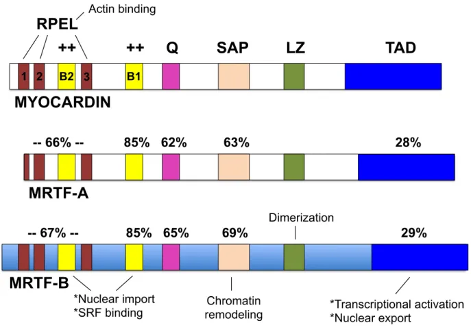
Myocardin and the MRTFs contain several conserved domains that mediate their transcriptional activity (Figure 1.3). First, myocardin, MRTF-A, and MRTF-B all possess a B1 domain located between the RPEL and glutamine-rich region near the N-terminus of the proteins. The B1 domain mediates the interaction with SRF (73). All three factors share an SAP domain, whose function is less defined. However, in other proteins, the SAP domain controls nuclear dynamics and organization (101). A leucine zipper (LZ) domain, located at the center of each transcription factor, mediates homo- and
heterodimerization between myocardin and the MRTFs. Finally, at the C-terminal region, myocardin contains a transcriptional activation domain (TAD) required for complete transactivation of SRF target genes (102). Interestingly, the TAD is weakly conserved between myocardin and the MRTFs, suggesting disparate mechanisms of regulation between the two transcription factors.
Previous studies from our lab indicated that S1PR2 signals through the RhoA GEF, LARG, to drive SMC differentiation and inhibit migration (103). Other studies demonstrated that LARG knockout mice exhibit decreased sensitivity to salt-induced hypertension (104). Further, combined deletion of LARG and PDZ Rho-GEF (PRG) in mice results in vascular branching defects, indicating that combinatorial interactions between RhoA GEFs influence vascular morphogenesis and/or overlapping function between two or more GEFs exists (105). Additional experiments from the Mack Lab identified an important role for the RhoA effectors, mDia1 and mDia2, in SMC
Figure 1.3. Domain structure and conservation of myocardin and the MRTFs. Conserved domains in the mouse myocardin, MRTF-A, and MRTF-B transcription factors include RPEL domains, Basic regions (B1 and B2), a Q-rich region, SAP
domain, Leucine Zipper (LZ) domain, and a transactivating domain (TAD). MRTF-A and MRTF-B share near identical sequence conservation with each other.
TAD
LZ
SAP
Q
++
++
B1 B2
1 2 3
RPEL
MYOCARDIN
MRTF-A
MRTF-B
-- 67% -- 85% 65% 69% 29% -- 66% -- 85% 62% 63% 28%
Chromatin
remodeling *Transcriptional activation *Nuclear export
Dimerization
*Nuclear import *SRF binding
reporter prevented re-expression of SM-MHC and calponin 7 days after carotid artery ligation in a mice. Interestingly, ligated arteries from DNmDia mice displayed reduced neo-intimas compared to wild-type littermates, most likely due to its inhibitory effects on SMC migration and polarity. Of note, a subset of SM22-Cre+/DNmDia+ mice exhibited a runted, hairless phenotype, patent ductus arteriosus, and embryonic hemorrhagic
phenotypes (107). In addition to conventional RhoA signaling, our lab and others demonstrated that nuclear RhoA signaling is a novel mechanism regulating SMC differentiation. mDia2, which shuttles between cytoplasmic and nuclear compartments, stimulates nuclear actin polymerization as well as MRTF-dependent SMC transcription. Furthermore, forced expression of NLS-tagged LARG significantly enhanced promoter activities of SMC-specific genes by upregulating RhoA signaling in the nucleus (108). Post-translational modifications of SRF, myocardin, and MRTF-A
In addition to directly regulating chromatin dynamics, post-translational
modifications (PTMs) also affect transcription factor function and downstream target gene expression (109). Some of these PTMs include phosphorylation, ubiquitylation, methylation, acetylation, and oxidation. Myocardin acetylation by p300 enhances its association with SRF, stabilizes the myocardin-SRF-CArG complex, and upregulates smooth muscle and cardiac gene expression (110, 111). Myocardin is also
phosphorylated by GSK3β and ERK, which both inhibit myocardin-dependent smooth muscle transcription (112, 113). Phosphorylation of SRF at T159 by PKA inhibits its interaction with CArG boxes, thereby diminishing downstream SMC-specific promoter activity (114).
cytoplasmic G-actin (115). More recent reports indicate that MRTF-A is phosphorylated at several other residues, each of which has varied effects on its nuclear localization (116). Interestingly, sumoylation of MRTF-A represses its ability to transactivate smooth muscle promoters, while SUMO modification activates myocardin at cardiac genes (117, 118). Clearly, tight post-translational regulation of SRF and the myocardin family of transcription factors is an important mechanism controlling smooth muscle gene expression in response to different stimuli and contexts. It will be necessary to identify additional PTMs regulating activity of smooth muscle transcription factors and their effects on SMC differentiation.
Regulation of SMC differentiation by Notch/RBPJ
Although myocardin and the MRTFs are by far the most potent drivers of SMC differentiation, additional transcription factors regulate smooth muscle gene expression. The transcription factor RBPJ is ubiquitously expressed and binds to consensus
GTGGG sequences in Notch target genes (119-121). Notch/RBPJ signaling is required for arterial specification during development and maintains blood vessel integrity by inducing SMC differentiation (122, 123). Work in zebrafish revealed that Notch signaling was essential for arterial specification. Specifically, inhibition of Notch diminished
the human disease, CADASIL, in which patients suffer from transient ischemic attacks due to subcortical infarcts. Histological examination of post-mortem tissues from CADASIL patients revealed loss of cerebrovascular SMC, very similar to that seen in Notch3 deficient mice (127, 128).
Notch is activated when a cell expressing jagged or delta-like ligand engages the Notch receptor expressed on the surface of a neighboring SMC. Upon notch activation, gamma secretase cleaves the notch intracellular domain (NICD), which translocates into the nucleus to bind to RBPJ. Under unstimulated conditions, RBPJ binds
mastermind-like (MAML), which represses notch target genes. However, with notch activation, NICD displaces MAML and increases RBPJ/Notch-dependent transcription. While this model implies that RBPJ activates gene expression, independent studies indicate that the effect of RBPJ on SMC differentiation is highly context specific (129, 130). For example, one study demonstrated that NICD overexpression and jagged-1 stimulation increased SM-MHC expression in SMC, while another study from the same lab reported that HERP-1, a notch target gene, was induced after arterial balloon injury and repressed myocardin-dependent smooth muscle transcription by physically
inhibiting SRF-CArG binding (131-133). Furthermore, our lab recently showed that RBPJ binds to methylated GC repressors in the SM-MHC promoter to inhibit
transcription after vascular injury (120). All together, these studies suggest that Notch can function as both a repressor as well as an activator of SMC differentiation
depending on its specific context.
Regulation of SMC differentiation by the TEADs
of Hippo leads to phosphorylation of the Mst1/2 serine/threonine kinases, which activate another set of serine/threonine kinases, called Lats1/2. Active Lats1/2 phosphorylate and inhibit Yap/Taz, which prevents their nuclear accumulation and subsequent
activation of the TEAD transcription factors. Thus, when the Hippo pathway is activated, TEADs inhibit target gene expression. Only when Hippo is inactive are Yap/Taz able to translocate to the nucleus to bind to the TEADs to relieve target gene repression. The Hippo signaling cascade has been implicated in a variety of cellular functions, including regulating cell size, proliferation, cell-cell communication, stem cell renewal, and
differentiation (135-137).
Members of the TEAD family, composed of TEADs 1-4, are expressed to varying degrees in smooth, cardiac, and skeletal muscle tissues and regulate expression of muscle-specific genes (138). TEAD1 is highly smooth muscle-specific and has been shown to regulate SMC differentiation, although the exact direction of effect is not understood (139, 140). All TEAD proteins contain a highly conserved N-terminal TEA DNA-binding domain, which recognizes consensus motifs. In particular, TEAD1
drive expression of muscle-specific genes (145, 146). It is thought that unique
combinations between TEAD1 and other transcription factors contribute to SMC-specific gene expression and may explain the different effects of TEAD1 on smooth muscle differentiation at different developmental stages.
Regulation of SMC differentiation by Nkx-3.2 and GATA-6
Nkx-3.2 and GATA-6 form a tripartite complex with SRF to modulate smooth muscle-specific differentiation. Interestingly, when either the Nkx, GATA, or CArG DNA binding motif is mutated within the alpha1 integrin luciferase promoter construct,
transcription activity is significantly reduced, indicating cooperativity between all three of these factors (147-149). Furthermore, Nkx-3.2, GATA-6, and SRF have overlapping expression in the arterial SMCs, which differs from the cardiac-specific expression patterns of Nkx-2.5 and GATA-4 (150). These studies indicate that transcription factors other than myocardin and the MRTFs likely function in an SRF/CArG-dependent
manner to further distinguish smooth versus cardiac muscle differentiation. Regulation of SMC differentiation by TGFβ
sac (153). TGF-β also plays a pivotal role in development of aortic aneurysms, in which aortic dilatation is due to degeneration of the artery’s medial layer. Although aortic aneurysms are relatively rare, they are associated with a strikingly high mortality rate. By far, the most direct line of evidence linking SMCs and aortic aneurysms are human mutations within the TGFβ signaling axis that lead to varying degrees of pathology. To date, mutations have been identified in TGFBR1, TGFBR2, TGFB2, SMAD3, and SMAD4, which are found in patients with aortic aneurysm syndromes (e.g., Loeys-Dietz Syndrome) (154-159). Of note, smooth muscle gene expression is downregulated in aortic aneurysm samples and is observed in a mouse model of early aortic aneurysm formation, indicating that failed SMC differentiation may contribute to disease (160).
The SMAD family of transcription factors, specifically SMAD2, 3, and 4, are critical for SMC differentiation and normal vascular development (152). The SMAD transcription factors (hereon referred to as SMADs) are downstream of TGF-β signaling. Briefly, cleaved TGF-β ligand binds to type II TGF-β receptor, which leads to
phosphorylation of the type I receptors. Subsequently, phosphorylated type I TGF-β receptor recruits and phosphorylates SMAD2 and SMAD3, which promotes their
interaction with SMAD4. The SMAD2/3/4 complex translocates into the nucleus, where it binds to a GTCT DNA element, but with low affinity. Given such weak binding, the SMADs interact with other transcription factors to drive robust TGF-β-dependent gene expression. For example, SMAD3 interacts with deltaEF1 to transactivate TGF-β-target genes. Importantly, some of the effects of TGF-β are SRF-CArG-dependent, and
transcriptional activation by TGF-β (163-165). In addition to these direct effects, TGF-β indirectly upregulates SMC differentiation by increasing the expression of genes that themselves enhance SMC transcription. For example, TGF-β increases expression of the RhoA GEFs, Net-1 and GEF-H1, which activate RhoA-dependent SMC-gene transcription (166, 167).
Regulation of SMC differentiation by extracellular matrix and cell stretch
Various extracellular matrix proteins signal through integrin receptors to differentially regulate SMC differentiation and phenotype (168-171). Freshly isolated SMCs grown on fibronectin are more synthetic and proliferative, while collagen IV and laminin maintain the SMC in its differentiated, contractile state (172). This response is best exemplified during the SMC’s response to injury, where breakdown of extracellular matrix proteins and re-expression of fibronectin stimulates SMC dedifferentiation and proliferation (173, 174). In brief, upregulation of extracellular matrix-integrin signaling by fibronectin leads to activation of the non-receptor tyrosine kinases, focal adhesion kinase (FAK) and Src, which activate the ERK signaling pathway (175). ERK activation downregulates SMC differentiation. In contrast, collagen IV upregulates SMC
differentiation by increasing expression of myocardin as well as enhancing enrichment of SRF at CArG boxes in SMC-specific gene promoters (176). The exact signaling mechanisms downstream of laminin and collagen IV that lead to increased SMC transcription, however, are less understood.
Stretch of the blood vessel, particularly in hypertension, results in increased contractile gene expression in the SMC. Additionally, prolonged stretch induces
role of RhoA in the stretch response is demonstrated by inhibiton of stretch-induced upregulation of SMC-specific gene expression in cells treated with the Rho inhibitor, C3 transferase, and to a lesser extent with the ROCK inhibitor, Y-27632 (180). Additionally, there is some evidence that FAK, ERK, and Akt are involved in the stretch response. Specifically, FAK and ERK phosphorylation increases in response to portal vein stretch, and FAK activity was required for the differentiation of mesenchymal stem cells
subjected to stretch (181, 182).
Please refer to Figure 1.4 for a summary of the transcription mechanisms regulating SMC differentiation under normal conditions as well as in the phenotypically modulated disease-associated state.
Epigenetic mechanisms regulating SMC differentiation
Histone acetylation and methylation
Epigenetic regulation of gene expression is characterized by mechanisms that reversibly modify DNA bases, histone tails, or transcription factors, without changes to the DNA sequence. Thus, two cells that are genotypically identical can have completely opposite phenotypes due to very different epigenetic mechanisms controlling their gene expression. One of the ways epigenetic mechanisms regulate transcription is by
affecting the accessibility of critical transcription factors to DNA (183). In its native conformation, genomic DNA is wrapped around core histones to form a nucleosome, which is the basic unit of chromatin. Histone modifications, which alter histone charge and thus interaction with other histone components and DNA, affect nucleosome density and chromatin organization. Less dense, more unpacked nucleosome configurations allow transcription factors to contact the DNA to influence gene
Figure 1.4. Contrasting the transcription mechanisms that regulate SMC
differentiation in the healthy versus phenotypically modulated SMC. Left) Multiple environmental cues and signaling pathways converge to regulate expression of mature SMC markers (e.g., SM-MHC). These include cell stretch, extracellular matrix, and GPCR activation, which signal through RhoA. TGFβRII and Notch activation are also potent stimulators of SMC differentiation. Right) Signaling mechanisms regulating dedifferentiation in SMC in disease states such as atherosclerosis. Injury stimulates downstream Ras/Raf/MEK/ERK pathways directly and indirectly by activating PDGF-BB signaling. Note that FAK inhibition of RhoA is transient and later leads to RhoA
activation. Expression of early response growth genes is upregulated, leading to
proliferation and fibronectin production, which further propogates phenotypic switching.
Nucleus
Cytoplasm
RhoA
ROCK mDia1/2
F F F F
RBPJ
CArG
SRF Stretch α β
FAK
Collagen IV, Laminin
Gq 12/13 G
GPCRs (S1PR2, etc.) GTCT TEAD1 GTGGG YAP/TAZ Smad 2/3 P Smad 4
TβRII
TGFβ MRTFs NICD Notch3 Jag-1, Dll4
Differentiation markers (e.g., SM-MHC) CArG
CArG
SRF Elk-1 P
Inhib. of differentiation markers
GC Repr.
KLF4
Early growth (e.g., c-fos) and prolif. genes
MRTF-A G G G SRF P PKA
α β
FAK Fibronectin RhoA PDGFRβ PDGF-BB
Ras/Raf
MEK ERK HDACs P INJURYHealthy SMC
Phenotypically
II, to initiate transcription.
Histone acetylation and methylation occur mainly on lysines and are catalyzed by histone acetyltransferases and histone methyltransferases, respectively. These histone marks are reversed by histone deacetylases and demethylases, respectively, as their names suggest. Generally, histone acetylation leads to a more “open” chromatin configuration (referred to as euchromatin), while methylation results in both open or closed (heterochromatin) chromatin states, depending on which histone and histone tail residue is modified. H3K4 and H3K79 methylation are associated with transcriptionally active, open regions in SMC-specific genes, while H3K9, H3K27, and H4K20
methylation mark heterochromatinized gene regions (184). Several histone/chromatin modifiers that regulate SMC differentiation have been identified, and these will be discussed below.
Histone modifications are tightly connected to the SRF/CArG interaction (185). Seminal work by the Owens Lab indicated that myocardin interacted with H3K4
dimethylation and enhanced SRF binding to CArG boxes in SMC gene promoters. This same study revealed that SRF enrichment at CArGs led to H3K9 acetylation and H3K79 dimethylation, both features of active transcription, since CArG mutant promoters did not display these features of open chromatin (63). Separate studies found that
myocardin induced acetylation of histones by recruiting the histone acetyltransferase, p300 (111). Thus, epigenetic mechanisms directly enhance SRF/myocardin binding to DNA, which in turn recruits histone-modifying enzymes that deposit specific marks to further promote accessibility of key transcription factors.
Chromatin and epigenetic modifiers in SMC
transcription. Histone lysine methylation is catalyzed by SET (Su(var)3 to 9, Enhancer of Zeste, Trithorax) domain-containing lysine methyltransferases. Seven families of histone methylransferases exist: SUV39, SET1, SET2, RIZ, SMYD, EZ, and SUV 4-20. Some methyltransferases, including SET7/9 and SET8, do not belong to any of these families. The most studied is the SUV39 family, which includes G9a, SUV39H1, SUV39H2, SETDB1, and SETDB2 and specifically methylate H3K9. Lysine
methyltransferases can mono-, di-, or tri-methylate a single lysine residue, and the degree of methylation for each methyltransferase is different (186). Additionally, each methyltransferase may methylate more than one lysine per histone. For example, SMYD2 methylates H3K4 and H3K36 (187). Adding to this complexity, methylation at one histone lysine residue may prevent or promote methylation at an adjacent lysine (183). Furthermore, there is additional crosstalk between methylation and other histone modifications, including acetylation and phosphorylation. Histone methylation is
reversed by various histone demethylases, each of which recognizes different lysines on specific histones. The H3K9-specific demethylase Jmjd1a is expressed in multiple tissues, including SMCs, and decreases H3K9 methylation at CArG-containing smooth muscle promoters. Interestingly, our lab showed that Jmjd1a interacts with all three myocardin family members, and Jmjd1a overexpression resulted in significant transactivation of myocardin/MRTF-dependent transcription of SMA and SM22 promoters. Conversely, Jmjd1a knockdown in SMC inhibited MRTF-dependent gene expression, which coincided with upregulation of H3K9 dimethylation at multiple SMC-specific promoters (188).
in regulating histone methylation at smooth muscle promoter regions. Inhibiting either SET7/9 or G9a in mice reduces renal fibrosis and expression of smooth muscle
markers, such as SMA (189, 190). Furthermore, SET7/9 induces expression of smooth muscle genes in mouse embryonic stem cells (191). In addition to their canonical roles in methylating histones, histone methyltransferases can also methylate non-histone proteins. For example, YY1, a transcriptional repressor of SMC differentiation, is
methylated by SET7/9. YY1 methylation at K173 and K411 increased its binding affinity to p53, RAD1, and ABL1 promoters (192). Whether or not SET7/9 methylation of YY1 affects binding to smooth muscle-specific promoters has yet to be determined. Also, further studies are needed to determine if YY1 methylation affects its interaction with SRF, since YY1 is a known SRF-interacting protein.
Brg1 and Brm, which are components of the ATP-dependent chromatin remodeling SWI/SNF complex, are required for myocardin/MRTF-dependent gene expression in SMC. Brg1, SRF, and MRTF-A form a transcriptional complex, and Brg1 facilitates the ability of both myocardin and MRTF-A to increase binding of SRF to CArG boxes (193, 194).
DNA methylation and demethylation are significant epigenetic mechanisms regulating smooth muscle gene transcription (195). The TET family of DNA
demethylases cause gene activation by oxidizing methylated-cytosine. Base excision repair converts (i.e. demethylates) methyl-cytosine to unmethylated-cytosine. TET2 associates with CArG elements and is necessary for SMC differentiation. TET2 expression correlates with SMC phenotype and is upregulated in mature, contractile SMCs but downsregulated after vascular injury and in atherosclerotic lesions.
of smooth muscle gene expression (195, 197). DNase-I Hypersensitivity
DNase-I Hypersensitivity is employed to identify open chromatin regions throughout the genome. Several software programs can be used to call the DNase Hypersensitive Sites (DHS) as peaks and thus indicate regions that are accessible to transcription factors (198, 199). In this manner, DHS can be used in combination with other relevant bioinformatic datasets (ChIP-seq, 3C, conservation, etc.) to denote
critical promoter and enhancer regions as well as long-range chromatin interactions that control cell type-specific gene expression (200).
Non-coding RNAs in SMC differentiation
Nearly 75% of the human genome is transcribed, but only 3% is actually translated into (i.e. “encodes”) protein (201). Over the last two decades, non-coding RNAs (ncRNAs) have emerged as significant regulators of cell function, affecting processes including proliferation, apoptosis, and differentiation (201-204). Although there are several categories of ncRNAs, the two most studied are the microRNAs (miRNAs) and long non-coding RNAs (lncRNAs). miRNAs are short single-stranded RNAs about 22 nucleotides in length that generally silence gene expression by binding to the 3’UTR of mRNA to block translation (205). Importantly, a single miRNA can target multiple genes, which is once reason why miRNAs can have such drastic effects on cell phenotype. Several miRNAs that affect SMC differentiation and/or phenotype have been identified, including miR-221 and miR-222, which inhibit vascular smooth muscle differentiation and increase SMC proliferation (206, 207). Knockout of these two
SMC differentiation, the most studied miRNAs are the miR-143/145 cluster. miR-143 and miR-145, which are transcribed from the same gene locus, are highly expressed in SMC. Numerous studies characterizing the expression pattern of the miR-143/145 cluster in vivo indicate that their expression is highly specific to vascular SMCs (209). miR-143/145 repress proliferation by inhibiting KLF4 and Elk-1 and activate
differentiation by stabilizing myocardin (210). The miR-143/145 duo is thus a rare example of miRNAs that selectively enhance rather than repress their target.
Interestingly, this miRNA cluster is regulated via an SRF-dependent mechanism, and thus can increase its own expression via a feed-forward mechanism by directly targeting and increasing myocardin levels. Furthermore, miR-145 represses several
actin-remodeling proteins, which results in substantial cytoskeletal reorganization (211, 212). Given that actin polymerization is directly linked to MRTF nuclear translocation and subsequent activation of SRF-dependent SMC genes, this is yet another pathway by which mir-145 affects SMC differentiation.
the introns of the coding genes they overlap, while intergenic lncRNAs are transcribed from genomic loci between genes (214).
Several lncRNAs in SMCs have been identified. One of the first lncRNAs to be identified was ANRIL at the CDKN2B-AS1 locus, which contains several DNA variants associated with cardiovascular disease (215) Interestingly, mutations in ANRIL lead to increased SMC proliferation and are associated with higher rates of coronary artery disease (CAD) (216). It is hypothesized that SNPs affecting ANRIL expression also control the expression of nearby cell-cycle control genes, CDKN2A and CDKN2B, and that aberrant SMC proliferation contributes to CAD (217). The most well characterized lncRNA in SMC, however, is MYOSLID (MYOcardin-induced Smooth muscle LncRNA, Inducer of Differentiation). MYOSLID was identified in a screen for lncRNAs that were significantly upregulated by myocardin overexpression in human coronary SMC.
MYOSLID contains 3 CArG boxes in its promoter. In addition to myocardin, MYOSLID is also regulated by TGFβ/SMAD signaling. Importantly, MYOSLID is localized to the cytoplasm, and thus does not directly affect SMC-specific gene transcription. Instead, MYOSLID is required for actin polymerization as well as SMAD2 phosphorylation, which regulate downstream SRF- and TGFβ-dependent smooth muscle genes, respectively. As partial evidence of this, knockdown of MYOSLID significantly reduces expression of SMA, CNN, and SM22 in human coronary SMC (218).
ENCODE Consortium, UCSC Genome Browser, and GTEx Database
Browser contains extremely useful bioinformatic datasets of transcriptionally interesting features deposited by different labs. One advantage of this tool is that information from multiple cell types is available, which is useful for making determinations regarding cell-type specificity of respective gene regions. Briefly, selected features that can be used to make predictions about active promoter/enhancer regions include H3K27Ac, H3K4me1, and DHS identified in various cell types. Furthermore, the UCSC Broswer integrates data from predictive biobases, such as miRNA binding sites from TargetScan as well as tissue specific gene expression from GTEx. All of these features allow the user to
analyze the multitude of transcription mechanisms that potentially regulate expression of a gene of interest.
Another useful web-based tool, the Genotype Tissue Expression (GTEx)
database (www.gtexportal.org), catalogues RNA-seq data from 53 different tissues as well as the genotype associated with each sample so eQTLs can be calculated for given genes. Users can view the top genes expressed in a specific tissue, search for eQTLs based on a gene or SNP ID, as well as view gene expression across all tissue types. One recent feature is the “histology image viewer,” where the user can view various tissue samples, each with an attached pathology note.
Genetic and molecular basis for blood pressure regulation1 RhoA signaling and hypertension
While monogenic diseases affecting renal salt-handling contribute to
hypertension, the fundamental cause of high blood pressure is increased peripheral vascular resistance. Increased vascular resistance is a direct result of increased SMC
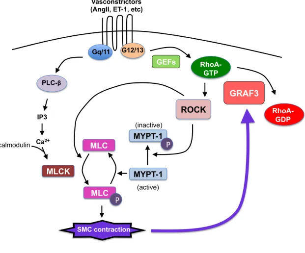
contractility, which is regulated by myosin light chain (MLC) phosphorylation. Activation of G-protein coupled receptors (GPCRs) by vasoconstrictors (angiotensin II, endothelin-1, etc.) leads to RhoA activation and a rise in intracellular calcium. RhoA signals
through its effectors, ROCK and mDia1/2, to increase actin polymerization, which has a direct effect on vessel tone. Calcium-calmodulin-dependent phosphorylation of MLC by myosin light chain kinase (MLCK) is the predominant mechanism regulating SMC contractility. ROCK inhibits myosin light chain phosphatase, thereby leading to sustained MLC phosphorylation and SMC contractility (Figure 1.5). Given the
importance of RhoA in regulating SMC contractility, it is no surprise that perturbation in upstream mediators or downstream effectors of RhoA in SMC affects blood pressure. Deletion of the SMC-specific RhoGAP GRAF3 (ARHGAP42) results in significant hypertension in mice due to increased RhoA activity and vascular resistance (219). In mouse models of hypertension, the RhoGEF p115 mediates angiotensin II-dependent RhoA activity and SMC contractility, while LARG is required for salt-induced
hypertension (104, 220). While it is clear that RhoA directly increases SMC contractility and blood pressure, the degree to which RhoA-dependent upregulation of contractile gene expression in SMC contributes to hypertension is not as apparent.
Public health relevance of hypertension
Hypertension is a devastating disease associated with significant morbidity and mortality due to detrimental pressure-related effects on the kidneys, heart, lungs, brain, and peripheral vasculature. Hypertension affects roughly 80 million people
Figure 1.5. Signaling mechanisms regulating SMC contraction. Vasoconstrictors bind to GPCRs, leading to calcium/calmodulin-dependent activation of myosin light chain kinase (MLCK), which phosphorylates myosin light chain (MLC). MLC is also activated by Rho-coiled kinase (ROCK). Phosphorylated myosin light chain incrases SMC contraction. In reponse to GPCR stimulation, GEFs promote RhoA-GTP, which activates ROCK. ROCK inhibits myosin phosphatase (MYPT-1), thereby leading to sustained MLC phosphorylation. RhoA is inactivated by the RhoGAPs, GRAF3. GRAF3 expression is upregulated with sustained cell stretch to limit the amount of SMC
contraction. P, phosphorylation.
Gq/11 G12/13
PLC-β
IP3
Ca2+
calmodulin
MLCK
MLC MLC
P
MYPT-1 MYPT-1
P
(inactive)
(active)
ROCK RhoA-
GTP
GEFs
RhoA- GDP
SMC contraction
GRAF3
hypertension in the United States, estimates project that reasonable BP control is achieved in only about half of hypertensive patients. This reality coupled with recent projections that the incidence of hypertension will increase to about 41% in the US by 2030, indicate the urgent need for better screening and treatment modalities (222). Improvements in the detection and management of hypertension will undoubtedly be accomplished through a better understanding of the complex etiology of this disease.
One way to better predict patient response to therapy is to gain a more comprehensive understanding of the genes and genetic variants that influence BP regulation. Recent projections indicate that up to 60% of BP variation can be explained by genetic factors, but that no single gene exerts a principal effect. Thus, BP is
considered to have a complex non-Mendelian mode of inheritance. Indeed a
combination of classic positional cloning strategies in families with numerous affected members combined with more recent population-based GWAS studies have led to the identification of 25 rare mutations and 53 SNPs that are predicted to contribute to BP control (223). The aim of this section of is to highlight variants that impinge on the expression or activity of members of the RhoA signaling axis.
RhoA-related forms of monogenic hypertension
signaling in vascular SMC may also play a role in Gordon’s Syndrome patients. Exclusion of exon 9 abrogates the Cullin-3 dependent interactions between RhoBTB and the E3 ligase and as RhoBTB serves as a chaperone to recruit RhoA to this degradation complex, expression of exon 9-deficient Cullin-3 leads to aberrant RhoA accumulation (225, 226).
SNPs/EQTLs in RhoA-signaling molecules
Because Rho kinases are major RhoA effector proteins and because both animal and human studies have shown that treatment with Rho-kinase inhibiting compounds lowers BP, a number of case-controlled studies were designed to determine if genetic variants in these genes might influence the development of human hypertension (Figure 1.6). One group examined the effect of ROCK2 genetic variations on BP in 168 pairs of mono- and dizygotic twins. In this study, four variants were identified in ROCK2, the most notable of which was a nonsynonymous SNP in exon 10 that resulted in a substitution of Thr with Asn at amino acid 431. Importantly, the Asn substitution was associated with increased systemic vascular resistance and BP and was predicted to account for 3-5% of the BP variance between these patients (227). Another study in which 18 tag SNPs within the ROCK2 locus were genotyped in 586 normotensive
controls and 607 hypertensive Caucasian patients identified a haplotype defined by four SNPs (rs965665, rs10178332, rs6755196, rs10929732) that was recessively associated with a lower risk of hypertension (p=0.003). However, a subsequent study in a separate population of 1344 Chinese patients with coronary artery disease and hypertension and 1267 ethnically and geographically matched controls did not find an association
Figure 1.6. Pharmacologic and genetic regulation of the RhoA signaling axis. Schematic ndicating the sites of action of pharmacological inhibitors (bold) of RhoA signaling molecules. Polymorphisms (SNPs/eQTLs) that could influence RhoA signaling are also shown. AJ: Adherens junction; A2R: Angiotensin type II receptor; ARBs:
Angiotensin receptor blockers; ACEIs: Angiotensin converting enzyme inhibitors; ASAH1: Acid ceramidase; SPHK1: Sphingosine kinase 1.
Points of pharmacologic or genetic activation/inhibition of RhoA signaling molecules
ARBs, ACEIs PLEKHA7 rs381815, rs12806040, rs112467382
RhoA
GEFs GAPs ArhGAP42: rs633185, rs604723ROCK 1
Geranyl-geranyl group StatinsROCK 2
PS1PR
ASAH1SPHK1 rs8176328, rs2247856 rs1049874,
rs1071645
miR-133 miR-145?
Azainadole-1 Fasudil,
Y-27632, GSK269962A, SB-772077 SLx-2119, SAR407899 cingulin paracingulin Rhotekin-2 CUL3 GDIs RhoBTB P rs1530440 nearby RTKN2
rs1530440 nearby RhoBTB; Also, mutations in Exon 9 of CUL in separate study