A Serendipitous Mutation Reveals the
Severe Virulence Defect of a
Klebsiella
pneumoniae fepB
Mutant
Michelle Palacios,aChristopher A. Broberg,aKimberly A. Walker,a Virginia L. Millera,b
Department of Microbiology and Immunology, University of North Carolina, Chapel Hill, North Carolina, USAa; Department of Genetics, University of North Carolina, Chapel Hill, North Carolina, USAb
ABSTRACT Klebsiella pneumoniaeis considered a significant public health threat be-cause of the emergence of multidrug-resistant strains and the challenge associated with treating life-threatening infections. Capsule, siderophores, and adhesins have been implicated as virulence determinants ofK. pneumoniae, yet we lack a clear un-derstanding of how this pathogen causes disease. In a previous screen for virulence genes, we identified a potential new virulence locus and constructed a mutant (smr) with this locus deleted. In this study, we characterize thesmrmutant and show that this mutation renders K. pneumoniae avirulent in a pneumonia model of infection. The smr mutant was expected to have a deletion of three genes, but subsequent genome sequencing indicated that a much larger deletion had occurred. Further analysis of the deleted region indicated that the virulence defect of thesmrmutant could be attributed to the loss of FepB, a periplasmic protein required for import of the siderophore enterobactin. Interestingly, a ΔfepB mutant was more attenuated than a mutant unable to synthesize enterobactin, suggesting that additional pro-cesses are affected. As FepB is highly conserved among the members of the family Enterobacteriaceae, therapeutic targeting of FepB may be useful for the treatment of Klebsiellaand other bacterial infections.
IMPORTANCE In addition to having a reputation as the causative agent of several types of hospital-acquired infections, Klebsiella pneumoniae has gained widespread attention as a pathogen with a propensity for acquiring antibiotic resistance. It is ca-pable of causing a range of infections, including urinary tract infections, pneumonia, and sepsis. Because of the rapid emergence of carbapenem resistance among Kleb-siella strains, there is a dire need for a better understanding of virulence mecha-nisms and identification of new drug targets. Here, we identify the periplasmic trans-porter FepB as one such potential target.
KEYWORDS Klebsiella, RamA, enterobactin, pneumonia, siderophore, yersiniabactin
K
lebsiella pneumoniae is a Gram-negative bacterium commonly classified as an opportunistic nosocomial pathogen capable of causing a variety of infections, including urinary tract infections, pneumonia, and sepsis (1–5). It is often found as a commensal resident of the gastrointestinal tract, and this is believed to be a primary source of infection (2, 6–8). Recently,K. pneumoniaealso has been shown to be capable of causing community-acquired infections such as pyogenic liver abscesses, meningitis, and endophthalmitis (9–11). The increasing prevalence of antibiotic-resistant strains only serves to compound the clinical importance ofK. pneumoniaeand the difficulty of treating those infected with extended-spectrum-lactamase-resistant or carbapenem-resistant strains (12–16). Resistance to carbapenems is of particular concern, as they are used as drugs of last resort to treat Gram-negative infections (12, 17).During infection, sequestration of iron by the host limits the availability of free iron,
Received1 August 2017Accepted1 August 2017 Published23 August 2017
CitationPalacios M, Broberg CA, Walker KA, Miller VL. 2017. A serendipitous mutation reveals the severe virulence defect of a Klebsiella pneumoniae fepBmutant. mSphere 2:e00341-17.https://doi.org/10.1128/mSphere .00341-17.
EditorSarah E. F. D'Orazio, University of Kentucky
Copyright© 2017 Palacios et al. This is an open-access article distributed under the terms of theCreative Commons Attribution 4.0 International license.
Address correspondence to Virginia L. Miller, [email protected].
Host-Microbe Biology
and as a result, bacteria produce their own chelators to scavenge iron. Iron acquisition is an essential component of most bacterial pathogens, as iron is required for cellular and metabolic activities (18). Siderophores are small secreted molecules with a high affinity for ferric iron; these are classified on the basis of the chemical nature of the Fe3⫹
coordination (19). The catecholate-type siderophore enterobactin is produced by most K. pneumoniaestrains (20, 21). However, community-acquired isolates and those that cause invasive disease typically encode additional siderophore systems (salmochelin, yersiniabactin, aerobactin) (22). Salmochelin is a C-glucosylated enterobactin produced by some isolates of Salmonella, Escherichia coli, and Klebsiella, and its synthesis is dependent on enterobactin. Mutants unable to produce enterobactin are also unable to produce salmochelin (23, 24). TheiroAlocus encodes enzymes necessary to modify enterobactin, as well as proteins required for salmochelin transport (25). The yersini-abactin locus is found in many invasive K. pneumoniae isolates and encodes a phenolate-type siderophore that was first identified as part of a pathogenicity island in Yersinia (26). Interestingly, in a genome-wide association study of a broad range of K. pneumoniaeisolates, yersiniabactin was found to be the most prevalent virulence-associated locus and was found to be a predictor of infection versus carriage (22). Aerobactin is yet another siderophore produced by a smaller fraction ofK. pneumoniae strains than either enterobactin or yersiniabactin (22). Although aerobactin has a lower affinity for Fe3⫹than enterobactin or yersiniabactin, it is frequently produced by
isolates from pyogenic liver abscesses (27).
To date, the identified virulence factors ofK. pneumoniaeprimarily include capsule, lipopolysaccharide (LPS), fimbriae, and siderophores, and these factors also have been identified as virulence factors in the strain used for the studies presented here (4, 28–34). Several high-throughput studies have been done with mouse models to identify additional bacterial virulence factors (34–40). Two of these screens were signature-tagged mutagenesis (STM) screens for factors affecting gastrointestinal col-onization and/or infection of the urinary tract (36, 37). These studies identified adhesins, LPS, and capsule. Another screen for gain of function when Klebsiella genes were expressed inE. coli identified a response regulator, AcrA, and LPS (40). A screen for genes expressed in vivo during septicemia identified genes involved in the use of siderophores (aerobactin and enterobactin) (39), and an STM screen in a model of liver abscess formation identified adhesins and regulators (38). Two of these studies focused on the identification of bacterial genes needed for survival in the lung; one approach used STM, and the other used transposon insertion site sequencing (34, 35). These screens identified capsule, LPS, siderophores, and transcriptional regulators. All of these screens also identified genes predicted to contribute generally to growth, as well as genes of unknown function.
Overall, there has been a lack of overlap in identified genes among the different screens conducted with lung, urinary tract, liver infection, and gastrointestinal coloni-zation models. This may be due to the fact that none of the screens were saturating, or it could be indicative of mechanisms that compensate for the loss of individual genes. These findings are further complicated by the use of different infection models and different pathogen and host strain backgrounds. While typically focused on the goal of identifying previously unknown bacterial factors contributing to disease, these screens primarily identified known virulence factors of K. pneumoniae, as well as metabolic functions generally contributing to growth.
K. pneumoniae strain. These studies found no role for ramAor nearby genes for virulence in a pneumonia model of infection. However, a serendipitous secondary mutation was identified, and further analysis of this mutation indicates that FepB, a periplasmic protein required for transport of enterobactin and salmochelin, is essential for virulence. Surprisingly, there were interesting differences in virulence between enterobactin synthesis mutants and the ΔfepBmutant.
RESULTS
The smr mutant is severely attenuated in a mouse model of pneumonia. A
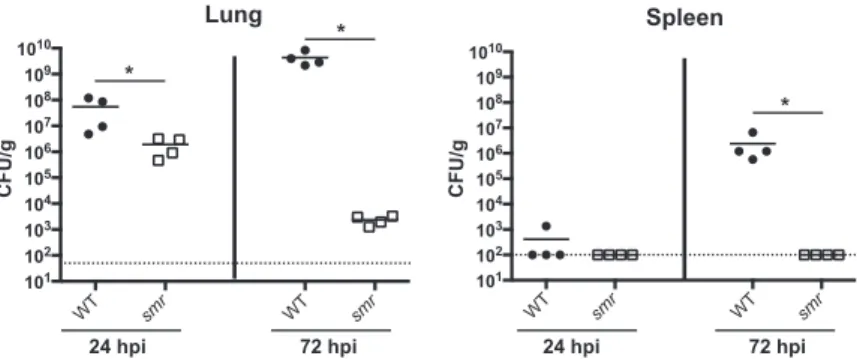
previous screen of strain KPPR1 transposon mutants identified genes required for colonization and survival in the lungs of infected mice (34). Thirteen mutants contain-ing disruptions withinramAor an adjacent gene,orf82, failed to be recovered from the lungs and spleens of infected mice. RamA is a transcriptional regulator linked to Salmonellasurvival in RAW 264.7 macrophages and virulence in BALB/c ByJ mice (41, 42). This led us to hypothesize that the ramA locus is important for the ability of K. pneumoniaeto infect the lungs. To test this, we constructed thesmr(spontaneous multidrug resistance) mutant, whereramAand the two flanking genes (orf82andromA) were targeted for deletion, and tested this strain in a mouse model of pneumonia (Fig. 1). Thesmrmutant caused slightly lower bacterial burdens at 24 h postinoculation (hpi) than KPPR1 (wild type [WT]). At 72 hpi, nearly 5 logs fewer CFU were recovered from mice infected with thesmrmutant than from WT-infected mice. The spleens of mice infected with the WT strain had nearly 107CFU/g of tissue, while thesmrmutant
was rarely detectable in the spleen at 72 hpi, reflecting a dissemination or systemic survival defect. Together, these data indicate that thesmrmutant is essentially avirulent in this infection model.
Deletions of individual genes in the targetedsmrlocus do not recapitulate the
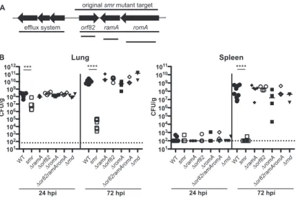
phenotype of thesmrmutant.To identify the gene(s) responsible for the phenotype of the smrmutant, we made in-frame deletions of each of the three genes (ΔramA, ΔromA, and Δorf82) in thesmrlocus and tested them in our pneumonia model (Fig. 2A). The phenotype of all three mutant strains resembled that of the WT, suggesting that the loss of a single gene was not sufficient to affect virulence (Fig. 2B). We concluded that neitherramA,orf82, norromA, individually contributed to virulence in this model or was responsible for the phenotype of thesmrmutant.
In examining the region more closely, we noted that an RND (resistance-nodulation-division superfamily) efflux pump system was encoded just upstream oforf82and that thesmrdeletion could have impacted the promoter driving the expression of this locus (Fig. 2A). RND efflux systems have been shown to play roles ranging from resistance to human antimicrobial peptides inPseudomonasto flagellar motility inBurkholderia(46).
WT smr WT smr
101 102 103 104 105 106 107 108 109 1010
WT smr WT smr
101 102 103 104 105 106 107 108 109 1010
Lung Spleen
*
* *
24 hpi 72 hpi 24 hpi 72 hpi
CFU/g CFU/g
FIG 1 Thesmrmutant is attenuated in a mouse model of pneumonia. Mice were inoculated i.n. with 2⫻ 104CFU of either the WT strain (KPPR1S; black circles) or the Δsmrmutant (VK82; white squares). At 24
or 72 hpi, mice were euthanized and their lungs and spleens were homogenized and plated for bacterial enumeration. Each symbol represents one mouse. The dotted line indicates the limit of detection, and symbols on the dotted line indicate that CFU counts were below the limit of detection. Data are from an individual representative experiment. Mann-Whitney tests were performed for statistical analysis.*,P⬍
Thus, we constructed two additional mutants, one with therndgenes and the other withorf82,ramA, andromAdeleted but with the putativerndpromoter intact (Δrndand Δorf82 ramA romA). The Δrndmutant colonized mice as efficiently as the WT strain (Fig. 2B). Intriguingly, the second mutant lacking the same three genes as thesmr mutant (Δorf82 ramA romA) also had no virulence defect.
Sequencing of the smr mutant reveals a large deletion.As targeted genetic
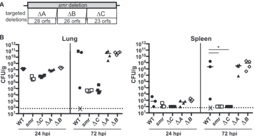
mutations in thesmrlocus failed to recapitulate thesmrphenotype, we hypothesized that the smr mutant contained a secondary mutation. Whole-genome sequencing revealed that the deletion in thesmrmutant was larger than intended. Instead of the targeted deletion oforf82,ramA, andromA, a single segment of 87,290 bp spanning 78 putative open reading frames was deleted.
A component of the enterobactin transport system contributes to virulence.To
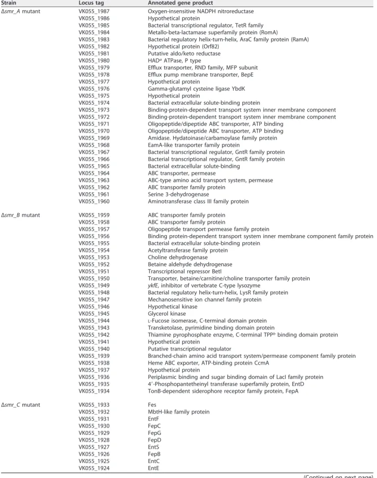
identify the factor(s) responsible for the virulence defect of the smr mutant, we constructed three mutants (Δsmr_A, Δsmr_B, and Δsmr_C) each with a deletion of approximately one-third of the genes deleted in thesmrmutant (Fig. 3A). The putative orf genes in each mutant are listed in Table 1. In our pneumonia model at 24 and 72 hpi, both Δsmr_Aand Δsmr_Bmutant-infected mice had bacterial burdens compa-rable to those of mice infected with the WT (Fig. 3B). However, the mice infected with Δsmr_Cmutant had⬎1 log fewer CFU/g at 24 hpi and nearly 6 logs fewer CFU/g at 72 hpi than mice infected with the WT. Thus, the Δsmr_C mutant recapitulated the phenotype of thesmrmutant, whereas the Δsmr_Aand Δsmr_Bmutants behaved like the WT strain.
Located within the region deleted in the Δsmr_Cmutant are genes necessary for the synthesis, export, and import of the siderophore enterobactin. We therefore hypothe-sized that a component of the enterobactin transport system was responsible for the virulence defect of thesmrmutant. We did not believe that the siderophore itself was responsible, as an ΔentB mutant, which is unable to synthesize enterobactin and salmochelin, is only modestly attenuated in this mouse pneumonia model (33). The enterobactin receptor FepA also was not implicated, as FepA is encoded within the region deleted in the Δsmr_Bmutant.
101 102 103 104 105 106 107 108 109 1010 1011 1012
original smr mutant target
efflux system orf82 ramA romA
CFU/g
24 hpi 72 hpi
101 102 103 104 105 106 107 108 109 1010 1011
CFU/g
24 hpi 72 hpi
*** **** ****
Lung Spleen
A
B
WTsmr
ΔramAΔorf82ΔromA
Δor82ramAromA
Δrnd WTsmr
ΔramAΔorf82ΔromA
Δor82ramAromA
Δrnd Δrnd Δrnd
Δor82ramAromA Δor82ramAromA
ΔromA
ΔromA Δorf82
Δorf82 ΔramA
ΔramA smr
smr WT
WT
FIG 2 Schematic ofsmrtargeted region andin vivophenotypes of mutants. (A) Schematic depicting open reading frames within or adjacent to thesmrtarget region (not to scale). Lines indicate the regions deleted in the mutants indicated. (B) Mice were inoculated i.n. with 2⫻104CFU of the WT strain (KPPR1S;
Siderophore transport involves several membrane proteins. For enterobactin, EntS and TolC are required for export, whereas FepA, FepDGC, and Fes are required for import. In addition, the periplasmic protein FepB is required for the import of both enterobactin and salmochelin. Because previous studies had implicated siderophore transport components in virulence (47), we targeted specific components of the enterobactin siderophore transport system and tested loss-of-function (ΔentS, Δfes, ΔfepB, andfepD::pKAS46) mutants in our pneumonia model (Fig. 4A). We included a different enterobactin synthesis (ΔybdB2 entABEC[referred to as Δentsyn]) mutant to confirm our previous findings obtained with the ΔentBmutant (33). We found that only the ΔfepBmutant recapitulated the phenotype of thesmrmutant, as demonstrated by the attenuation in the lungs and the lack of dissemination at 24 and 72 hpi (Fig. 4B). Consistent with previously studies, neither the Δentsynmutant (Fig. 4B) nor the ΔentB mutant (Fig. 5) recapitulated thesmrphenotype (33). In addition, loss offepBdid not affect the expression of the yersiniabactin system (Fig. 6), consistent with results previously obtained with an enterobactin synthesis mutant (33). Thus, the periplasmic transport protein FepB contributes to virulence in a manner distinct from that of enterobactin and salmochelin uptake alone.
A variety of different approaches were used to complement the ΔfepBmutant, but all were unsuccessful. Plasmid-based approaches failed to complement growth under iron-depleted conditions, despite the constitutive expression offepB(data not shown). We also attempted to repair the deletion, but this strain could not be obtained, for reasons we do not understand. Problems withfepBcomplementation are not unprec-edented and were also reported for aSalmonella fepBmutant (47). To ensure that the observed phenotype of the ΔfepB mutant was not a consequence of secondary mutations, a secondfepBmutant (fepB2) was constructed and found to recapitulate the virulence and growth phenotypes of the originalfepBmutant (Fig. 5). Additionally, we sequenced across the deletion junction of both of the ΔfepBmutants and obtained the expected sequence, suggesting that a larger deletion of the region surroundingfepB had not occurred (data not shown). Expression of the genes adjacent tofepB,entCand entS, was assessed by quantitative reverse transcription-PCR. Expression ofentCand entSwas not detected in the ΔfepBmutant but was in the WT (data not shown). EntC and EntS may be needed for growth under low-iron conditions, and their lack of expression provides a possible explanation for failed complementation intrans.
How-WTsmr C A B WTsmr C A B
101
102
103
104
105
106
107
108
109
1010
1011
1012
X
WTsmr C A B WTsmr C A B
101
102
103
104
105
106
107
108
109
1010
1011
1012
X
*
Lung Spleen
CFU/g CFU/g
24 hpi 72 hpi 24 hpi 72 hpi
smr deletion
28 orfs 26 orfs 23 orfs
ΔA ΔB ΔC
targeted deletions A
B
FIG 3 The smrmutant phenotype is recapitulated by a smaller targeted deletion. (A) Schematic depicting targeted subregions of thesmrmutant (not to scale). (B) Mice were inoculated i.n. with 2⫻104
TABLE 1 Genes deleted in breakdown mutants
Strain Locus tag Annotated gene product
Δsmr_Amutant VK055_1987 Oxygen-insensitive NADPH nitroreductase VK055_1986 Hypothetical protein
VK055_1985 Bacterial transcriptional regulator, TetR family VK055_1984 Metallo-beta-lactamase superfamily protein (RomA)
VK055_1983 Bacterial regulatory helix-turn-helix, AraC family protein (RamA) VK055_1982 Hypothetical protein (Orf82)
VK055_1981 Putative aldo/keto reductase VK055_1980 HADaATPase, P type
VK055_1979 Efflux transporter, RND family, MFP subunit VK055_1978 Efflux pump membrane transporter, BepE VK055_1977 Hypothetical protein
VK055_1976 Gamma-glutamyl cysteine ligase YbdK VK055_1975 Hypothetical protein
VK055_1974 Bacterial extracellular solute-binding protein
VK055_1973 Binding-protein-dependent transport system inner membrane component VK055_1972 Binding-protein-dependent transport system inner membrane component VK055_1971 Oligopeptide/dipeptide ABC transporter, ATP binding
VK055_1970 Oligopeptide/dipeptide ABC transporter, ATP binding VK055_1969 Amidase. Hydatoinase/carbamoylase family protein VK055_1968 EamA-like transporter family protein
VK055_1967 Bacterial transcriptional regulator, GntR family protein VK055_1966 Bacterial transcriptional regulator, GntR family protein VK055_1965 Bacterial extracellular solute-binding
VK055_1964 ABC transporter, permease
VK055_1963 ABC-type amino acid transport system, permease VK055_1962 ABC transporter family protein
VK055_1961 Serine 3-dehydrogenase
VK055_1960 Aminotransferase class III family protein
Δsmr_Bmutant VK055_1959 ABC transporter family protein VK055_1958 ABC transporter family protein
VK055_1957 Oligopeptide transport permease family protein
VK055_1956 Binding protein-dependent transport system inner membrane component family protein VK055_1955 Bacterial extracellular solute-binding protein
VK055_1954 Acetyltransferase family protein VK055_1953 Choline dehydrogenase VK055_1952 Betaine aldehyde dehydrogenase VK055_1951 Transcriptional repressor BetI
VK055_1950 Transporter, betaine/carnitine/choline transporter family protein VK055_1949 ykfE, inhibitor of vertebrate C-type lysozyme
VK055_1948 Bacterial regulatory helix-turn-helix, LysR family protein VK055_1947 Mechanosensitive ion channel family protein
VK055_1946 Hypothetical kinase VK055_1945 Glycerol kinase
VK055_1944 L-Fucose isomerase, C-terminal domain protein VK055_1943 Transketolase, pyrimidine binding domain protein
VK055_1942 Thiamine pyrophosphate enzyme, C-terminal TPPbbinding domain protein
VK055_1941 Hypothetical protein
VK055_1940 Putative transcriptional regulator
VK055_1939 Branched-chain amino acid transport system/permease component family protein VK055_1938 Heme ABC exporter, ATP-binding protein CcmA
VK055_1937 Hypothetical protein
VK055_1936 Periplasmic binding and sugar binding domain of LacI family protein VK055_1935 4=-Phosphopantetheinyl transferase superfamily protein, EntD VK055_1934 TonB-dependent siderophore receptor family protein, FepA
Δsmr_Cmutant VK055_1933 Fes
VK055_1932 MbtH-like family protein VK055_1931 EntF
VK055_1930 FepC VK055_1929 FepG VK055_1928 FepD VK055_1927 EntS VK055_1926 FepB VK055_1925 EntC VK055_1924 EntE
ever, this alone cannot explain the attenuation in vivo, as a ΔentS mutant was not attenuated and a ΔentCmutant (enterobactin synthesis) had a more modest attenua-tion level than the ΔfepBmutant (Fig. 4A) (33). Thus, we conclude that deletion offepB results in a phenotype distinct from that of other enterobactin system mutants.
AfepBmutant resembles a⌬entB⌬ybtSdouble mutant.We previously showed
that a ΔentB⌬ybtSmutant that is deficient in all siderophore production was severely attenuated (33). In comparing the defect of the ΔfepBmutant strain to those of other siderophore mutants, we noticed that the phenotype of the ΔfepBmutant was similar to that of the ΔentB⌬ybtSmutant. Because the attenuation of the ΔfepBmutant was much greater than that of the ΔentBmutant, we hypothesized that the role of FepB is not limited to enterobactin import and that it might be involved in an additional iron acquisition system. To gain a better understanding of the relationship between the phenotypes of these mutants, we tested the ΔfepBmutant together with the ΔentB ⌬ybtS mutant to determine if its virulence defect resembles that of a ΔentB⌬ybtS mutantin vivoand included a ΔentBmutant as a control (Fig. 5). The ΔfepBand ΔentB ⌬ybtSmutants had similar attenuation levels, which were more severe than that of the TABLE 1 (Continued)
Strain Locus tag Annotated gene product
VK055_1923 EntB VK055_1922 EntA
VK055_1921 Proofreading thioesterase in enterobactin biosynthesis, YbdB2 VK055_1920 Carbon starvation CstA family protein
VK055_1919 Helix-turn-helix family protein VK055_1918 Hypothetical protein
VK055_1917 Plasmid stabilization system family protein VK055_1916 Short-chain dehydrogenase family protein
VK055_1915 Iron-containing alcohol dehydrogenase family protein VK055_1914 ABC transporter family protein
VK055_1913 Branched-chain amino acid transport system/permease component family protein VK055_1912 Periplasmic binding and sugar binding domain of LacI family protein
VK055_1911 LVIVD repeat family protein
aHAD, haloacid dehalogenase.
bTPP, thiamine pyrophosphate.
entCEBA fepB entS fepDGC entF fes
101 102 103 104 105 106 107 108 109 1010 1011
CFU/g
24 hpi 72 hpi 24 hpi 72 hpi
** *
*
101 102 103 104 105 106 107 108 109 1010 1011
CFU/g
*
* **
Lung Spleen
A
B
WTsmrΔfepB
ΔentsynΔentSΔ fes
fepD::pKAS46 WTsmrΔfepB
ΔentsynΔentSΔ fes
fepD::pKAS46
WTsmrΔfepB
ΔentsynΔentSΔ fes
fepD::pKAS46 WTsmrΔfepB
ΔentsynΔentSΔ fes
fepD::pKAS46
FIG 4 FepB is responsible for thesmrmutant’s phenotype. (A) Schematic of the enterobactin genes located in the Δsmr_Cregion. (B) Mice were inoculated i.n. with 2⫻104CFU of the WT (KPPR1S; black
ΔentBmutant. This finding raises the question of whether FepB may be required for iron acquisition via systems other than enterobactin and salmochelin.
To address the role of FepB in iron uptake and to determine if the virulence defect could be due to reduced iron acquisition, we used anin vitrogrowth model. The ΔfepB, ΔentB, and ΔentB⌬ybtSmutants were grown in defined medium with or without the iron-chelating agent 2,2=-dipyridyl (DP). All of the strains had similar growth rates in the absence of DP, indicating that the mutants grow normally when iron levels are sufficient (Fig. 7A). However, in the presence of DP, the growth of the ΔfepBand ΔentB⌬ybtSmutants was severely restricted (Fig. 7B). Interestingly, the growth of the ΔentBmutant was restricted compared to that of the WT strain, but the triple sidero-phore (ΔentB⌬ybtS) mutant and the ΔfepBmutant grew even more slowly than the ΔentB mutant. These data suggest that FepB contributes to growth in an iron-dependent manner that is distinct from its known role in enterobactin and salmochelin uptake.
Yersiniabactin import is unaffected in a⌬fepBmutant.The ΔfepBmutant had a
stronger phenotype than an enterobactin/salmochelin synthesis mutant, and it resem-bled that of a triple siderophore mutant in both virulence and growth under iron limitation. Yersiniabactin is the only known siderophore produced by the ΔentBmutant
101 102 103 104 105 106 107 108 109 1010 1011
101 102 103 104 105 106 107 108 109 101010 11
WT
ΔfepBΔentB
ΔentBybtSΔfepB2 WT
ΔfepBΔentB
ΔentBybtSΔfepB2
WT
ΔfepBΔentB
ΔentBybtSΔfepB2 WT
ΔfepBΔentB
ΔentBybtSΔfepB2
CFU/g CFU/g
24 hpi 72 hpi 24 hpi 72 hpi
* *
Lung Spleen
FIG 5 A ΔfepBmutant resembles a triple siderophore mutantin vivo. Mice were inoculated i.n. with 2⫻ 104CFU of the WT (KPPR1S; black circles) or the ΔfepB(VK412; open squares, small closed circles), ΔentB
mutant (VK087; open diamonds), or ΔentBybtS(VK089; black squares) mutant. At 24 or 72 hpi, mice were sacrificed and their lungs and spleens were homogenized and plated for bacterial enumeration. Each symbol represents one mouse. The dotted line indicates the limit of detection, and symbols on the dotted line indicate that CFU counts were below the limit of detection. The data are from an individual representative experiment. Mann-Whitney tests were performed for statistical analysis.*,P⬍0.05.
0 20000 40000 60000 80000 100000
RFU/OD
600
WT
ΔfepB WT ΔfepB
No DP 200μM DP
***
ns
ybt-gfp
FIG 6 ybtAexpression is unchanged in the ΔfepBmutant. The WT strain and a ΔfepBmutant containing the yersiniabactin synthesis gene,ybtA, promoter cloned into the pPROBEgfpreporter plasmid were grown overnight, subcultured to an OD600of 0.2, and grown in LB medium for 6 h with or without
but not the ΔentB⌬ybtSmutant. Thus, we wanted to assess if the ΔfepBmutant is defective in yersiniabactin uptake. To do this, we performed a cross-feeding experiment to determine if the growth defect of the ΔfepBmutant under iron-limited conditions could be restored in the presence of yersiniabactin by coculturing the ΔfepBmutant with a yersiniabactin-producing strain. We predicted that if FepB is required for yersiniabactin import, a feeder strain producing yersiniabactin would be unable to restore the growth of the ΔfepBmutant. In this assay, test strains were spread onto M9 medium supplemented with 0.4% glucose and 0.2% Casamino acids (M9-CAA) agar containing DP and feeder strains were then spotted onto the surface of the plates. The WT and ΔentB, ΔentB⌬ybtS, and ΔfepBmutant strains were used as test strains, and the WT and the ΔentB(capable of producing yersiniabactin) and ΔybtS(does not produce yersiniabactin) mutants were used as feeder strains. As expected, the ΔybtSmutant was not able to complement the growth defect of the ΔfepBmutant, as the ΔfepBmutant should not be able to use the enterobactin produced by this strain (Fig. 8A). The WT and the ΔybtSmutant were able to complement the growth of the ΔentB⌬ybtSmutant, as expected (Fig. 8B). Importantly, the ΔentBmutant and the WT were able to restore the growth of the ΔentBmutant (as expected), as well as the ΔfepBmutant. This finding suggests that yersiniabactin can still be imported by a ΔfepBmutant.
To determine if the complementation of the ΔfepB mutant’s growth defect by a yersiniabactin-producing strain in the cross-feeding experiment was due to yersiniabac-tin production rather than the production of other secreted bacterial products, we performed a similar experiment by spotting purified apo-yersiniabactin instead of feeder strains. As described above, test strains (WT strain and ΔfepBand ΔentB⌬ybtS mutants) were spread onto M9-CAA agar containing DP. Various concentrations of apo-yersiniabactin were applied to paper discs that were placed on the agar plate to test for growth restoration and thus the ability to utilize yersiniabactin (Fig. 8C). The WT strain was able to grow even without yersiniabactin supplementation. The ΔentB⌬ybtS and ΔfepBmutants did not grow around the vehicle control (distilled H2O [dH2O]) disc.
However, upon the addition of yersiniabactin, the growth defect of the ΔentB⌬ybtS
0 2 4 6
0.01 0.1 1 10
hours
O
D
600
no DP
WT
fepB entB
entBybtS
0 2 4 6
0.01 0.1 1 10
hours
O
D
600
100μM DP
A
B
mutant was restored in a concentration-dependent manner; this is an expected result because this strain is still able to import exogenous yersiniabactin. Addition of apo-yersiniabactin also restored the growth of the ΔfepBmutant (Fig. 8C). Together, these data suggest that FepB is not required for yersiniabactin importin vitroand that the virulence defect of the ΔfepBmutant is due to a mechanism unrelated to yersiniabactin import.
Capsule production is not responsible for the ⌬fepB mutant’s phenotype.
Capsule is considered a primary virulence factor ofK. pneumoniae(reviewed in refer-ence 4). Therefore, to test if there was a change in capsule production that could contribute to the ΔfepB mutant’s phenotype, we measured its uronic acid content. When the ΔfepBmutant and the WT strain were grown in Luria-Bertani (LB) medium at 37°C, the same conditions used for the inoculum used in mouse experiments, there was no difference in capsule production (Fig. 9A). Similarly, when mucoviscosity was measured (another assay for capsule phenotypes), we saw no measureable difference between the WT and the ΔfepBmutant (Fig. 9B).
Because iron levels can affectK. pneumoniaecapsule production (48), we decided to test if capsule production is altered in the ΔfepBmutant under low-iron conditions. All four siderophore system (ΔfepB, ΔentB, ΔybtS, and ΔentB⌬ybtS) mutants had a modest, nonsignificant reduction in capsule production (Fig. 9C). The mucoviscosity of the siderophore system mutants was also lower than that of the WT (Fig. 9D). Importantly, there was no difference between the capsule production levels of the ΔfepBand ΔentB mutants. How FepB affects virulence is not clear, but it does not appear to be related
Test Strain Feeder Strain
Wild-type Ent
-
Ybt+
Ent+
Ybt-Wild-type ΔfepB
ΔentB
ΔentBΔybtS
+
+
+
+
+
+
+
+
+
+
+
−
WT
Δ
fepB
Δ
entB
Δ
ybtS
1μM1μM
1μM
100μM
100μM
100μM
dH2O
dH2O
dH2O
WT
Ent+
Ybt-Ent- Ybt+
A B
C
FIG 8 Addition of yersiniabactin restores the growth defect of the ΔfepBmutant under iron-limited conditions. Test strains were grown in M9-CAA and spread plated onto M9-CAA agar containing 100M DP. (A) Plate testing of the ΔfepBmutant (spread plated). Feeder (WT and ΔentBand ΔybtSmutant) strains were then spot plated to test for complementation (growth restoration around the feeder spot). (B) Summary of results represented as⫹for growth and – for no growth of the WT strain, the ΔfepB
mutant, the ΔentBmutant, or the ΔentB⌬ybtSdouble mutant. (C) Addition of purified yersiniabactin (1 mM or 100M) or the dH2O vehicle to the WT strain, the ΔfepBmutant, or the ΔentB⌬ybtSdouble
to the amount of capsule produced (Fig. 9A and C)) or the mucoviscosity of the capsule (Fig. 9B and D), as the uronic acid content and sedimentation of the ΔfepBmutant were comparable to those of the enterobactin synthesis mutant, which is only modestly attenuated.
DISCUSSION
The repertoire of confirmed K. pneumoniae virulence factors has changed little during the past 2 decades (2, 4). Although a number of large screens forK. pneumoniae virulence determinants have been performed (34–40), unfortunately, there have been few follow-up analyses of the results of these screens. In a screen of signature-tagged mutants in a pneumonia model of infection, we identified a locus that includedramA as potentially important for virulence (34), and a recent study suggested that overex-pression oframAaffects virulence and leads to LPS modifications (45). In this study, we constructed a mutant (smr) with this locus deleted and found that it was cleared from the lungs following intranasal inoculation and that it was unable to spread systemically. Why deletion oframAor the surrounding genes did not result in a virulence defect in the lungs and/or spleen when 11 insertions in this region were identified in the STM screen remains a mystery (34). One possibility is that in the STM screen, each insertion mutant was screened essentially in competition with 95 other mutants, most of which behave like the WT strain. Therefore, aramAmutant may have a competitive disad-vantage when at a ratio of ~1:100 with the WT but will not exhibit a defect when inoculated on its own. RamA has been implicated in the regulation of pathways important for multidrug resistance (43, 44), and thus, it may still be important in the context of antibiotic treatment or in a strain background that is not hypervirulent. Subsequent analysis of thesmrmutant indicated that the virulence defect was due not to deletion of theramAlocus but rather to the deletion offepB, a gene encoding
0 50 100 150 200 250
0.0 0.2 0.4 0.6
WT
ΔfepB WT ΔfepB
**** ****
Uronic Acid Mucoviscosity
μ
M/OD
600
OD
600
0 50 100 150 200
0.0 0.2 0.4 0.6
WT
ΔfepB
cpsB::Tn5Kn2
ΔentB ΔybtS
ΔentBybtS
WT
ΔfepB
cpsB::Tn5Kn2
ΔentB ΔybtS
ΔentBybtS
***
*** ****
μ
M/OD
600
OD
600
cpsB::Tn5Kn2
Uronic Acid 200μM DP
cpsB::Tn5Kn2
200μM DP Mucoviscosity
A B
C D
FIG 9 Capsule phenotype of the ΔfepBmutant. Overnight cultures of the WT strain, the ΔfepBmutant, and a capsule-deficient strain (cpsB::Tn5Kn2) were subcultured to an OD600of 0.2 and grown in LB
a protein required for enterobactin and salmochelin import (49–51). ThefepBmutant had a more severe growth defect in iron-limited medium and a more severein vivo defect than an enterobactin synthesis (ΔentB) mutant; the ΔentBmutant would also be deficient in salmochelin production. The contributions of the siderophores enterobac-tin, salmochelin, and yersiniabactin toKlebsiellavirulence have been examined previ-ously, and individually, they were found to contribute only minimally to infection (32, 33, 52). The data presented here reveal that while enterobactin/salmochelin may be dispensable for the virulence of a strain also able to produce yersiniabactin in a K. pneumoniae lung infection model, the enterobactin/salmochelin importer FepB is necessary to establish infection. Furthermore, under bothin vitroandin vivoconditions, the ΔfepBmutant resembles a ΔentB⌬ybtSmutant, which is unable to produce any of the three siderophores encoded by this strain (enterobactin, salmochelin, and yersini-abactin). Together, these observations suggest that FepB contributes to virulence and growth under iron limitation in an unanticipated way.
Siderophores are synthesized in the cytoplasm and require machinery for export and subsequent import following iron sequestration. Enterobactin is synthesized by EntABCDEF and is exported to the periplasm via the inner membrane protein EntS and subsequently through the outer membrane via the membrane channel protein TolC (53). Once bound to ferric iron, enterobactin (enterobactin-Fe3⫹) binds the outer
membrane siderophore receptor FepA and is translocated into the periplasm by a TonB-dependent mechanism. In the periplasm, enterobactin-Fe3⫹ then binds the
periplasmic chaperone FepB and is shuttled to the inner membrane, where it interacts with the inner membrane transport complex FepDGC and is ultimately released into the cytoplasm (50, 54, 55). Salmochelin utilizes a similar export apparatus but is imported via the bacterial outer membrane receptor IroN, and then FepB shuttles it to FepDGC (56). Export and yersiniabactin import appear to be similar, although several steps in yersiniabactin transport remain to be elucidated (53). Specifically, no periplas-mic protein (FepB equivalent) has been identified in the yersiniabactin import system. Because of the similarities in the phenotypes of the ΔfepB and triple siderophore mutants and because no FepB equivalent has been identified in the yersiniabactin import system, we initially hypothesized that FepB may be involved in yersiniabactin import. However, our results show that a ΔfepBmutant can still utilize yersiniabactin for growthin vitro, and thus, the role of FepB in growth under iron limitation and virulence remains unclear. A recent crystal structure of FepB indicates that it can form a trimer (57) and thus possibly could coordinate a target other than enterobactin-Fe3⫹, but this
has yet to be demonstrated.
A contribution of the periplasmic enterobactin transporter FepB to pathogenesis also was observed inSalmonella enterica(47).Salmonellaproduces both enterobactin and salmochelin, and both siderophores require FepB for import (25). However, Nagy et al. found that afepBmutant had lower colonization levels in mice than afepA-iroN double mutant (encoding the outer membrane receptors for enterobactin and salmo-chelin) in a gastric model of infection (47, 58). This is comparable to our results obtained withK. pneumoniaeand suggests that the role of FepB in virulence extends beyond siderophore transport. The fact that this phenomenon has been reported in two Gram-negative pathogens hints that this may be a conserved mechanism in other bacterial species. One possible explanation for this observation is that in a ΔfepB mutant, enterobactin is not recycled properly and accumulates extracellularly and perhaps this is detrimental to the bacteria, given that enterobactin can enhance copper toxicity (59). However, in this scenario, the Δsmr_C mutant (which is a ΔentBΔfepB double mutant and has other genes [listed in Table 1] deleted) should not have this phenotype, as it would be unable to produce enterobactin. However, the data pre-sented here suggest that this is not the case, as the Δsmr_C mutant has a virulence defect comparable to that of a ΔfepBmutant.
disease associated with the hypervirulence phenotype (22). In an analysis of a broad sampling of over 300 strains, only 33% of an individual strain’s genome is part of the coreKlebsiellagenome, and the remaining 67% is composed of “accessory” genes that vary significantly from strain to strain (22). Until recently, the gene profiles necessary to cause the different types of infections associated withK. pneumoniaewere not clear. However, recent bioinformatics analyses of large strain collections, combined with information on the type of infection, have revealed that some specific gene profiles are associated with colonization versus infection versus invasive disease. For example, the presence of rmpA(a regulator of capsule), as well as the genes required for the production and use of the siderophores aerobactin, salmochelin, and yersiniabactin, was highly associated with strains isolated from infections versus carriage alone (22). Interestingly, an additional five loci were associated with invasive infections (versus noninvasive infections or carriage), includingfepB. This is consistent with the require-ment we observed forfepBto cause disseminated infection in mice and what has been observed inSalmonella(47).
With antibiotic resistance on the rise, the development of new therapeutics to combat infection by multidrug-resistant bacteria is an urgent need (60). Siderophore systems present an attractive target for drug development because of the conservation of these systems among Gram-negative pathogens (61). Immunization with the yers-iniabactin receptor FyuA or the siderophores themselves (yersyers-iniabactin and aerobac-tin) was protective when tested in a murine model of E. coliurinary tract infection (62–64). FepB may be an especially attractive target to consider for drug development, as it is required for disseminated infections and is found in a wide variety of bacteria. In addition to being potential targets for drug development, siderophores represent an attractive system to exploit as a drug delivery mechanism to overcome the permeability barrier of the outer membrane. In essence, the siderophore can be used as a “Trojan horse” to target a siderophore-drug conjugate to the siderophore-iron transport sys-tems (61). This would allow the delivery of drugs to the periplasm and potentially to the cytoplasm. From the work presented here and withSalmonella, one such periplasmic target could be FepB itself. Drug-siderophore conjugates have been developed, and a catechol-cephalosporin conjugate, cefiderocol (S-649266), was found to have lower MIC90s than the antibiotics cefepime, piperacillin-tazobactam, and meropenem when
tested against several Gram-negative bacteria, including multidrug- and carbapenem-resistant strains (65–67). Cefiderocol displayed antibacterial properties when testedin vivoand is currently being tested in a phase 3 clinical trial against carbapenem-resistant Gram-negative infections in humans (66, 68). Thus, investigations probing the mecha-nisms of siderophore transport can provide the basis for promising new therapeutics.
MATERIALS AND METHODS
Ethics statement.Mouse experiments were conducted in accordance with theGuide for the Care and Use of Laboratory Animalsof the National Institutes of Health (69). All animal studies were approved by the Institutional Animal Care and Use Committee at the University of North Carolina (UNC) at Chapel Hill (protocols 11-127 and 14-110). All efforts were made to minimize suffering. Animals were monitored daily following inoculation and were euthanized upon exhibiting signs of morbidity.
Bacterial strains and culture conditions.The bacterial strains and plasmids used in this study are described in Table 2. The WT parental strains are KPPR1, a Rifrderivative of ATCC 43816 (34), and KPPR1S,
a Strrderivative of KPPR1; they have identical growth characteristicsin vitroandin vivo.K. pneumoniae
strains were grown aerobically in LB medium or M9-CAA overnight at 37°C. Where indicated, 100 or 200M DP (Sigma-Aldrich, St. Louis, MO) was added to M9 or LB medium, respectively, to deplete the available iron. Antibiotics were added to the medium as appropriate at the following concentrations: kanamycin, 50g/ml (Kan50); rifampin, 30g/ml (Rif30); streptomycin, 500g/ml (Strep500). Bacterial
growth was monitored by measuring the optical density at 600 nm (OD600).
in LB medium (no antibiotics) and then plated on LB agar with Strep500to select for transconjugants that
had excised the plasmid. Kansclones were screened by PCR to verify the loss of the targeted gene(s).
An insertional disruption of thefepDCGoperon was constructed in KPPR1S (fepD::pKAS46) by plasmid integration into thefepDgene. A DNA fragment generated by PCR with primers MP313 and MP314 (Table 3) was cloned into pKAS46. The resulting plasmid, pKAS46fepD::kan, was conjugated into KPPR1S as described above. Colonies with integration of the plasmid on the chromosome were identified by plating on LB agar with Rif30and Kan50.
Isogenic mutants of KPPR1 (ΔromAand Δsmr) were generated by allelic exchange with pKO3 as previously described (73). pKO3 is a vector that allows the use of sucrose as a positive selection for the loss of the vector. DNA fragments were amplified by PCR with the primer sets indicated in Table 3 and cloned into pKO3, generating plasmids pKO3ΔromAand pKO3Δsmr.
Whole-genome sequencing of thesmrmutant.Total DNA from thesmrmutant (VK82) was isolated with a genomic DNA purification kit (Qiagen), and the sample was submitted to the UNC High-Throughput Sequencing Facility for sequencing. An Illumina HiSeq 2000 instrument generated 2⫻75-bp paired-end reads. A mapped genome assembly was produced with the “Map Reads to Reference” tool in CLC Genomics Workbench v7.5.1 by using the published KPPR1 genome as the template (74). Thesmr
mutant and KPPR1 parent strain genomes were then compared with the “Basic Variant Detection” tool in CLC Genomics Workbench to identify mutations in the smrstrain. Mutations were visualized by aligning these genomes with Mauve (75).
TABLE 2 Bacterial strains and plasmids used in this work
Strain or plasmid Description Reference
E. coli
DH5␣ F⫺80dlacZΔM15 Δ(lacZYA-argF)U169 deoP recA1 endA1 hsdR17(r
K⫺mK⫺) Invitrogen S17-1pir TprStrrrecA thi pro hsdR hsdM⫹RP4-2-Tc::Mu Km Tn7pir(lysogen) 72
K. pneumoniae
KPPR1 Rifrderivative of ATCC 43816 34
KPPR1S Strrderivative of KPPR1 This work
VK060 KPPR1cpsB::Tn5Kn2 34
VK082 smrmutant This work
VK087 KPPR1 ΔentB 33
VK088 KPPR1 ΔybtS 33
VK089 KPPR1 ΔentB⌬ybtS 33
VK131 KPPR1 ΔromA This work
VK174 KPPR1S ΔramA This work
VK266 KPPR1S Δorf82ΔramAΔromA This work
VK269 KPPR1S Δrnd This work
VK270 KPPR1S Δorf82 This work
VK274 KPPR1S Δsmr_A This work
VK275 KPPR1S Δsmr_B This work
VK276 KPPR1S Δsmr_C This work
VK320 KPPR1S Δfes This work
VK321 KPPR1S ΔybdB2 entABEC(Δentsyn) This work
VK411 KPPR1S ΔentS This work
VK412 KPPR1S ΔfepB This work
VK413 KPPR1SfepD::pKAS46 This work
VK555 KPPR1S ΔfepB2 This work
Plasmids
pKAS46 vector Kanamycin resistance, suicide vector,rpsL⫹ 70 pK03 vector sacB, temperature-sensitive origin of replication 73
pPROBE vector Kmr,gfpexpression vector 77
pKO3ΔromA romAflanking region in pKO3 This work
pKO3ΔramKO smrflanking region in pKO3 This work
pKAS46ΔramA ramAflanking region in pKAS46 This work
pKAS46Δorf82 orf82flanking region in pKAS46 This work
pKAS46Δorf82ramAromA orf82 ramA romAflanking region in pKAS46 This work
pKAS46Δrnd rndflanking region in pKAS46 This work
pKAS46ΔfepB fepBflanking region in pKAS46 This work
pKAS46Δsmr_A smr_Aflanking region in pKAS46 This work
pKAS46Δsmr_B smr_Bflanking region in pKAS46 This work
pKAS46Δsmr_C smr_Cflanking region in pKAS46 This work
pKAS46Δfes fesflanking region in pKAS46 This work
pKAS46ΔentS entSflanking region in pKAS46 This work
pKAS46ΔybdB2entABEC ybdB2 entABEC(Δentsyn) flanking region in pKAS46 This work
pfepD::pKAS46 Disruption offepD This work
Murine model of pneumonia.Five- to 8-week-old, female C57BL/6 mice (Jackson Laboratories) were anesthetized by intraperitoneal injection with 200l of a mixture of ketamine (6.66 mg/kg) and xylazine (10.6 mg/kg). Overnight bacterial cultures were diluted in phosphate-buffered saline (PBS), and 20l was inoculated intranasally (i.n.) in two 10-l aliquots for a total of ~2 ⫻104 CFU/mouse as previously
described (34). At 24, 48, or 72 hpi, mice were euthanized by a lethal injection of 200l of sodium pentobarbital (150 mg/kg). Organs were removed, homogenized in PBS, serially diluted, and plated to quantify the number of CFU/g of tissue.
Mucoviscosity assay.Mucoviscosity was determined as previously described (35, 76). Briefly, over-night cultures were grown in LB medium, subcultured to an OD600of 0.2 in fresh medium, and grown
at 37°C. After 6 h, cultures were normalized to 1.0 U of OD/ml and centrifuged for 5 min at 1,000⫻g
and the OD600of the supernatant was measured. TABLE 3 Primers used in this study
Primer Sequencea(5=to 3=) Description
MP66 TGACTAGATATCGCTGATTACCGAAGCGGACTG 5=flank forward ΔramA MP67 TGCATATCTAGAGGAAATCGTCATATGCTCTCT 5=flank reverse ΔramA MP68 TGCATATCTAGACACTGAGGCGCGCCTCTCCTG 3=flank forward ΔramA MP69 TCGATAGCGGCCGCCGACTGGCGCTGTACATCGCG 3=flank reverse ΔramA MP114 TGACTAGATATCTCGCCCGAGGGCGTCGTAAAC 5=flank forward Δorf82 MP71 TGCATATCTAGACTCGAGCGGTAAACCAGGAGA 5=flank reverse Δorf82 MP72 TGCATATCTAGACAGTGGATGTTTCATGTCATG 3=flank forward Δorf82 MP115 TCGATAGCGGCCGCGGGATGAACCGTATCAACGGC 3=flank reverse Δorf82 MP124 TGACTAGATATCCGATTTTGCCTGCTATGCGCA 5=flank forward Δrnd MP125 TGCATATCTAGACATCGGCGGGGGTAAGCGCGG 5=flank reverse Δrnd MP126 TGCATATCTAGAGTTCACCCGGTCGCCCAGCGG 3=flank forward Δrnd MP127 TCGATAGCGGCCGCGCCACGGCAGGTCTGGCAGCA 3=flank reverse Δrnd MP103 TGACTAGATATCGGCGTCGTAAACTTTGGGTTA 5=for Δorf82 ramA romA MP104 TGCATATCTAGATTCCAGTGGATGTTTCATGTC 5=rev Δorf82 ramA romA MP105 TGCATATCTAGACTGACCAGACAAAAGCCCCCA 3=for Δorf82 ramA romA MP106 TCGATAGCGGCCGCCGACAGCTGGCACATTTCGTT 3=rev Δorf82 ramA romA MP171 TCGATAGCGGCCGCCTGTGCGCTCCCTGCGCCATG 5=flank forwardsmrΔA MP172 TGCATATCTAGACTGGCGAAGTAGGGGAGGGGG 5=flank reversesmrΔA MP173 TGCATATCTAGAACCGAGATTTAATCTCTCCAC 3=flank forwardsmrΔA MP174 TGACTAGATATCTCCAACTTTTGGGGTGCAGTC 3=flank reversesmrΔA MP175 TGACTAGATATCCCATGCGCTTGCGCGGGCCTA 5=flank forwardsmrΔB MP176 TGCATATCTAGAGCTTACGATATTTCCAATCCG 5=flank reversesmrΔB MP177 TGCATATCTAGATGCGCCTCATTAAGCGGGTCC 3=flank forwardsmrΔB MP178 TCGATAGCGGCCGCAATGACAGAATGTTAAGGACA 3=flank reversesmrΔB MP179 TGACTAGATATCTGCGCCTCATTAAGCGGGTCC 5=flank forwardsmrΔC MP180 TGCATATCTAGAAGTCACGCTATACATAGGGTT 5=flank reversesmrΔC MP181 TGCATATCTAGAGCGCACCCTGGCGGAGCCACT 3=flank forwardsmrΔC MP182 TCGATAGCGGCCGCATTAACGACAGGTTGCGCGAA 3=flank reversesmrΔC
MP282 TGACTAGATATCAGAATTTAACAACACCGAAAC 5=flank forward ΔybdB2 entABEC MP192 TGCATATCTAGAACCGCGGTGCTGGGCTAAGAA 5=flank reverse ΔybdB2 entABEC MP193 TGCATATCTAGAAGCCAGTGACGTTTCCATATC 3=flank forward ΔybdB2 entABEC MP194 TCGATAGCGGCCGCGCAACCTCGCTCCACTGGCGC 3=flank reverse ΔybdB2 entABEC MP195 TGACTAGATATCGGATATAGAGCTCGGAAGGCT 5=flank forward ΔfepB
MP196 TGCATATCTAGAGAAGTTCACGTCATCGCATCC 5=flank reverse ΔfepB MP197 TGCATATCTAGACTGTTCGGCTAACGCGGGCTG 3=flank forward ΔfepB MP198 TCGATAGCGGCCGCCGCTGGCGCTTGTCGGCGTGC 3=flank reverse ΔfepB MP199 TGACTAGATATCGCGCTCTGCTGGTGCTCCAGC 5=flank forward ΔentS MP200 TGCATATCTAGAATTGTCAACGAAAGTTAAGTA 5=flank reverse ΔentS MP201 TGCATATCTAGAGGATTGTCGGTTCATTACAGC 3=flank forward ΔentS MP202 TCGATAGCGGCCGCCGGGTGAGCGTCTGCATCAGC 3=flank reverse ΔentS MP207 TGACTAGATATCGCGCGGCAACCAGCGGTAAAC 5=flank forward Δfes MP208 TGCATATCTAGAGGCCAACGCGAACCGATTATT 5=flank reverse Δfes MP244 TGCATATCTAGATGCGCCTCATTAAGCGGGTCC 3=flank forward Δfes MP231 TCGATAGCGGCCGCAATGACAGAATGTTAAGGACA 3=flank reverse Δfes MP313 TGACTAGATATCCCTTAGCCGCCGCGCTTA 5=forwardfepD::kan MP314 TGCATATCTAGATTGCGGGTGAGCGTCTGC 3=reversefepD::kan ramKOA5=INsmaI TCCCCCGGGACCGCTTTGACGGTCAT 5=flank forwardsmr
ramKOA3=IN2 CGCGGTAGATTCCAAACATA 5=flank reversesmr
ramKOB5=IN ATCCTGACCAGACAAAAGCCCCATCC 3=flank forwardsmr ramKOB3=INSma TCCCCCGGGGACAGCTGGCACATTTC 3=flank reversesmr romA5=inXba GCTCTAGAGCCAGTCCGCTTCGGTAA 5=flank forward ΔromA romA5=in CGACTTTCATCGCTTTCCTAATA 5=flank reverse ΔromA
romA3=in CGTCATATGCTCTCTCCTCTGAT 3=flank forward ΔromA
romA3=inXbaI GCTCTAGAGCACAGCTTAGCCAGGTG 3=flank reverse ΔromA
Extraction and quantification of capsule.Uronic acid was extracted and quantified as previously described (28). Briefly, overnight cultures were grown in LB medium, subcultured to an OD600of 0.2 in
fresh medium, and grown at 37°C. After 6 h, 500l of culture was added to 100l of 1% Zwittergent– 100 mM citric acid and incubated at 50°C for 20 min. Cells were pelleted, and 300l of the supernatant was added to 1.2 ml of absolute ethanol, incubated at 4°C for 20 min, and centrifuged for 5 min at maximum speed. The pellet was resuspended in 200l of dH2O, added to 1.2 ml of 12.5 mM sodium tetraborate in
sulfuric acid, and incubated for 5 min at 100°C. A 20-l volume of 0.15% 3-phenylphenol was added, and the absorbance at 520 nm was measured. The glucuronic acid content was determined from a standard curve of glucuronic acid (Sigma-Aldrich, St. Louis, MO) and expressed in micromoles per OD unit.
Measurement of promoter activity.Expression of the yersiniabactin-encoding locus was assessed
in vitro with a transcriptionalgfp reporter containing the sequence 500 bp upstream of theybtA
promoter cloned into pPROBE (33, 77). The bacteria were grown overnight at 37°C in LB medium, subcultured to an OD600of 0.2, and grown for 6 h with or without 200M DP. All strains were assayed
in triplicate. Fluorescence was detected with a Synergy HT microplate reader (BioTek Instruments, Winooski, VT) and measured in relative fluorescence units per OD600unit.
In vitro growth curves.To monitor bacterial growth, bacterial strains were grown overnight in M9-CAA at 37°C, subcultured to an OD600of 0.05 in fresh medium in 250-ml flasks, and grown with
aeration for 6 h at 37°C. OD600 readings were recorded at the intervals indicated. Medium was
supplemented with 100M DP to examine bacterial growth under iron-limiting conditions.
Cross-feeding assay.To determine if secreted siderophores could restore the growth of siderophore mutants in iron-depleted medium, a cross-feeding assay was performed as previously described, with minor modifications (78). Bacteria were grown overnight at 37°C in M9-CAA. Approximately 1⫻105CFU
of each test strain was spread onto M9-CAA agar plates containing 100M DP. Feeder strains were then spotted (2.5l of overnight culture) onto the agar, and the plates were incubated at 37°C overnight.
To determine if purified yersiniabactin could restore the growth of siderophore mutants in iron-depleted medium, test strains were spread on M9-CAA agar as described above. Iron-free yersiniabactin (EMC Microcollections, Germany) was resuspended in ethanol, and 10l of either 1 mM or 100M yersiniabactin (diluted in dH2O) was spotted onto filter disks on the plate to assess
yersiniabactin-dependent growth complementation.
Statistical analysis.Statistical analyses were performed with GraphPad Prism, version 6.0 (GraphPad, San Diego, CA).
ACKNOWLEDGMENTS
We thank Deborah Ramsey for construction of KPPR1S, Matt Lawlor for construction of the Δsmrmutant, and Chris O’Connor for construction of the ΔromAmutant.
This work was supported by UNC Infectious Disease Pathogenicity training grant 5T32AI007151 to C.A.B. M.P. was supported in part by UNC Initiative for Maximizing Student Diversity (IMSD) award 5R25GM055336 from the NIGMS and a Howard Hughes Medical Institute (HHMI) Med into Grad Scholar grant to the UNC at Chapel Hill.
REFERENCES
1. Clegg S, Murphy CN. 2016. Epidemiology and virulence ofKlebsiella pneu-moniae. Microbiol Spectr 4:1–17.https://doi.org/10.1128/microbiolspec.UTI -0005-2012.
2. Podschun R, Ullmann U. 1998.Klebsiellaspp. as nosocomial pathogens: epidemiology, taxonomy, typing methods, and pathogenicity factors. Clin Microbiol Rev 11:589 – 603.
3. Broberg CA, Palacios M, Miller VL. 2014.Klebsiella: a long way to go towards understanding this enigmatic jet-setter. F1000Prime Rep 6:64.
https://doi.org/10.12703/P6-64.
4. Paczosa MK, Mecsas J. 2016.Klebsiella pneumoniae: going on the offense with a strong defense. Microbiol Mol Biol Rev 80:629 – 661.https://doi .org/10.1128/MMBR.00078-15.
5. Shon AS, Bajwa RPS, Russo TA. 2013. Hypervirulent (hypermucoviscous)
Klebsiella pneumoniae: a new and dangerous breed. Virulence 4:107–118.
https://doi.org/10.4161/viru.22718.
6. Martin RM, Cao J, Brisse S, Passet V, Wu W, Zhao L, Malani PN, Rao K, Bachman MA. 2016. Molecular epidemiology of colonizing and infecting isolates ofKlebsiella pneumoniae. mSphere 1:e00261-16.https://doi.org/ 10.1128/mSphere.00261-16.
7. Lerner A, Adler A, Abu-Hanna J, Cohen Percia S, Kazma Matalon M, Carmeli Y. 2015. Spread of KPC-producing carbapenem-resistantEnterobacteriaceae: the importance of super-spreaders and rectal KPC concentration. Clin Mi-crobiol Infect 21:470.e1– 470.e7.https://doi.org/10.1016/j.cmi.2014.12.015. 8. Montgomerie JZ. 1979. Epidemiology ofKlebsiellaand hospital-associated
infections. Rev Infect Dis 1:736 –753.https://doi.org/10.1093/clinids/1.5.736. 9. Pope JV, Teich DL, Clardy P, McGillicuddy DC. 2011.Klebsiella
pneu-moniaeliver abscess: an emerging problem in North America. J Emerg Med 41:e103– e105.https://doi.org/10.1016/j.jemermed.2008.04.041. 10. Kashani AH, Eliott D. 2013. The emergence ofKlebsiella pneumoniae
endogenous endophthalmitis in the USA: basic and clinical advances. J Ophthalmic Inflamm Infect 3:28https://doi.org/10.1186/1869-5760-3-28. 11. Siu LK, Yeh KM, Lin JC, Fung CP, Chang FY. 2012.Klebsiella pneumoniae
liver abscess: a new invasive syndrome. Lancet Infect Dis 12:881– 887.
https://doi.org/10.1016/S1473-3099(12)70205-0.
12. Robilotti E, Deresinski S. 2014. Carbapenemase-producingKlebsiella pneumoniae. F1000Prime Rep 6:80.https://doi.org/10.12703/P6-80. 13. Khan SN, Khan AU. 2016. Breaking the spell: combating multidrug resistant
“superbugs.” Front Microbiol 7:174. https://doi.org/10.3389/fmicb.2016 .00174.
14. Munoz-Price LS, Poirel L, Bonomo RA, Schwaber MJ, Daikos GL, Cormican M, Cornaglia G, Garau J, Gniadkowski M, Hayden MK, Kumarasamy K, Livermore DM, Maya JJ, Nordmann P, Patel JB, Paterson DL, Pitout J, Villegas MV, Wang H, Woodford N, Quinn JP. 2013. Clinical epidemiology of the global expansion ofKlebsiella pneumoniaecarbapenemases. Lancet Infect Dis 13:785–796. https://doi.org/10.1016/S1473-3099(13) 70190-7.
15. Mathers AJ, Peirano G, Pitout JDD. 2015. The role of epidemic resistance plasmids and international high-risk clones in the spread of multidrug-resistantEnterobacteriaceae. Clin Microbiol Rev 28:565–591.https://doi .org/10.1128/CMR.00116-14.