i
The effects of 28 days of beta-alanine supplementation on the physical working capacity at heart rate threshold (PWCHRT).
Mary Nina Woessner
A thesis submitted to the faculty of the University of North Carolina at Chapel Hill in partial fulfillment of the requirements for the degree of Master of Arts in the department of Exercise and Sport Science in the College of Arts and Sciences (Exercise Physiology).
Chapel Hill 2013
Approved By:
Abbie E. Smith-Ryan, Ph.D.
Eric D. Ryan, Ph.D.
iii ABSTRACT
MARY NINA WOESSNER: The effects of 28 days of beta-alanine supplementation on the physical working capacity at heart rate threshold (PWCHRT).
(Under the direction of Abbie Smith-Ryan, Ph.D.)
Beta-alanine (BA) supplementation has proven to be an effective means of
delaying fatigue. The purpose of this study was to determine the effect of 28 days of BA
supplementation on the physical working capacity at heart rate threshold (PWCHRT), a
submaximal aerobic fatigue measure. Testing included eight-nine total visits: an
enrollment day, resting EKG, physical screening, peak oxygen consumption (VO2peak),
and two PWCHRT assessments over four days. Thirty subjects (mean ± SD; age 21.0 ± 2.1
years; body mass 72.7 ± 14.5kg; height 170.1 ± 7.9 cm) were randomly assigned to BA
(n=15) or placebo (PL, n=15) groups. Significant differences existed between BA and PL
for PWCHRT (p=0.000; BA∆=+24.2, PL∆=+11.2), but not for VO2peak (p=0.222), TTE (p=
0.562), or VT (p=0.134). Change scores with 95% confidence intervals showed
significant improvements in PWCHRT and TTE for BA only. BA may increase heart rate
ACKNOWLEDGEMENTS
First I would like to acknowledge and thank everyone who helped me through any
aspect of my thesis project (faculty, friends and family). My advisor, Dr. Abbie
Smith-Ryan, was an incredible resource and support throughout the entire process. I greatly
appreciate the many hours of editing, testing and advising that she put towards me and
my thesis. I would also like to thank Dr. Eric Ryan for both editing my documents and
overseeing some of the data collection testing. I am also incredibly thankful to have had
the support and knowledgeable insight of Sharon Malley, and am grateful to her
commitment to this project as well. Additionally, Dr. Anthony Hackney was critical in
the completion of my thesis, and I would like to thank him for all the hours he put
towards performing the physical screenings for my study. Last, but not least, I want to
thank all of my classmates for their encouragement, assistance, and support throughout
v
TABLE OF CONTENTS
Chapter
I. INTRODUCTION……….………..…1
Peripheral Fatigue……….………...1
Physical Working Capacity Tests………....2
Beta-Alanine Supplementation………....3
II. LITERATURE REVIEW………...……….8
Central and Peripheral Fatigue and Heart Rate………..………12
Cooke et al., 1988, “The inhibition of rabbit skeletal muscle contraction by hydrogen ions and phosphate.” Robergs et al., 2004, “Biochemistry of exercise-induced metabolic acidosis.” Davis, 1995, “Central and peripheral factors in fatigue.” Kent-Braun, 1999, “Central and peripheral contributions to muscle fatigue in humans during sustained maximal effort.” Robinson et al., 1966, “Control of heart rate by the autonomic nervous system. Studies in man on the interrelation between baroreceptor mechanisms and exercise.” Beta-Alanine Supplementation……….17
Zoeller et al., 2006, “Effects of 28 days of beta-alanine and creatine monohydrate supplementation on aerobic power, ventilatory and lactate thresholds, and time to exhaustion.” Artioli et al., 2009, “Role of beta-alanine supplementation on muscle carnosine and exercise performance.”
Stout et al., 2008, “The effect of beta-alanine
supplementation on neuromuscular fatigue in elderly (55-92 Years): a double-blind randomized study.”
Hill et al., 2007, “Influence of beta-alanine supplementation on skeletal muscle carnosine concentrations and high intensity cycling capacity.”
Harris et al., 2006, “The absorption of orally supplied beta-alanine and its effect on muscle carnosine synthesis in human vastus lateralis.”
Smith et al., 2011, “Exercise-induced oxidative stress: the effects of beta-alanine supplementation in women.”
Physical Working Capacity Tests……….25
Stout and Cramer et al., 2006, “Effects of twenty-eight days of beta-alanine and creatine monohydrate supplementation on the physical working capacity at neuromuscular fatigue threshold.”
Wagner and Housh, 1993, “A proposed test for determining physical working capacity at the heart rate threshold.” Devries et al., 1987, “A method for estimating physical working capacity at the fatigue threshold (PWCFT).”
Weir et al., 1997, “Effect of an aerobic training program on physical working capacity at heart rate threshold.”
Mielke et al., 2008, “Estimated times to exhaustion at the PWC
VO2, PWC HRT, and VT.”
III. METHODOLOGY…………..………..32
IV. MANUSCRIPT………..37
vii
Methods……….39
Results………43
Discussion………..45
V. CONCLUSION...………...………48
VI. TABLES...……….49
VII. FIGURES…...………53
LIST OF TABLES
1. Demographic Characteristics. Values reported as mean ±
standard deviation (SD)………...49
2. Average daily calorie intake data from three day food log
(mean ± SD) [PRO=Protein, CHO=Carbohydrate]………...…....50
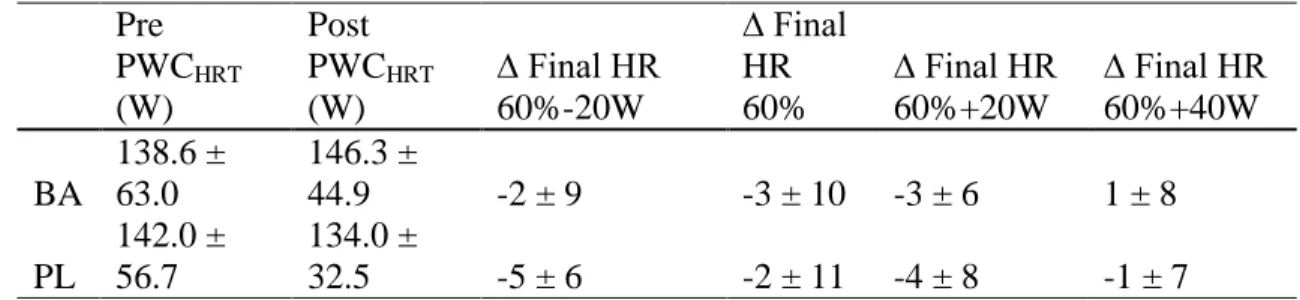
3. Baseline and post-supplementation physical working capacity at heart rate threshold(PWCHRT) values for beta-alanine (BA) and placebo (PL). Final heart rate (bpm) and change scores at each
of the four workloads. Values reported as mean ± SD…….………...51
ix
LIST OF FIGURES
1. Figure 1A shows the heart rate slopes plotted for each of the four pre-determined power outputs. Figure 1B shows the same slope coefficients plotted against the power outputs with a regression line in order to determine the PWCHRT (the
y-intercept).……….………..…53
2. Change scores (post-pre) for final HR (bpm) for the four workloads (± 95%CI) and change score for PWCHRT (W). *Denotes significance.……….………..54
3. Change Scores (post-pre) ± 95% CI for VO2 and VT……….…………..55
4. Change Scores (post-pre) ± 95% CI for TTE …….………..56
CHAPTER I
INTRODUCTION
The ability to delay fatigue may play a crucial role in performance outcomes. The
point of fatigue onset itself is difficult to pinpoint as there are multiple factors, both central and peripheral, that attribute to the body’s inability to produce a maximum force.
Central fatigue results from the brain reducing neural activation to the working muscles
during exercise, subsequently causing a decrease in power output, motivation, and the
up-regulation of the neurotransmitter serotonin (1). Peripheral fatigue is defined by
mechanisms that affect the muscle directly and that are distal to the neuromuscular
junction (1, 2). While mechanisms of peripheral fatigue include depletion of substrate
availability, excitation-contraction coupling failure and oxidative stress, during high
intensity exercise one of the largest contributors to fatigue is metabolite accumulation,
specifically hydrogen ions (H+) (3). During exercise, an increase in metabolism leads to excess accumulation of H+ by-products from the hydrolysis of ATP (3). An increase [H+]
combined with a corresponding decreasing in intramuscular pH, has been identified as
one of the primary causes of peripheral fatigue (3). Correspondingly, research has
demonstrated that delaying the accumulation of H+ through buffering mechanisms can delay fatigue onset (4-7).
The human body has natural systems to buffer metabolite accumulation and the
2
monitored by the bicarbonate buffer system by combining the free H+ with bicarbonate to form a the weaker carbonic acid (8). While this system works to keep the blood pH from
rising, eventually the H+ accumulation overwhelms the system and the pH drops. Another endogenous mechanism for buffering the H+ accumulation is muscle carnosine. Muscle
carnosine acts to maintain acid-base homeostasis by being a H+ acceptor (9). Carnosine is synthesized in the body naturally by L-histidine and beta-alanine (BA). Research has
shown that supplementing with BA can significantly increase muscle carnosine levels and consequently increase the muscle’s buffering capabilities and delay fatigue onset. (4,
9-11).
To pinpoint fatigue onset, various performance tests were developed utilizing fatigue
variables such as: electromyography (EMG) activity, ventilatory threshold and heart rate
threshold, to name a few. Originally developed in 1981 by Moritani et al. (12), the
physical working capacity (PWC) test was designed to determine the point of muscular
fatigue onset. This category of tests are based on the initial work by Monod and Sherrer
(13), who developed a relationship between total work done and time to fatigue. The
earliest PWC test was based on the physical working capacity at fatigue threshold
(PWCFT), where the rate of rise in EMG activity as a function of time was calculated at
four different power outputs. The slopes were plotted against the workloads and the linear
plot was extrapolated to the point of zero slope which was considered the fatigue
threshold (12, 14). Studies have previously shown the PWC test to be sensitive to
changes from training and supplementation (15, 16). Another PWC test utilizing heart
rate as a fatigue variable may provide additional means for quantifying BA’s buffering
The physical working capacity at heart rate threshold (PWCHRT) test was developed
by Wagner and Housh (17) to determine the maximum workload an individual can
maintain on a cycle ergometer without a significant increase in HR. The test involves
four eight minute cycle rides at different workloads, while measuring the change in HR
over time. An aerobic training study by Weir et al. (18) demonstrated that the PWCHRT is
sensitive to adaptations as a result of aerobic training. Another study by Mielke et al. (19)
aimed to validate the PWCHRT by comparing it to PWCVO2 and ventilatory threshold
(VT). Findings indicated that the power output at the PWCHRT, PWCVO2 and VT
thresholds were not significantly different, validating the PWCHRT as an indirect measure
of fatigue.
Utilizing HR as a fatigue measure in the current study provides an additional variable
to quantify fatigue, contributing to the existing body of BA research. A study by Smith et
al. (20) examined the effects of BA supplementation on women and found that following
28 days of supplementation, HR and ratings of perceived exertion (RPE) were lower
during a 40 minute treadmill run compared with placebo subjects. This suggests that BA
could have a positive effect on HR and RPE during submaximal exercise.
Beta-alanine research has shown supplementation has a positive effect on delaying
fatigue, by way of increasing intramuscular carnosine levels and thereby augmenting H+ buffering (10, 15, 16). Results from a study by Stout et al.(16) indicated that 90 days of
BA supplementation in an older population can effectively increase the PWCFT. Another
study by Stout et al. (15) showed that 28 days of BA supplementation in a similar
population can significantly increase the PWCFT. These studies indicate that physical
4
and that BA is effective at increasing fatigue thresholds. No studies to date have assessed
the effects of BA supplementation on the PWCHRT.
In addition to previous literature exploring BA’s effect on fatigue thresholds, several
studies have also examined its influence on aerobic fitness measures such as VO2peak,
ventilatory threshold (VT) and total time to exhaustion (TTE). Due to BA’s main
influence being on delaying acidosis, studies have consistently shown it to have no effect
on VO2peak (a measure than does not mainly rely on anaerobic sources of energy)(10, 11,
15). Research has given conflicting results for BA’s effect on TTE with some indicating
BA increasing TTE (10, 21) and some indicating no effect (11). VT has demonstrated
increases with BA supplementation (10, 21) indicating that BA has a potential positive
influence on aerobic physiological measures that occur at moderate intensities or when
anaerobic systems begin to play a role (TTE in BA literature is typically taken during
higher intensity exercises or during VO2peak tests).
While the focus of BA research involves mechanisms pertaining to peripheral fatigue,
with the highest levels of carnosine located intramuscularly, there are concentrations
within the brain and heart tissue as well (22). It is suggested that BA supplementation
may increase carnosine levels within the brain, thereby affecting the onset of central
fatigue, as well as peripheral fatigue in the active muscles. It is also well established that
an increase in H+ leads to a decrease in pH which stimulates afferent pain receptors (23). The presence of pain could inhibit a person’s willingness and/or motivation to continue
exercise, thereby initiating volitional fatigue. Therefore, an increase in carnosine can
peripheral factors (24) , it is not the aim of the present study to differentiate between the
two.
The primary purpose of this study was to determine the effects of 28 days of BA
supplementation on the PWCHRT. A secondary purpose was to determine if the PWCHRT
can be used as a predictor for VO2peak.
Purpose
1. The primary purpose of this study was to determine the effect of 28 days of
beta-alanine supplementation, a non-essential amino acid, on the physical working
capacity at heart rate threshold (PWCHRT).
2. A secondary purpose was to assess the effect of beta-alanine supplementation on
aerobic measures- VO2peak, TTE and VT.
Research Questions
1. Does four weeks of beta-alanine supplementation increase PWCHRT?
2. Does four weeks of beta-alanine supplementation increase VO2peak, TTE and VT?
Hypotheses
1. PWCHRT will significantly increase following 28 days of beta-alanine
supplementation.
2. VO2peak will not significantly increase following 28 days of beta-alanine
6
3. TTE will significantly increase following 28 days of beta-alanine supplementation
4. VT will significantly increase following 28 days of beta-alanine supplementation
Delimitations
1. Thirty-eight recreationally active men and women were recruited to participate in
this study.
2. Participants were between the ages of 18-35.
3. The duration of the study per subject was approximately six weeks including four
weeks of supplementation.
4. During a VO2peak oxygen consumption test, VO2peak was measured.
5. A physical working capacity at heart rate threshold test was completed in order to
calculate PWCHRT.
6. Participants had not consumed any performance enhancing supplements
containing beta-alanine or creatine within the last three months.
Limitations
1. Subject recruitment was from exercise classes on campus and through fliers located in fitness areas on UNC’s campus. Therefore subject population was not
truly random.
2. Due to the time-commitment, participant withdrawal occured.
Assumptions
1. Subjects provided accurate health, medical, nutritional and physical activity
history.
3. Subjects adhered to guidelines stipulating no change in normal nutritional and
exercise patterns.
4. Subjects complied with the supplementation protocol.
Operational Definitions
VO2peak– A VO2max is the maximal capacity of an individual’s ability to utilize oxygen
during exercise and can be used as a measure of physical fitness. It is determined to be
the point at which oxygen consumption does not increase despite an increase in
workload. All VO2max tests performed on a cycle ergometer are considered to be peak
tests (VO2peak).
Physical working capacity at heart rate threshold (PWCHRT)- This assessment is used to
identify the power output associated with the onset of fatigue, as determined by heart
rate. Theoretically the PWCHRT represents the highest power output that can be
maintained for an extended period of time without signs of fatigue, as identified by an
increase in heart rate.
8
CHAPTER II
LITERATURE REVIEW
Fatigue can be defined as an inability to continually produce maximum force, or
inability to maintain a given exercise intensity (8). While fatigue can be identified by a
decrement in force, often the cause of fatigue involve smaller factors that simply lead to a disturbance in the body’s ability to maintain homeostasis (8). Fatigue is attributed to
combined central and peripheral attributes. Central fatigue describes the influence within
the brain and spinal cord, whereas peripheral fatigue refers to mechanisms that affect the
muscle directly, distal to the neuromuscular junction (1, 2). Mechanisms of peripheral
fatigue are numerous and can be traced to excitation-contraction coupling failure,
depletion of substrate availability, oxidative stress and metabolite accumulation (25).
During intense exercises, increases in ATP hydrolysis yields an accumulation of
inorganic phosphate (Pi) (3). The hydrolysis of ATP also results in the release of a proton
in the form of a hydrogen ion (H+). While Pi acts as a buffer to the increasing H+
accumulation, the increase in intracellular Pi is much less than the total amount of ATP
hydrolysis. Due to the hydrolysis of ATP, there is also an accumulation of ADP, which
combines with Pi to further produce ATP and lactate (3). While other factors of
metabolite accumulation act on muscular pH and play a role in peripheral fatigue, H+ accumulation is the focus of the current study. An increased [H+], and a corresponding
(4). Animal studies have shown a decrease in pH from 7 to 6 leads to a significant
decrease in isometric tension in the muscle, resulting in a reduced force production (5).
Literature has shown that delaying the accumulation of H+ may concomitantly delay the onset fatigue (4-7).
Central fatigue is typically defined as a person’s inability to maintain voluntary force
due to the subconscious brain reducing neural activation to contracting muscles during a
fatiguing exercise (1, 26). Fatigue in many instances results from an individual’s
unwillingness to continually produce adequate central drive to maintain a given force or
power output (1). One identified source of decreased motivation (central drive) is the
up-regulation of the neurotransmitter serotonin during exercise (1). An increase in serotonin,
and corresponding decrease in dopamine levels, is associated with central fatigue. The
combination of central and peripheral fatigue has been shown to account for the total loss
of force during sustained muscle contractions (25). The autonomic nervous system
(ANS), is composed of both a sympathetic nervous system (SNS) and parasympathetic
nervous system (PNS) that act as complimentary systems. Both the SNS and PNS
influence heart rate by varying impulses in nerve fibers in the sinoatrial node (27). The
heart is dually innervated by the PNS and ANS, and is up- and down-regulated by the
two respectively. The central control of heart rate can be demonstrated by the anticipatory
rise in heart rate seen in athletes prior to race events. Thus without any muscle
contraction or energy, there is a distinct increase in heart rate. In addition, during exercise
the CNS stimulation modulates the ANS, and the peripheral sensors then regulate the
effects according to muscle needs. Previous literature has demonstrated that central
10
chronic fatigue patients (28). At lower VO2peak values, but higher ratings of perceived
exertion (RPE), chronic fatigue patients previously demonstrated significantly lower
heart rate, indicating a centrally mediated increase in effort perception. This increased perception of effort led to subjects’ unwillingness or inability to achieve maximal
performance. Extending this to normal fatigue processes during intense or long duration
exercise, central factors may play a limiting role in a person’s ability or willingness to
achieve high intensity exercise. The central command theory suggests that at the onset of
exercise centrally controlled signals set the cardiovascular response, while peripheral
feedback loops are responsible for the fine tuning adjustments as exercise continues.
Despite the overlap between the two systems, it is clear that both central and peripheral
factors influence heart rate during exercise (26).
Nutritional supplementation may be an effective way to delay fatigue onset.
Recently, beta-alanine (BA), a non-essential amino acid has been shown to effectively
delay neuromuscular fatigue (10, 11). The direct effect of BA supplementation is to
increase carnosine levels within the muscle (4, 9). Beta-alanine supplementation has been
shown to significantly increase intramuscular carnosine levels by as much as 60% (9) and
also improve exercise performance by delaying the drop in pH (29). To date, research suggests carnosine’s most effective role is delaying peripheral muscle fatigue by
maintaining acid-base homeostasis through buffering H+ (9) . With a dissociation content
(pKa) of 6.83 , carnosine is highly effective at accepting excess H+ in the muscle in order to maintain intramuscular pH (30). Carnosine is synthesized from L-histidine and BA, but
due to the high concentration of L-histidine within the plasma, beta-alanine has been
While beta-alanine’s positive effect on peripheral fatigue via an increase in
skeletal muscle carnosine are well documented (4, 9, 32), its direct effect on central
fatigue has not yet been examined. While the highest concentrations of carnosine are in
skeletal muscle, carnosine has also been located within the brain (22). High
concentrations of carnosine are found in the olfactory lobe, which has been shown to
influence both central and peripheral characteristics (22). One of the side effects
experienced acutely with BA supplementation, paresthesia (or prickling sensations on the
skin), is linked to BA activated strychnine-sensitive glycine receptor sites located within
the spinal cord (33, 34). It is still undetermined what effect carnosine could have on the
central nervous system, and, more specifically, delaying central fatigue.
Various fatigue tests have been utilized to examine the effectiveness of BA
supplementation. The physical working capacity test has been used to show significant
increases in physical working capacities following supplementation(10, 15, 16). The
physical working capacity tests were originally developed by Moritani et al. 1981 and
Devries et al. 1982 to measure the point of onset of muscular fatigue (12, 14). The
researchers utilized electromyographic (EMG) fatigue curves to identify an EMG fatigue
threshold. Prior to this, however, Monod and Scherrer (1963) found a relationship
between total work done at varying workloads and the duration of time to fatigue. The
slope of this relation was termed critical power and represented the power output a
muscle could maintain without fatigue (13). However, these original measures required
subjects to perform supramaximal tests, such as a VO2max. Therefore, in 1987, Devries et
al. completed a follow-up study to submaximally estimate physical working capacity at
12
represents the highest power output that can be maintained for an extended period of time
without evidence of fatigue (19). Utilizing submaximal loads and the same principles
developed by Devries et al. (1987), the physical working capacity at heart rate threshold
test (PWCHRT) was developed by Wagner and Housh (17) in 1993 in order to identify the
highest cycle workload an individual can maintain without a significant increase in heart
rate over an extended period of time (17). Studies utilizing the PWCHRT have found it to
be highly sensitive to the effects of aerobic training programs (18), as well as an accurate
estimate of the maximal power output that can be produced without a significant rise in
heart rate (17).
The purpose of this study is to determine the effect of four weeks of BA
supplementation on the physical working capacity at heart rate threshold in men and
women. By utilizing heart rate as the main measure of fatigue, this study will be
indirectly examining BA’s effect on both central and peripheral fatigue. Previous
research on BA has shown that it is effective in delaying fatigue peripherally by
increasing muscle carnosine, but its effect on central mechanisms is still unknown. The
physical working capacity tests offer an easy, cost sensitive way to quantify a fatigue
threshold. The following annotated article review will examine previous research
describing fatigue, heart rate response to exercise, BA, and physical working capacity
Central and Peripheral Fatigue and Heart Rate
Cooke, Franks, Luciani, and Pate 1988 (5)
The purpose of this study was to gain a more comprehensive understanding of the
effects of phosphate and hydrogen ions on parameters of contraction by varying their
concentrations in rabbit psoas muscles. Dissected psoas fibers from the rabbits were fully
activated utilizing Ca2+ at a constant temperature of 10ºC. The velocities of induced contractions were measured utilizing load clamps. Fiber ATPase was also monitored
utilizing enzyme coupling of ADP production to reduced NADH, and then measuring the
depletion of NADH. Results showed that increasing phosphate concentration from 3 to
20mM at a pH of 7.0 decreases ATPase activity, does not affect maximum contraction
velocity (Vmax), and causes a 25% decrease isometric tension (P0). Changing the pH from
7 to 6, while maintaining the 3mM concentration of phosphate, showed a 45% decrease
inP0. These results are similar to those of other studies which indicated that a decrease in
muscular pH from 7 to 6 results in a decrease in isometric tension. The main conclusion
drawn from this study is that most of the fiber contraction inhibition seen in fatiguing
muscles is explained by product inhibition (H+ and Pi).
Robergs, Ghiasvand, and Parker 2004 (3)
The focus of this review article was to compile the previous and current literature
available on metabolic acidosis to form a comprehensive review. Once thought to be a
major source of metabolic acidosis, lactate is known to play an important role in initial
buffering of H+ accumulation to the lactate dehydrogenase reaction (pyruvate to lactate)
14
The lactate has a further use as it can be transported from the producing cell to be used as
a substrate for metabolism in other skeletal cells, the liver or kidneys. The real source of
H+ accumulation has been more correctly identified as ATP hydrolysis. A phosphate (Pi) is produced and has the ability to buffer the free proton that is released from the water.
During ATP hydrolysis, the ADP and Pi go on to act as substrates for glycolysis to
produce more ATP, which leaves the extra proton to accumulate if the other buffering
systems cannot meet the necessary removal rates. Another source of increased H+ could
be from an accumulation of NADH + H+ produced via the glyceraldehydes 3-phosphate dehydrogenase reaction step of glycolysis. This could occur when the rate of substrate
efflux from glycolysis is greater than the mitochondrial activity to accept the products.
The conclusions of this review were that lactate facilitates proton removal from the
muscle, lactate production is essential to supporting ATP regeneration from glycolysis,
and metabolic acidosis results from an increased reliance on non-mitochondrial ATP
turnover (glycolysis, etc).
Davis, 1995 (1)
The purpose of this review article was to explore the causes central fatigue,
specifically the role that brain serotonin (5-HT) plays in leading to central fatigue. This
article proposes that central fatigue plays a crucial role in limiting overall exercise
performance. Increased 5-HT is suggested to be the main contributor to central fatigue in
prolonged exercise due to its known effects on arousal, lethargy and sleepiness.
Increases in brain 5-HT are due to increases in blood-borne tryptophan (TRP) delivery to
(f-TRP) that is transported to the brain. The transport mechanism occurs with other amino
acids, specifically the branched-chain amino acids (BCAAs). Synthesis of brain 5-HT
increases when the ratio of f-TRP to total plasma BCAAs increases. Animal studies have
indicated an association between increase levels of brain 5-HT and fatigue during
continuous prolonged exercise. Another study utilizing rats found that drug-induced
increases in brain 5-HT lead to faster onset of fatigue whereas drug-induced decreases in
brain 5-HT lead to a delay in fatigue onset.
The article also explores the effectiveness of nutritional supplementation to delay
central fatigue. Studies have indicated that consumption of the large doses of BCAAs
needed to significantly change the ration of f-TRP to BCAAs would also increase plasma
ammonia which could lead to toxic effects on the brain and negative changes to
metabolism. A study utilizing carbohydrate supplementation found that supplementing
with 6 or 12% carbohydrate-electrolyte drink solutions lead to greatly reduced plasma
f-TRP levels as compared to water supplementation, and that the carbohydrate-electrolyte
supplement was successful in delaying fatigue. This study contributes to the current body
of fatigue literature by highlighting the role that central fatigue can play in limiting
exercise performance, and by indicating the importance of further research on
mechanisms of central fatigue.
Kent-Braun, 1999 (7)
The purpose of this study was to estimate the contribution of peripheral and
central factors to human skeletal muscle fatigue. Nine people (five men, and four women,
16
active were recruited for this study. Subjects performed a baseline MVC of the
dorsiflexor muscles, then a sustained isometric MVC for 4 minutes with a titanic
stimulation at the end of the 4 minutes. An EMG was used to quantify muscle activation
during maximal voluntary isometric contractions (MVC) of the ankle dorsiflexor
muscles. Fatigue was quantified by the change in force during the MVC. Measures of
intramuscular metabolism were also made during the exercise utilizing magnetic
resonance spectroscopy.
The results of the study showed a significant difference between the titanic and
voluntary fatigue (15%), and an overall 78% fall in MVC. Data indicated that
approximately 20% of the fatigue developed during the sustained MVC was due to
central fatigue, and the other 80% was due to intramuscular sources such as an increase
proton concentration. There was a decrease in central activation ratio (CAR), defined as
the peak MVC/peak total force, which is indicative of a slight central activation failure
during exercise.
The study also found significant changes in the intramuscular metabolism during
exercise. The fall in pH was correlated with the observed fall in force, and the lack of
peripheral activation failure is indicative of metabolic inhibition leading to fatigue. Other
studies cited have indicated that there is a linear relationship between [H+] and fatigue.
Robinson, Epstein, Beiser, and Braunwaldet, 1966 (27)
The purpose of this study was to identify the roles of the efferent pathways that control heart rate during supine exercise by observing the effects of β-adrenergic
years) were recruited for this study: four normal volunteers and one patient who had
undergone a successful mitral commissurotomy. All subjects were men who were in good
physical condition, but were not trained athletes. After familiarization with the
equipment, the subjects were placed on a tilt table in the supine position and their heart
rate (HR) was recorded via ECG. The subject was then tilted to 45º and 80º and HR was
again recorded. The subjects were then moved to a supine bicycle where their HR was
recorded at rest as well as during the fourth minute of exercise at each series of work
levels. Oxygen uptake was recorded continuously throughout. The protocol was
performed 3 more times: one where the cardiac sympathetic nerves were blocked, one
where the parasympathetic nerves were blocked, and one where both were blocked.
Sympathetic blockade has been found to lower HR during exercise, suggesting
sympathetic stimulation is responsible for cardiac acceleration. Results of the study
indicated that the increase in HR during moderate exercise was due to withdrawal of
parasympathetic stimulation. At higher workloads, increases in HR were more due to
sympathetic stimulation. These results were determined by comparing the
double-blockade response to the single double-blockade responses of HR.
Beta-Alanine Supplementation
Derave, Ozdemir, Harris, Pottier, Reyngoudt, Koppo, Wise, and Achten, 2007 (32)
The purpose of this study was to determine whether 28 days of BA
supplementation would increase muscle carnosine levels in the calf and improve
performance in a 400 meter sprint. Fifteen male (age 18.4 ± 1.5 yrs) track-and-field
18
study was a placebo-controlled, double-blind study where 7 athletes consumed a placebo
(maltodextrin) for the duration of the study, and 8 consumed six divided doses of 400mg
capsules BA CarnoSyn® time release capsules.2.4g/day for the first 4 days, 2.6g/day for the next 4 days, and 4.8g/day for the remaining days. A week prior to supplementation,
pre-testing was performed. A proton magnetic resonance spectroscopy (MRS) was
utilized to determine initial muscle carnosine concentrations. Isokinetic and isometric
fatigue tests using a Biodex isokinetic dynamometer was utilized to evaluate the
contractile performance of the knee extensors. The isokinetic test was performed on the
right leg that consisted of 5 sets of 30 maximal voluntary isokinetic knee extensions at a
velocity of 180º/second. Subjects had a one minute recovery between each set of 30
repetitions. Peak torque data was measured and used to calculate an average peak torque
for each set of exercise. The left leg was then use for isometric testing. The knee was
fixed at 45º, and the maximum voluntary contraction (MVC) torque was recorded. The
highest of three trials was recorded as their final MVC. Finally, subjects completed a 400
meter sprint on a separate day, following a self-constructed warm-up routine lasting ~45
minutes.
The results of this study showed substantial increases in muscle carnosine (+47%
in the soleus, +37% in the gastrocnemius). Since the subjects were highly trained athletes,
these increases were similar to, but slightly lower than other studies that utilized
untrained subjects. Even subjects with high initial levels of muscle carnsoine saw
increases in concentration, which suggests a lack of a supplement ceiling. The results of
this study also showed that in spite of improvements in muscle torque following
knee extension torque was significant increased in all 5 bouts following supplementation,
and increased in the first 2 bouts following a placebo. This suggests that BA
supplementation can attenuate fatigue in the later stages of exercise. There was no
significant effect on isometric endurance.
Zoeller, Stout, O’Kroy, Torok, and Mielke, 2006 (11)
The purpose of this study was to determine the effect of four weeks of BA
supplementation alone or in combination with creatine monohydrate (Cr) on endurance
performance. Fifty-five men (age 24.5 ± 5.3 years) were recruited for this study. Subjects
were randomly assigned to one of four supplementation groups: placebo (PL, 34g of
dextrose; n=13), creatine (Cr; 5.25g of creatine monohydrate plus 34g of dextrose; n=12),
beta-alanine ( Carnosyn®, NAI, San Marcos, CA) (B-Ala; 1.6g beta-alanine plus 34g of dextrose; n=14), or beta-alanine plus creatine (Phosphagen EliteTM, EAS, Golden, CO)
(CrBA; 5.25g of creatine monohydrate; 1.6g beta-alanine plus 34g of dextrose; n=16).
For pre-testing, subjects performed a graded exercise test (GXT) on a cycle ergometer to
determine VO2peak, ventilatory threshold (VT), and lactate threshold (LT). For the GXT
protocol, initial power was set to 30 watts, and was increased by 30 watts every 2 minutes
as the subjects maintained a pedal cadence of 70rpm. The test was stopped when the
subject could no longer maintain the rpm, or until volitional fatigue. To confirm the test
was a VO2peak, subjects had to meet the following criteria: a plateau in heart rate (HR)
values or attainment of HR within 10% of age-predicted HRmax, a plateau in oxygen
20
than 1.15. Subjects then completed 28 days of their assigned supplementation protocol
and finished with post-testing consisting of the same GXT. Results of this study
demonstrated that the Cr group showed improvements in power at VT and total time to
exhaustion (TTE), while B-Ala only had improvements in power output at LT. In
contrast, the combination group, CrBA showed increases in power output at LT, VO2 at
LT, VO2 at VT, power output at VT, and percent VO2 at VT. This data indicates that
supplementation with the combination of creatine and beta-alanine may dely the onset of
fatigue, via LT and VT measures during incremental cycle exercise in men.
Artioli, Gualano, Smith, Stout, and Lancha 2009 (31)
The purpose of this review paper was to discuss the current information on
carnosine and beta-alanine metabolism and to highlight the effects of beta-alanine
supplementation on exercise performance. For purposes of the present study, the
important findings will be limited to the effects of beta-alanine supplementation.
Beta-alanine has been shown to effectively increase muscle carnosine after only 28 days of
supplementation. Harris et al. in 2006 (9) demonstrated that 800mg is the maximum
tolerable single dose of beta-alanine, but that this dose could be repeated throughout the
day if there was a three hour time window between dosings. Later studies showed that
using controlled release capsules could be utilized in order to double the dose from
800mg to 1600mg. A study by Zoeller et al. in 2007 (11) reported that beta-alanine
improves the anaerobic threshold. Stout et al. in 2007 (10) showed a significant increase
of 13% on ventilatory threshold in women supplementing with beta-alanine. The
conclusion from these studies was that the increased buffering capacity created by
smaller lactate accumulation. Another study by Stout et al. in 2006 (15) utilized 55 men
with a mean age of 24.5 to demonstrate beta-alanine’s ability to delay fatigue by
increasing the physical working capacity at fatigue threshold after 28 days of beta-alanine
supplementation. Overall, studies utilizing exercise protocols that elicited extreme
acidosis demonstrated increases in performance linked with beta-alanine
supplementation. Protocols that elicit less of an acidosis response, (such as those having a
single bout of exercise lasting less than 60 seconds, or those using small muscle groups)
resulted in little or no effect from beta-alanine supplementation. Another focus of this
review was the adverse effects of beta-alanine supplementation. The only known one is
the symptoms of paresthesia, or tingling sensations, that are triggered by ingesting a high
single dose of beta-alanine, and tend to disappear within an hour. Controlled time-release
capsules or smaller dosages can avoid the symptoms altogether. The conclusions of this
review were that 28 days was a significant amount of time to increase muscle carnosine;
beta-alanine supplementation can improve fatigue thresholds; and in order to avoid
paresthesia, controlled time-release capsules should be utilized.
Stout, Graves, Smith, Hartman, Cramer, Beck, and Harris, 2008(16)
The purpose of this study was to determine the effects of 90 days of beta-alanine
supplementation on the onset of neuromuscular fatigue when measured by the physical
working capacity at fatigue threshold (PWCFT) test in elderly men and women.
Twenty-six untrained men and women were recruited for this study. For pre-testing, subjects
completed a PWCFT. Subjects were fitted to a cycle ergometer and began pedaling at 50 rpms with an initial power output determined by the researchers based on participant’s
22
minutes. After each two minutes there was a rest interval long enough to return heart rate
(HR) to within 10bpm of the arrival HR. During each workload, EMG samples were
recorded on the vastus lateralis. PWCFT was determined to be the highest power output
that resulted in a non-significant slope value for the EMG amplitude verse time
relationship. Following pre-testing, subjects were randomly assigned to either the
beta-alanine group (BA, 2.4 grams/day of beta-beta-alanine, CarnoSyn®, n=12) or the placebo group (PL, 2.4 grams/day of microcrystalline cellulose, n=14). All subjects ingested one
capsule (containing 800mg) three times a day for 90 days. Within three to five days
following supplementation subjects completed a post-supplementation PWCFT. Results
showed a significant increase in PWCFT (+28.6%) in the BA group, but no significant
change in the PL group. The results of this study indicate that 90 days of beta-alanine
supplementation in the elderly can significantly increase PWCFT.
Hill, Harris, Kim, Harris, Sale, Boobis, Kim, and Wise, 2007 (4)
The purposes of this study were to further evaluate the effect of four weeks of
beta-alanine supplementation on the accumulation of muscle carnosine, to determine if
changes were fiber specific, and to see if the effects altered results of a high intensity
cycling capacity test. Twenty-five male subjects who were physically active but not in
structured training programs were recruited. Following a cycle performance test, subjects
were split into two groups; one that supplemented with beta-alanine (CarnoSyn®) for either 4 or 10 weeks (n=13), and one that had a matching placebo (PL).Beta-alanine (BA)
was divided into 8 daily doses with total dosage per day increasing each week (4.0g-6.4g
muscle biopsy of the vastus lateralis was administered 1-2 days following the cycle test,
and 1-2 days following the 4 and 10 week tests. For muscle fiber assessment, 20-40
single muscle fibers were dissected from the 0 and 10 week biopsies of subjects in the
beta-alanine group. Results showed muscle carnosine concentrations were significantly
increased at both the 4 (19.9 ± 1.9 mmol/kg dm) and 10 (30.1 ± 2.3 mmol/kg dm) week
markers for the BA group with no significant changes observed in the control. Muscle
fiber content changes were not significantly different between type IIa and type I muscle
fibers. Total muscle carnosine content was greater in type IIa fibers, but the increases
over the 10 weeks were not significantly different. For the cycling test, total work done
(TWD) was significantly increased following 4 (+13%) and 10 weeks (16.2%) of BA
supplementation, with no significant change seen in the control group. The findings of
this study support BA supplementation as an effective enhancer of muscle carnosine at
both 4 (+58%) and 10 (+80.1%) weeks. Most importantly, this study showed a
relationship between greater muscle carnosine content and increased power output via the
changes seen in TWD in the cycle tests. The authors conclude that this is one of the first
studies linking improvements in whole body exercise capacity to an increase in
intracellular H+ buffering content.
Harris, Tallon, Dunnett, Boobis, Coakley, Kim, Fallowfield, Hill, Sale and Wise, 2006 (9)
The purpose of this study was to investigate the absorption of BA and the effect
of supplementation on muscle carnosine synthesis. Six healthy male subjects (age 33.5 ±
9.9 years) were recruited for this study. All subjects underwent four of the five
24
ingesting chicken broth containing 40mg/kg of body weight (bwt) of BA. For treatments
B, C, D, and E subjects ingested 3ml/kg bwt of a drink containing either 0, 10, 20, or 40
mg/kg bwt of beta-alanine (CarnoSyn®), followed by 5ml/kg bwt of water. Blood samples were taken at 10 minute intervals for the first 90 minutes following
supplementation and then at 120, 180, 240 and 360 minutes. Complications with subjects
administered 40mg/kg bwt of beta-alanine arose when they rapidly experienced
symptoms of flushing (prickly sensations) which began at 20 minutes and lasted an hour.
As a result of this, two subjects refused the dose and were given 10 and 20 mg/kg bwt
instead. Symptoms were seen with the 20mg/kg bwt again, but to a much lesser extent.
There was a significant increase in plasma BA following the chicken broth ingestion, but
the increase was only half the increase seen in subjects consuming 40mg/kg bwt of
beta-alanine. Important findings in this study indicated that administration of BA above
10mg/kg bwt was associated with severe flushing symptoms. The chicken broth which
resulted in similar BA concentrations to those elicited by 20mg/kg bwt supplementation,
did not lead to any symptoms.
Smith, Stout, Kendall, Fukuda, and Cramer, 2011 (20)
The purpose of this study was to determine the effects of BA supplementation on
oxidative stress in women. Twenty-four moderately trained (3-7 days per week of
aerobic, resistance or recreational activities) women were used in this study. Each subject
had a total of four laboratory visits. On visit one subjects completed an initial run to
determine VO2max and peak velocity (PV). No more than 3 days later, subjects came in
subjects ran at 70-75% of their PV for 40 minutes. Post run blood samples were collected
at two and four hours post-exercise. Upon completion of all pre-testing, participants were
randomly assigned to a placebo (PL) group (800mg/tablet of maltodextrin, 2 tablets 3
times daily) or a BA group (800 mg/tablet, 2 tablets 3 times daily; CarnoSyn®). Subjects
then supplemented for 28 days, reporting to the lab after two weeks to report intake and
side effects. Subjects then returned to the lab for post-testing in the form of a VO2 max
and the 40 minute oxidative stress run. Results of this study showed that, while
non-significant, 28 days of BA supplementation still had a slight influence on aerobic
performance defined by total time to exhaustion (TTE) and ventilatory threshold (VT).
Supplementation had no significant effect on the oxidative stress markers- antioxidant
capacity, superoxide dismutase, glutathione, and 8-Isoprostane. Rating of perceived
exertion decreased (non-significantly) in the BA group at 30 and 40 minutes of the
oxidative stress run. Ventilatory threshold also increased non-significantly in the BA
group suggested an increased buffering capacity and improvement in submaximal
performance.
Physical Working Capacity Tests
Stout, Cramer, Mielke, O’Kroy, Torok, and Zoeller, 2006 (15)
The purpose of this study was to determine the effects of 28 days of beta-alanine
(BA) and creatine monohydrate (CrM) supplementation on the physical working capacity
at neuromuscular fatigue threshold (PWCFT). Fifty-one men (age 24.5± 5.3 years), who
had not ingested creatine or other dietary supplements for the last 12 weeks, were
26
to use as the measure for neuromuscular fatigue. Subjects were asked to ride an
electronically braked cycle ergometer at a pedal frequency of 70rpm starting at a
resistance of 60 watts (W). The power output was increases by 30W every 2 minutes until
the pedal cadence could not be maintained. At every 2 minute interval six 10-second
EMG samples were recorded. The PWCFT was calculated later by averaging the highest
and lowest power output that had non-significant slope values for the EMG amplitude vs.
time relationship. Following pre-testing, subjects were randomly assigned to one of four
treatment groups: 1) placebo (PLA; 34g dextrose; n=13), 2) creatine (CrM; 5.25g CrM
plus 34g dextrose; n=12), 3) b-Ala (1.6g b-Ala plus 34g dextrose; n=12), 4) b-Ala plus
CrM (CrBA; 5.25 g of CrM plus 1.6g b-Ala plus 34g dextrose; n=14). Subjects took the
supplements 4 times per day for 6 days, and then twice per day for the last 22. Subjects
completed the same tests in post-testing as in pre-testing. Results of this study showed
that b-Ala supplementation could delay the onset of neuromuscular fatigue by
significantly increasing the PWCFT (+14.5%). The combination of CrBA did not have an
additive effect to the delay in fatigue, but did significant increase the PWCFT (+11%),
while Cr alone had no significant effect. These findings suggest that beta-alanine
supplementation, with or without creatine, may delay the onset of neuromuscular fatigue
during incremental cycle ergometry.
Wagner and Housh, 1993 (17)
The purpose of this study was to introduce the physical working capacity at heart
rate threshold (PWCHRT) test that could be used to estimate the maximal power during
Eight sedentary men (age 22 ± 2 years) were recruited for this study. The subjects had
nine laboratory visits, each separated by at least 48 hours. On the first four visits subjects
completed four different workloads for determination of the PWCHRT, and on the last five
subjects completed hour long rides in order to validate the PWCHRT. For determination of
PWCHRT subjects completed four different workloads ranging from 105-200 watts (W).
Upon reporting to the lab, subjects were fitted to the electronically braked cycle
ergometer and completed a five minute warm-up at 70rpm with a resistance of 50W.
Following the warm-up, subjects completed the eight minute work bout at a workload
chosen so that they could complete the full eight minutes, but would also elicit a positive
slope in the heart rate verses time relationship. Heart rate was recorded every 15 seconds,
but only the last five minutes were used to allow for stabilization of the HR and to ensure
the rise in HR was due to the demands of the workload and not cardiac adjustment. After
all four workloads were completed, the power outputs were plotted as a function of the
slope for the HR versus time relationships. The PWCHRT was determined to be the y
intercept of the plot, or the point of zero slope. For validity of the test, one hour
continuous work bouts were performed at randomly assigned power outputs equivalent to
80, 100, 120, 140 and 160% of PWCHRT. Subjects were blinded from the workload until
completion of all workloads. Warm-up procedures were identical to the PWCHRT trials,
and following the warm-ups subjects completed an hour long ride at the prescribed
intensity. Statistical analysis included regression analyses using least squares method and
t tests were used to determine if there were significant differences between the mean
28
coefficients corresponding to workloads equal to or less than the PWCHRT had HR
increases less than 0.1bpm∙min-1. This minimal rise indicates that the PWCHRT can
provide an accurate estimate of the power output that can be maintained for an hour
without significant fatigue. The authors concluded that the PWCHRT would be an
effective way to assess the effects of interventions designed to increase work capacities.
DeVries, Tichy, Housh, Smyth, Tichy, and Housh, 1987 (35)
The purpose of this study was to evaluate the physical working capacity at fatigue
threshold (PWCFT) test on a cycle ergometer with fatigue being determined by EMG
fatigue curves in the quadriceps muscle. Thirty-two healthy men (age 23.4 ± 3.1 years)
with fitness levels ranging from sedentary to highly trained athletes were recruited for
this study. For determination of the PWCFT, subjects completed four pre-determined
workloads on the cycle ergometer ranging from 420 kp m/min to 1260 kp m/min. Each
workload lasted two minutes and was followed by a rest period long enough to bring the
heart rate back to within 10bpm of the initial heart rate. EMG was recorded on the
quadriceps throughout the two minute work bouts and PWCFT was determined to be the
lowest workload producing a slope of the EMG amplitude verse time relationship that
was significantly different than a zero slope. Subjects were also tested for lactate
threshold (OBLA) via blood draws within 1-2 minutes of each exercise bout, and
percentage heart rate range at PWCFT. Seventeen subjects were retested for PWCFT a
week following initial testing to determine reliability. Fifteen subjects were retested for
OBLA a week following initial testing as well. Results of this study showed that the
quartile of the subjects there was a small non-significant difference between the mean
power of OBLA and the mean power of PWCFT. However, for the fit quartile, there was a
significant difference (p≤ .05) between the two mean powers. The authors conclude that
the results suggest that fit subjects are limited by central factors, whereas in unfit
subjects, muscular (or peripheral) fatigue factors are the limiter. From this study it can be
concluded that the PWCFT is an effective and reliable way to evaluate the physical status
and to monitor training progress in individuals. It is ideally suited for individuals who are
unable to complete maximal workloads, as the PWCFT utilizes short, submaximal and
discontinuous workloads.
Weir LL, Weir JP, Housh, and Johnson, 1997(18)
The purpose of this study was to determine the effect of an eight week aerobic
training program on the physical working capacity at heart rate threshold (PWCHRT).
Nineteen college-aged men and women who had not participated in a regular aerobic
exercise program for at least one year were recruited for this study. For pre- and
post-testing, subjects completed both a VO2max and a PWCHRT on a cycle ergometer. For the
VO2max subjects completed a five minute warm-up at 30 watts (W). The first stage of the
test had a resistance of 50W and a required pedal cadence of 70rpm. The W was
increased by 30W every two minutes until voluntary exhaustion. The test was considered
a true VO2max if the subjects met two of the following criteria: a plateau of VO2 with an
increase in W, maximal heart rate within 15bpm of the age predicted max heart rate, or a respiratory exchange ratio (RER) ≥1.1. For determination of PWCHRT, subjects again
30
of 70rpm. Each subject then completed four eight minute workloads (on separate days)
ranging in resistance from 55-215W. The rate of rise in heart rate as a function of time for
each workload was determined for each subject. The PWCHRT was calculated by plotting
the workloads as a function of the slope for the heart rate verse time relationship and
taking the y-intercept. Following pre-testing, subjects were randomly assigned to either a
training or control group. The training group completed three exercise sessions a week
for eight weeks, while the control group had no training sessions and simply maintained
their normal activities. The training sessions consisted of a five minute warm-up on a
cycle, followed by 30 minutes of cycling at intensities between 70 and 90% of max heart
rate at 70rpm. Results of this study showed a significant increase in PWCHRT (30.1 W)
following training indicating that the PWCHRT is sensitive to an eight week aerobic
training program. The study also showed only a moderate relationship between VO2max
and PWCHRT (r2= 0.31). This indicates that VO2max and PWCHRT are controlled by different mechanisms and cannot be explained by similar variables. There was also a
decrease in heart rate slope coefficients following training. The authors speculate this
drop could be due to increased stroke volume, decreased sympathetic drive or an increase
in parasympathetic activity. Regardless of the mechanism, the decrease itself refutes
previous research suggesting that heart rate slopes remain stable with training.
Mielke, Housh, Malek, Beck, Hendrix, Schmidt, and Johnson, .2008 (19)
The purpose of this study was to further validate the PWCHRT and the PWC VO2
tests by utilizing the results of the tests to find an estimated time to exhaustion (ETTE)
ventilatory threshold (VT). Ten men and women with a mean age of 23 were recruited for
the study. Their physical activity status ranged from sedentary (not currently participating
in aerobic or resistance training) to moderately active (4-5 hours of exercise per week).
They performed an incremental test to exhaustion on an electronically braked cycle
ergometer in order to determine VO2peak and VT. VO2peak was identified as the highest
VO2 value in the last 30 seconds of the test if the subject met two of the termination
criteria: 1) 90% of age-predicted max HR, 2) respiratory exchange ratio >1.1, 3) a plateau
of oxygen uptake (defined as <150 ml/min increase in VO2 during the last 30 seconds).
VT was defined was identified as the VO2 value corresponding to the intersection of two
regression lines derived from points below and above the breakpoint in the VCO2 vs.
VO2 relationship. On a separate visit, subjects completed a physical working capacity test
which included four randomly ordered workloads to exhaustion at different power
outputs (98-246 watts), to determine the PWCHRT and the PWC VO2. Power curve
analyses were used to estimate the ETTE at PWCHRT, PWC VO2, and VT. Results
showed no significant mean differences between the three fatigue thresholds suggesting
that fatigue thresholds determined by HR and VO2 could be used for estimating VT. The
power-curve analyses also indicated, contrary to previous studies, that the subjects could
only maintain the power outputs for the PWCHRT, PWC VO2, and VT for 29 ± 6, 21 ± 3,
and 27 ± 11 minutes respectively. The authors speculate the differences in this study as
compared to previous ones could be due to the fatiguing protocol used in this one as well
as differences in subject training status. The findings of this study for the similar power
thresholds between PWCHRT, PWC VO2, and VT support the PWCHRT as being a valid
32 CHAPTER III
METHODS
Experimental Design
All subjects reported to the lab a total of eight or nine times. Prior to enrollment,
subjects were given an overview of the study and asked to sign an informed consent approved by the University’s Institutional Review Board. They were then given a
physical and a 12-lead resting electrocardiogram (EKG). Depending on the subject’s
availability the EKG and physical screen were sometimes on separate days (leading to
some subjects completing the study in 8 visits, and some taking 9). A familiarization trial
occurred for the peak oxygen consumption test (VO2peak) prior to performing the first
VO2peaktrial on a cycle ergometer, with 24-48 hours between sessions. The PWCHRT
testing occurred on visits four and five where subjects performed four randomized
workloads on two separate days. Following pre-testing, participants were randomly
assigned to supplementation group A or B (beta-alanine (BA) or placebo (PL)) and
underwent twenty eight days of supplementation. Following completion of
supplementation, subjects returned for three post-testing days consisting of a VO2peak, and
two separate days of PWCHRT.
Subjects
Thirty eight men and women between the ages of 18 and 31 were recruited for
Eight individuals that were enrolled were not included in the final analysis: one subject
was not cleared medically, one subject had taken beta-alanine within the last 3 months,
two suffered outside injuries within the pre-testing phase, and four subjects did not come
back following enrollment for unknown reasons. Participants were recreationally active
(accumulating 1-5hrs of exercise per week) and cleared through a physical and resting
EKG. Participants were excluded if they had taken performance enhancing supplements
within the previous 3 months, including beta-alanine and creatine. Before pre-testing and
at the start of post-testing subjects turned in a three day food log to account for any
dietary changes in consumption of non-essential amino acids throughout the duration of
the study. During the study the participants were asked to maintain their normal dietary
and exercise habits.
Peak Oxygen Consumption Test
Participants performed a total of three VO2peak tests, two pre-supplementation and
one post-supplementation, on an electronically-braked cycle ergometer (Corival Lode,
Gronigen, The Netherlands) in order to determine peak oxygen uptake. Upon arrival
subjects were fitted on a cycle ergometer with the handlebars adjusted to a comfortable
position and the seat height adjusted so that there was no more than an approximate 15
degree bend in the knee on full extension. Heart rate (HR) was monitored throughout the
test using a Polar telemetry system (Polar Electro Inc., Lake Success, NY) Pedal cadence
was maintained between 60-80 rpms, with an initial power output set at 20 watts (W).
The workload was increased by 1 W every 3 seconds until the participant could no longer
34
analyzed throughout the test breath by breath via open-circuit spirometry (Parvo Medics
TrueMax 2400, Salt Lake City, UT) to determine VO2 peak. This data was averaged over
15 second increments and the highest 15 second VO2 value elicited during the test was
considered VO2peak if two of the following criteria are met: 1) Either a plateau in the HR
or an HR within 10% of the age-predicted max HR, 2) a plateau in VO2 (an increase of no more than 150 ml∙min-1
), 3) a respiratory exchange ratio (RER) value greater than
1.15 (ACSM, 1991). The higher of the two pre-supplementation VO2peak tests was recorded as the subject’s VO2peak in order to allow for familiarization. There were 24-48
hours between each testing session.
Assessment of the physical working capacity at heart rate threshold (PWCHRT) For determination of PWCHRT, participants completed four different workloads on
a Lode electronically-braked cycle ergometer over a period of two days. Workloads were
randomly assigned each day with two workloads being completed on each of the two
days. Upon arrival, subjects were fitted with a Polar telemetry system and asked to sit
passively for five minutes in order to obtain resting HR. Once fitted for seat and
handlebar height, subjects completed a three minute warm-up at a self-selected pace and
resistance. Following the warm-up, the subjects completed the eight minute test at one of
the predetermined workloads (60% of Max Watts, 60%-20W, 60%+20W and 60%+40W)
.Heart rate was recorded every 30 seconds over the first three minutes, and then every 15
seconds for the last five minutes of the test (17). Only the last five minutes of HR data
were used for later computations as the first three minutes were allotted for the
cardiovascular measures to stabilize. When the eight minute test was completed, the
were asked to recover passively for 15 minutes or until they reached 10% of their post
warm-up HR. The second workload was completed in a similar fashion but without a
warm-up. Day two of testing occurred 24-48 hours after day one.
Supplementation
Following pre-testing, participants were randomly assigned to either the placebo
group (PL: 6.4g/day, 2 tablets 4 times per day maltodextrin) or the beta-alanine group
(BA: 6.4g/day, 2 tablets 4 times per day; CarnoSyn®, Natural Alternative Inc) using a
double-blind placebo controlled design (Groups A and B). Placebo and beta-alanine
supplements were identical in appearance and taste. Two capsules were consumed three
times per day (with at least two hours between each ingestion) for 28 days. Participants
were required to log daily supplement intake and check in with the study-coordinator
halfway into supplementation to report consumption and side-effects. Any remaining
product and dosing logs were returned at post supplementation.
Statistical Analysis
An analysis of covariance (ANCOVA) was utilized to assess PWCHRT outcomes
by treatment, corrected for baseline differences in the PWCHRT. Four separate two-way
mixed factorial ANOVAs [2 × 2; treatment (PL vs. BA) × time (pre vs. post)] were used
to analyze VO2peak, ventilatory threshold (VT) and total time to exhaustion (TTE). One
three-way mixed factorial ANOVA [ 4 × 2 × 2; treatment (PL vs. BA) × final HR at each
workload (60%VO2peak vs. 60% VO2peak-20W vs. 60% VO2peak+20W vs. 60%
36
PWC workloads. Ninety-five percent confidence intervals were also used to evaluate
PWCHRT mean differences, final HR differences, TTE and VT using Excel (Microsoft
Office Professional Plus 2010). All statistics were run utilizing SPSS Version 20 (IBM;
37 CHAPTER IV
MANUSCRIPT
INTRODUCTION
Beta-alanine (BA) supplementation has been shown to significantly increase
muscle carnosine levels (9, 32), thereby augmenting muscle buffering capacity (9).
Previous research suggests that exercise-induced decreases in intramuscular pH, due to an
accumulation of H+, can lead to decreases in power output and a corresponding increase
in fatigue onset (2). An endogenous mechanism for buffering H+ accumulation during exercise is attributed to muscle carnosine, which assists in maintaining acid-base
homeostasis by being a H+ acceptor (9). Carnosine is synthesized in the body naturally by L-histidine and BA (4, 9, 32). Due to naturally high plasma concentration of L-histidine,
BA acts as the rate limiting component of carnosine synthesis (9). Direct carnosine
supplementation in humans is inefficient due to the activity of carnosinase, which
immediately breaks down carnosine into its substituents upon ingestion (36, 37).
Therefore, BA supplementation is supported to be the most effective way to augment
muscle carnosine concentration.
Though the highest levels of carnosine lie within skeletal muscle, there are
sizeable concentrations within brain and cardiac tissues (22). The majority of existing BA
research has focused on peripheral contributions to fatigue (32, 38). However, there may
38
animal data has suggested augmented carnosine concentrations within the brain can lead
to significant positive changes in neurological control mechanisms (39, 40). If BA can
also increase carnosine levels within the brain in humans, it has the potential to assist in
delaying both peripheral and central aspects of fatigue; however this has yet to be
evaluated. One indirect measure of central fatigue is heart rate as it is controlled by both
central and peripheral factors (24). During exercise, an increase in H+ and a
corresponding decrease in pH can stimulate pain receptors(23). These painful afferent signals from the muscles inhibit the person’s willingness/motivation to continue
exercising (23). Heart rate increases correspondingly with the stress placed on the
cardiovascular system. So as workload increases, so does HR and eventually fatigue
onset occurs (23). In this case, peripheral factors, combined with central motivation, pain,
potential sympathetic control, and oxidative stress, may lead to a non-linear increase in
HR. If BA can increase carnosine levels within the brain and decrease acidosis, it has the
potential to assist in delaying fatigue both peripherally and centrally.
Physical working capacity (PWC) tests have been utilized to quantify fatigue
onset (12, 14, 17), as well as a tool to examine the influence of potential ergogenic aids,
such as creatine (11, 15), arginine (41), and BA (10, 15, 16). The PWC test has been
shown to identify a theoretical point of fatigue onset by regressing a time-distance linear
relationship (35, 38). Specifically, the physical working capacity at heart rate threshold
(PWCHRT) utilizes HR as its fatigue quantifier. Wagner and Housh developed the
PWCHRT in order to estimate a workload that can be sustained on a cycle ergometer for an
extended period of time without fatigue (a steady-state HR) (17). Previous studies have