ARTIGO ORIGINAL
Prevalence of Dental Caries in Patients with Type 1
Diabetes Mellitus Treated with Multiple Insulin Injections
and that of Individuals without Diabetes
Prevalência de Lesões de Cárie de Doentes com Diabetes
Mellitus
Tipo 1 Tratados com Múltiplas Administrações
de Insulina e de Indivíduos sem Diabetes
* Co-primeiros autores.
1. Área de Medicina Dentária. Faculdade de Medicina. Universidade de Coimbra. Coimbra. Portugal.
2. Serviço de Bioestatística e Informática Médica. Faculdade de Medicina. Universidade de Coimbra. Coimbra. Portugal. 3. Serviço de Endocrinologia, Diabetes e Metabolismo. Centro Hospitalar e Universitário de Coimbra. Coimbra. Portugal. 4. Instituto Biomédico de Investigação de Luz e Imagem. Faculdade de Medicina. Universidade de Coimbra. Coimbra. Portugal.
Autor correspondente: Diogo Machado. [email protected]
Diogo MACHADO*1, Ana COELHO*1, Anabela PAULA1, Francisco CARAMELO2, Francisco CARRILHO3,
Luísa BARROS3, Carla BATISTA3, Miguel MELO3, Manuel Marques FERREIRA1,4, Eunice CARRILHO1,4
Acta Med Port 2017 May;30(5):402-408 ▪ https://doi.org/10.20344/amp.8050
RESUMO
Introdução: A diabetes mellitus apresenta diferentes complicações micro e macrovasculares, sendo também um fator de risco para
diversas complicações orais. O objetivo do presente estudo é estabelecer uma relação entre a cárie dentária em doentes com diabetes mellitus do tipo 1 tratados com múltiplas injeções de insulina e em indivíduos sem diabetes. É ainda objetivo caracterizar os hábitos de higiene oral desta população.
Material e Métodos: Um estudo clínico e observacional de caráter analítico e transversal foi realizado. Trinta doentes com diabetes
mellitus do tipo 1 e 30 indivíduos sem diabetes foram observados e questionados sobre informações referentes à sua histórica médica. O exame oral foi realizado de acordo com as normas da Organização Mundial de Saúde, tendo-se utilizado o índice de cárie ICDAS. A análise estatística foi realizada e foi estabelecido um nível de significância de 5%.
Resultados: Os doentes com diabetes mellitus apresentaram um índice de cárie semelhante ao dos indivíduos sem diabetes.
Verificou-se, ainda, um índice de placa bacteriana superior nos doentes com diabetes mellitus. Apenas 10% dos doentes que referiam episódios de hipoglicemia noturna escovam os dentes após a ingestão de glicose.
Discussão: Apesar de existir alguma controvérsia na literatura quanto à prevalência de cárie em doentes com diabetes mellitus, os
resultados obtidos estão de acordo com a maioria dos estudos realizados até à data. No entanto, os doentes apresentam um maior índice de placa bacteriana, o que lhes pode conferir um maior risco para o desenvolvimento de determinadas patologias orais.
Conclusão: Não foi encontrada nenhuma associação estatisticamente significativa entre a diabetes mellitus do tipo 1 e o
desenvolvi-mento de cárie dentária.
Palavras-chave: Cárie Dentária; Diabetes Mellitus Tipo 1; Insulina; Higiene Oral; Saúde Oral
ABSTRACT
Introduction: In addition to macro and microvascular complications that are associated with the disease, hyperglycaemia is also a
risk factor for several oral complications. The aim of this study is to establish a relationship between dental caries in patients with type 1 diabetes mellitus treated with multiple insulin injections and that of individuals without diabetes. It is also an aim to characterize the oral hygiene habits of this population.
Material and Methods: An observational clinical study of analytical and cross-sectional nature was conducted. Thirty patients with
type 1 diabetes mellitus and 30 individuals without diabetes were observed and questioned about information regarding their medical history. Oral examination was conducted according to the standards of the World Health Organization and ICDAS was used for caries detection. Statistical analysis was performed and the significance level was set at 5%.
Results: Patients with diabetes mellitus showed similar caries levels to that of individuals without diabetes. Patients with diabetes
mellitus had a higher dental plaque index. Only 10% of the patients having episodes of nocturnal hypoglycaemia brush their teeth after glucose intake.
Discussion: Although there’s some controversy in the literature regarding the prevalence of caries in patients with diabetes mellitus,
the results are in agreement with a great number of studies. However, patients with diabetes mellitus have a higher plaque index which can be associated with a higher risk for developing certain oral pathologies.
Conclusion: No statistically significant association was found between type 1 diabetes mellitus and dental caries.
Keywords: Dental Caries; Diabetes Mellitus, Type 1; Insulin; Oral Health; Oral Hygiene
INTRODUCTION
Diabetes mellitus (DM) includes a group of metabolic disorders with disrupted glucose metabolism and is characterised by the presence of high plasma glucose levels (hyperglycaemia).1-4 The hyperglycaemia state is
related to an impaired action of insulin in peripheral tissues and in organs and/or to an inadequate pancreatic beta-cell insulin secretion.1,4,5 As a chronic disease it is associated
ARTIGO ORIGINAL
and with lesion, dysfunction and failure of different organs and tissues in the long-term, leading to micro andmacro-vascular complications.3,5-7
DM diagnostic criteria of the American Diabetes Association (ADA) and the World Health Organization (WHO) are based on fasting blood glucose levels higher than 126 mg/dL, as well as on glycated haemoglobin A1c (HbA1c) levels ≥ 6.5.1,3,5,8,9
The presence of beta-cell destruction leading to a decline in insulin release is found in type-1 diabetes mellitus (DM1), affecting 5-10% of all the patients with diabetes.1,2,10 Daily
insulin administration is therefore crucial for blood glucose regulation, as well as for patient’s survival.2,5,11,12 Daily
multiple insulin administration regimen is followed by most patients with DM1.11-13 Insulin therapy seems associated
with a significant body weight increase and hypoglycaemia and nutritional care is crucial for patients with diabetes.14
Hyperglycaemic crises inducing diabetic ketoacidosis (more frequent in DM1) and non-ketotic hyperosmolar hyperglycaemic state (more frequent in DM2) are the major acute complications of poorly controlled DM. In the long-term, diabetes is one of the major causes of blindness, kidney failure, lower limb amputation, coronary disease and stroke.8,13-16
Intra-oral manifestations of diabetes are more prevalent in DM2,17 including the presence of gingivitis, periodontal
disease, acidic pH change, degenerative disease of the salivary glands (with lipid accumulation and subsequently increased salivary viscosity and decreased salivary flow rate), xerostomia (associated with hyperglycaemia-induced dehydration), changes in salivary composition (related to increased glucose, IgA, calcium, alpha-amylase, lysozyme and lactoferrin and decreased magnesium levels), burning mouth syndrome, dysgeusia, glossodynia, recurrent
oral aphthous ulcers, actinic and angular cheilitis, oral lichen planus, skin hyperpigmentation, fissured tongue,
hairy tongue, infection by Candida albicans, enamel
hypocalcification, accelerated tooth eruption, ketosis breath
and tooth decay lesions.17-33 The association between DM1
and the presence of tooth decay lesions is still controversial, despite the studies carried out so far.33
This study aimed at the definition of the relationship between DM1 and tooth decay, based on the comparison of the oral health status in patients with and without diabetes mellitus. No differences in the prevalence of tooth decay lesions between both groups was the null hypothesis.
MATERIAL AND METHODS
In total, 60 patients (30 with DM1 and 30 patients with no diabetes – control group) attending the Department of
Endocrinology at the Centro Hospitalar da Universidade de
Coimbra (CHUC) were referred for dental treatment at the Dental Clinic of the Faculty of Medicine of the University of Coimbra (FMUC), upon approval by the Ethics Committee of the FMUC and complying with the Declaration of Helsinki. Patients diagnosed with DM1 for at least two years and following a multiple daily insulin administration regimen were included in the study. Patients with fixed orthodontic appliances were excluded from the study.
At admission, patients were asked about their medication and therapeutic regimen, the presence of any complications associated with heart, kidney, vascular and/or eye diseases, laboratory data regarding diabetes metabolic control (HbA1c), as well as the presence of family history of diabetes. Patients were divided into two groups, according with the level of metabolic control: controlled (HbA1c < 7.5%) and poorly controlled patients (HbA1c ≥ 7.5%), according with the results of the Diabetes Control and Complication
Table 1 - Classification criteria of plaque debris index at the dental surface as per Greene and Vermillion’s Simplified Oral Hygiene Index
Score Criteria
0 No debris or stain present
1 Soft debris covering no more than one third of the tooth surface, or presence of extrinsic stains without other debris regardless of surface area covered
2 Soft debris covering more than one third of the tooth surface, but no more than two-thirds of the exposed tooth surface 3 Soft debris covering more than two thirds of the exposed tooth surface
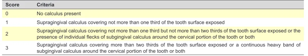
Table 2 - Classification criteria of the levels of calculus at the dental surface as per Green and Vermillion’s Simplified Oral Hygiene Index
Score Criteria
0 No calculus present
1 Supragingival calculus covering not more than one third of the tooth surface exposed
2 Supragingival calculus covering not more than one third but not more than two thirds of the tooth surface exposed or the presence of individual flecks of subgingival calculus around the cervical portion of the tooth or both
ARTIGO ORIGINAL Trialand its follow-up studies. 34,35
As regards patient’s oral hygiene habits, the frequency of attendance at dental clinics, teeth brushing frequency (number of times per day), toothbrush replacement frequency (number of times per year) and the use of mouthwash in oral hygiene were inquired. The patients with DM1 were also asked whether or not they used to brush their teeth upon recovery from an episode of nocturnal hypoglycaemia.
Dental examination has been carried out according with the WHO recommendations.
The levels of oral hygiene were assessed by using the Simplified Oral Hygiene Index (OHI-S) developed by Greene and Vermillion36 and the plaque score index at
the vestibular surface of tooth number 16, 26, 11 and 31 and at the lingual surface of tooth number 36 and 46 has been used for the determination of the OHI-S. In case any of the abovementioned teeth were missing, the surface of the adjacent teeth (second or third molar and contralateral central incisors) were assessed. OHI-S scores range from 0 to 3, according with the criteria shown in Table 1 and 2. The mean score from all those obtained corresponds to each patient’s OHI-S.
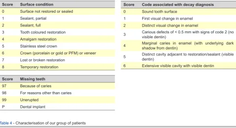
Dental condition has been assessed according with the International Caries Detection and Assessment System (ICDAS),37 consisting of a standardized system of clinical
assessment allowing for the detection, assessment,
diagnosis and monitoring of tooth decay lesions. The system is based on a two-digit code assigned to each tooth surface, in which the first one corresponds to whether or not any filling or fissure sealant are present and the second code regards whether or not any tooth decay lesions were found (Table 3).
The observer has been monitored by an examiner experienced in the use of the ICDAS and a high level of inter-observer and intra-observer agreement has been obtained (k = 0.87 and k = 0.97, respectively).
This was an observational, analytical and
cross-sectional study and IBM® SPSS® v.22.0 (IBM Corporation,
Armonk, New York, EUA) software has been used. Chi-square, Mann-Whitney and Fisher’s test and Spearman’s correlation coefficient have been used for data analysis; a 5% level of significance has been assumed.
RESULTS
No statistically significant differences were found between both groups as regards patient’s age and a statistically significant difference has been found regarding gender (Table 4).
Only two patients in the DM1 group have described the use of metformin.
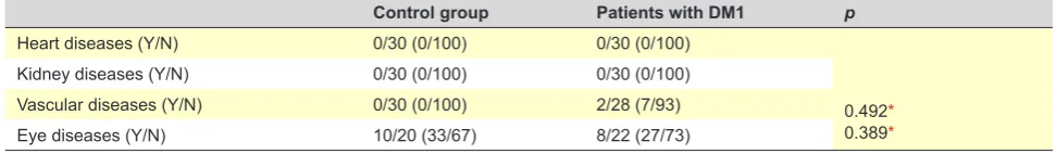
No statistically significant association has been found between the presence of heart, kidney, vascular and eye complications and the presence of diabetes (Table 5). Two
Table 3 - Classification of the International Caries Detection and Assessment System (ICDAS)
Score Surface condition
0 Surface not restored or sealed 1 Sealant, partial
2 Sealant, full
3 Tooth coloured restoration 4 Amalgam restoration 5 Stainless steel crown
6 Crown (porcelain or gold or PFM) or veneer 7 Lost or broken restoration
8 Temporary restoration
Score Code associated with decay diagnosis
0 Sound tooth surface 1 First visual change in enamel 2 Distinct visual change in enamel
3 Carious defects of < 0.5 mm with signs of code 2 (no visible dentin)
4 Marginal caries in enamel (with underlying dark shadow from dentin)
5 Distinct cavity adjacent to restoration/sealant (visible dentin)
6 Extensive visible cavity with visible dentin
Score Missing teeth
97 Because of caries
98 For reasons other than caries 99 Unerupted
P Dental implant
Table 4 - Characterisation of our group of patients
Control group Patients with DM1 p
Gender (M/F) 9/21 (30/70) 17/13 (57/43) 0.067#
Age ( ) 27.6 ± 11.3 27.1 ± 12.5 0.648##
#Chi-square test;##Mann–Whitney’s test x±s
ARTIGO ORIGINAL
DM1 patients presented with chronic venous insufficiency as a vascular complication; patients from both groups have described the presence of astigmatism and myopia and the presence of cataracts and/or diabetic retinopathy has not been described by any patient.
As regards the test group, a 37.5% percentage of patients with levels of HbA1c consistent with a good metabolic control has been found.
No statistically significant differences have been found as regards heart, kidney, vascular or eye pathologies
between controlled andpoorly controlled patients.
No statistically significant differences were found between both groups as regards the frequency of attendance at dental clinics, teeth brushing and toothbrush replacement frequency. As regards the frequency of attendance at dental clinics and the frequency of teeth brushing, patients in both groups attended on average once a year dental clinics and follow their oral hygiene twice a day, namely upon main meals (lunch and dinner). As regards toothbrush replacement frequency, patients in the control group replaced on average four times a year their toothbrushes, while the patients in the DM1 group did it three times a year. Only three DM1 patients (10%) described having brushed their teeth upon sucrose intake due to an episode of nocturnal hypoglycaemia.
Twelve patients both in DM1 and in control group have described the use of antiseptic mouthwash as an adjuvant to teeth brushing.
Poorer levels of oral hygiene have been found in DM1 patients when compared to the patients with no diabetes, shown by a higher plaque score index in DM1 group when compared to the control group (IP = 2.33 and 1.43, respectively, p = 0.002). In addition, no statistically significant differences were found between both groups regarding the number of decayed, missing and filled teeth (Table 6).
No significant correlation has been found in DM1 patients between teeth brushing frequency and the number
of missing (Fisher’s exact test, p = 0.472), decayed (Fisher’s exact test, p = 0.642) and filled teeth (Fisher’s exact test, p = 0.804).
A statistically significant association (Fisher’s exact test,
p = 0.037) between the presence of DM1 and the presence
of family history of diabetes has been found: patients with a family history of diabetes showed a higher risk of diabetes (odds ratio 3.5 (OR 95% CI [1.2, 10.2]). Eighteen DM1 patients and nine patients in the control group had a family history of diabetes.
As regards DM metabolic control, no statistically significant association has been found between the presence of controlled or poorly controlled diabetes and oral health status of patients with DM1 (missing, decayed and filled teeth).
DISCUSSION
Our study aimed at the definition of a relationship between DM1 and tooth decay, based on the comparison of oral health status in DM1 patients vs. patients with no diabetes.
The association between tooth decay and diabetes has been the subject of different studies and some of these have found that DM1 patients can show higher susceptibility for the development of tooth decay lesions when compared to patients with no diabetes. This higher susceptibility is based on risk factors considered as predisposing for the development of tooth decay, such as salivary flow and composition changes, changes in oral microflora, poor dietary and metabolic control and sub-optimal oral
hygiene.33 A higher risk of tooth decay was found in DM1
patients and can be explained by the synergistic action of the disease with patient’s poor metabolic control, leading to high concentrations of salivary glucose associated with the hyperglycaemic state and impaired neuro-regulatory mechanism of the salivary glands. The increased concentration of salivary glucose in the oral environment promotes bacterial growth and proliferation, increasing
Table 5 - Characterisation of our group of patients as per the number of missing, decayed and filled teeth
Control group Patients with DM1 p
Missing teeth 1.57 1.43 0.798*
Decayed teeth 4.13 3.83 0.261*
Filled teeth 2.07 2.73 0.513*
* Fisher’s exact test
Table 6 - Distribution of heart, kidney, vascular and eye complications
Control group Patients with DM1 p
Heart diseases (Y/N) 0/30 (0/100) 0/30 (0/100)
0.492*
0.389*
Kidney diseases (Y/N) 0/30 (0/100) 0/30 (0/100) Vascular diseases (Y/N) 0/30 (0/100) 2/28 (7/93)
Eye diseases (Y/N) 10/20 (33/67) 8/22 (27/73)
ARTIGO ORIGINAL the lactic acid production and reducing pH levels and salivary buffer capacity, considered as risk factors for the
development of tooth decay.21,30,33 Some studies have not
found any relationship between DM metabolic control and the development of tooth decay,18,21 other studies did not
find any significant relationship between both entities, while others have described a decreased risk for the development of tooth decay.20,23
Even though no statistically significant differences were found between the presence of other concomitant pathologies (including heart, eye, kidney and vascular diseases) and DM1 metabolic control, some studies have found that a 1% reduction in the level of HbA1c is associated with a very relevant reduction in the prevalence of macro and microvascular complications, showing the relevance of keeping a good DM1 metabolic control.12,20 In our
study, the absence of results with statistical significance is probably related to the small number of patients from each group (controlled vs. poorly controlled DM1 patients) upon stratification according with DM1 metabolic control. In addition, the disease duration in the group of patients with diabetes would have been relevant, which has not been considered.
A higher OHI-S score has been found in the DM1 group when compared with the control group, in line with other studies, which may have been associated with the poor metabolic control of the disease.20,21,33 Even though DM1
patients have shown higher OHI-S scores than patients with no diabetes, it has been described that plaque is less cariogenic due to the decreased sucrose intake in these patients, which is related to their diet.20,21
Only three patients (10%) described teeth brushing upon sucrose intake, regarding episodes of nocturnal hypoglycaemia, showing patient’s education, concern and knowledge on the relevance of oral hygiene upon carbohydrate intake.
No statistically significant association has been found between teeth brushing frequency, DM1 metabolic control and the development of tooth decay in the DM1 group, even though this group included a higher percentage of patients with poorly controlled diabetes. As described above, some studies have found an association between DM metabolic control and the development of decay, due to the high concentration of salivary glucose, which is the ideal substrate for bacterial growth and proliferation.18,19,21 Decreased teeth
brushing frequency cannot be independently associated with the development of tooth decay lesions, due to the multifactorial aetiology of the disease,6,38,39 even though it
has a very relevant role in the prevention of tooth decay and in keeping optimal oral health.
The results of our study are in line with others in which no consistent association has been found between tooth
decay and DM1. This is still a controversial association: some authors found it, while others did not find any relationship between them.18-21,23,28 On the one hand, the
higher prevalence of decay in patients with poorly controlled DM1 can be associated with an increased concentration of salivary glucose and salivary gland disorder, with subsequently increased salivary viscosity and decreased
pH and flow, leading to the colonisation by Streptococcus
mutans and Lactobacillus spp.19,33 On the other hand, the
reduction in tooth decay lesions found in patients with DM1 can be associated with decreased levels of salivary glucose due to diet control, with lower sucrose intake.19,33
Even though the frequency of attendance at nutrition clinics by DM1 patients has not been assessed, these patients usually attend Endocrinology and Nutrition outpatient clinics at the CHUC, which may in part explain the decreased number of tooth decay lesions that was found and may have been associated with a decreased concentration of salivary glucose, as described above.33
Even though no statistically significant differences have been found between both groups, different diabetes-related oral complications have been described, as well as different risk factors for the development of tooth decay lesions. Therefore, a careful follow-up of these patients is crucial in order to allow for prevention and early diagnosis. This was one of the main advantages of the study as a follow-up for treatment and prevention of diabetes oral manifestations has been carried out.
HbA1c levels, as well as DM1 metabolic control, the signs and symptoms at the time of any hypoglycaemic crisis, as well as the presence of anxiety, increased sweating, tachycardia, impaired consciousness and lethargy during the examination should be considered before any dental treatment.13,14 Patients must be asked to bring their
own glucose monitoring device, as well as their insulin pens and some sucrose-rich food (to be taken in case of hypoglycaemia).9,14 Dentists have a relevant role in the
observation, survey and record of any diabetes-related oral sign or symptom, as well as in primary counselling and referral to the specialist. The treatment of oral cavity diseases should also be considered in order to prevent or delay the development of diabetes-related oral complications.9,13
ARTIGO ORIGINAL
CONCLUSION
No higher prevalence of tooth decay lesions in patients with type-1 diabetes mellitus has been found in this study, when compared to patients with no diabetes, in line with the null hypothesis initially established.
Even though there is no scientific evidence regarding the association of the prevalence of tooth decay lesions and type-1 diabetes mellitus, the knowledge of both diseases is extremely relevant for dental treatment and further studies are recommended, as well as the involvement of larger groups of patients.
ACKNOWLEDGMENTS
The authors wish to acknowledge the Sociedade
Portuguesa de Diabetologia for having assigned the
Bolsa de Estudo Pedro Eurico Lisboa SPD/BAYER to the
study ‘Caracterização das Alterações no Biofilme e na Saliva de Diabéticos do Tipo I com Bomba de Insulina: Estudos in Vivo e in Vitro em Fibroblastos’, in which this study has been included, as well as to the Department of
Endocrinology, Diabetes and Metabolism of the Centro
Hospitalar e Universitário de Coimbra. The authors also
wish to acknowledge Helena Donato, Diretor of the Serviço
de Documentação of the Centro Hospitalar e Universitário
de Coimbra for having provided some relevant references
regarding the subject of the study.
HUMAN AND ANIMAL PROTECTION
The authors declare that the followed procedures were according to regulations established by the Ethics and Clinical Research Committee and according to the Helsinki Declaration of the World Medical Association.
DATA CONFIDENTIALITY
The authors declare that they have followed the protocols of their work centre on the publication of patient data.
CONFLICTS OF INTEREST
The authors declare that there were no conflicts of interest in writing this manuscript.
FINANCIAL SUPPORT
The authors declare that the “Bolsa de Estudo Pedro Eurico Lisboa SPD/BAYER” grant has been assigned to the study “Caracterização das Alterações no Biofilme e na Saliva de Diabéticos do Tipo I com Bomba de Insulina: Estudos in Vivo e in Vitro em Fibroblastos”, within which the present study is included.
REFERENCES
1. Canivell S, Gomis R. Diagnosis and classification of autoimmune diabetes mellitus. Autoimm Rev. 2014;13:403-7.
2. Atkinson MA. The pathogenesis and natural history of type 1 diabetes. Cold Spring Harb Perspect Med. 2012;2.
3. Thomas CC, Philipson LH. Update on diabetes classification. Med Clin North Am. 2015;99:1-16.
4. Cameron FJ, Wherrett DK. Care of diabetes in children and adolescents: controversies, changes, and consensus. Lancet. 2015;385:2096-106. 5. Atkinson MA, Eisenbarth GS, Michels AW. Type 1 diabetes. Lancet.
2014;383:69-82.
6. Pugliese A. The multiple origins of Type 1 diabetes. Diabet Med. 2013;30:135-46.
7. Levy L, Zeichner JA. Dermatologic manifestation of diabetes. J Diabetes. 2012;4:68-76.
8. Imam K. Clinical features, diagnostic criteria and pathogenesis of diabetes mellitus. Adv Exp Med Biol. 2012;771:340-55.
9. Association AD. Standards of Medical Care in Diabetes-2016 Abridged for Primary Care Providers. Clin Diabetes. 2016;34:3-21.
10. Vehik K, Ajami NJ, Hadley D, Petrosino JF, Burkhardt BR. The changing landscape of type 1 diabetes: recent developments and future frontiers. Curr Diab Rep. 2013;13:642-50.
11. Gajos G, Pilacinski S, Zozulinska-Ziolkiewicz D. Controversies in diabetes in 2013 - a brief update. Adv Clin Exp Med. 2013;22:777-84. 12. Lechleitner M, Hoppichler F. Insulin therapy. Wien Med Wochenschr.
2011;161:300-4.
13. Wray L. The diabetic patient and dental treatment: an update. Br Dent J. 2011;211:209-15.
14. Kidambi S, Patel SB. Diabetes mellitus: considerations for dentistry. J Am Dent Assoc. 2008;139:8S-18.
15. Maletkovic J, Drexler A. Diabetic ketoacidosis and hyperglycemic hyperosmolar state. Endocrinol Metab Clin North Am. 2013;42:677-95. 16. Marcovecchio ML, Chiarelli F. Microvascular disease in children
and adolescents with type 1 diabetes and obesity. Pediatr Nephrol. 2011;26:365-75.
17. Silva MF, Barbosa KG, Pereira JV, Bento PM, Godoy GP, Gomes DQ. Prevalence of oral mucosal lesions among patients with diabetes mellitus types 1 and 2. An Bras Dermatol. 2015;90:49-53.
18. Saes Busato IM, Bittencourt MS, Machado MA, Grégio AM, Azevedo-Alanis LR. Association between metabolic control and oral health in adolescents with type 1 diabetes mellitus. Oral Surg Oral Med Oral Pathol Oral Radiol Endod. 2010;109:e51-6.
19. Miralles L, Silvestre FJ, Hernández-Mijares A, Bautista D, Llambes F, Grau D. Dental caries in type 1 diabetics: influence of systemic factors of the disease upon the development of dental caries. Med Oral Patol Oral Cir Bucal. 2006;11:E256-60.
20. Siudikiene J, Machiulskiene V, Nyvad B, Tenovuo J, Nedzelskiene I. Dental caries and salivary status in children with type 1 diabetes mellitus, related to the metabolic control of the disease. Eur J Oral Sci. 2006;114:8-14.
21. Rai K, Hegde AM, Kamath A, Shetty S. Dental caries and salivary alterations in Type I Diabetes. J Clin Pediatr Dent. 2011;36:181-4. 22. Lalla E, Cheng B, Lal S, Tucker S, Greenberg E, Goland R, et al.
Periodontal changes in children and adolescents with diabetes: a case-control study. Diabetes Care. 2006;29:295-9.
23. Orbak R, Simsek S, Orbak Z, Kavrut F, Colak M. The influence of type-1 diabetes mellitus on dentition and oral health in children and adolescentes. Yonsei Med J. 2008;49:357-65.
24. Lal S, Cheng B, Kaplan S, Softness B, Greenberg E, Goland RS, et al. Accelerated tooth eruption in children with diabetes mellitus. Pediatrics. 2008;121:e1139-43.
25. Sima C, Glogauer M. Diabetes mellitus and periodontal diseases. Curr Diab Rep. 2013;13:445-52.
26. Preshaw PM, Alba AL, Herrera D, Jepsen S, Konstantinidis A, Makrilakis K, et al. Periodontitis and diabetes: a two-way relationship. Diabetologia. 2012;55:21-31.
ARTIGO ORIGINAL
diseases and dental caries in children with type 1 diabetes mellitus. Mediators Inflamm. 2015;2015:8.
29. Kaur G, Holtfreter B, Rathmann W, Schwahn C, Wallaschofski H, Schipf S, Nauck M, Kocher T. Association between type 1 and type 2 diabetes with periodontal disease and tooth loss. J Clin Periodontol. 2009;36:765-74.
30. Busato IM, Ignácio SA, Brancher JA, Moysés ST, Azevedo-Alanis LR. Impact of clinical status and salivary conditions on xerostomia and oral health-related quality of life of adolescents with type 1 diabetes mellitus. Community Dent Oral Epidemiol. 2012;40:62-9.
31. Pranckeviciene A, Siudikiene J, Ostrauskas R, Machiulskiene V. Severity of periodontal disease in adult patients with diabetes mellitus in relation to the type of diabetes. Biomed Pap Med Fac Univ Palacky Olomouc Czech Repub. 2014;158:117-23.
32. Indurkar MS, Maurya AS, Indurkar S. Oral Manifestations of Diabetes. Clin Diabetes. 2016;34:54-7.
33. Sampaio N, Mello S, Alves C. Dental caries-associated risk factors and type 1 diabetes mellitus. Pediatr Endocrinol Diabetes Metab. 2011;17:152-7.
34. Greene JC, Vermillion JR. The simplified oral hygiene index. J Am Dent Assoc. 1964;68:7-13.
35. Pitts NB, Ekstrand KR. International Caries Detection and Assessment System (ICDAS) and its International Caries Classification and Management System (ICCMS) - methods for staging of the caries process and enabling dentists to manage caries. Community Dent Oral Epidemiol. 2013;41:e41-52.
36. Frandsen CS, Dejgaard TF, Madsbad S. Non-insulin drugs to treat hyperglycaemia in type 1 diabetes mellitus. Lancet Diabetes Endocrinol. 2016 Sep;4:766-80.
37. Galleri L, Sebastiani G, Vendrame F, Grieco FA, Spagnuolo I, Dotta F. Viral infections and diabetes. Adv Exp Med Biol. 2012;771:252-71. 38. Christen U, von Herrath MG. Do viral infections protect from or enhance
type 1 diabetes and how can we tell the difference? Cell Mol Immunol. 2011;8:193-8.