S
Spprriinngg22001199,,VVooll88,,NNoo22 D
DOOII::1100..2222008888//IIJJMMCCMM..BBUUMMSS..88..22..111188
Circulating MiR-10b, MiR-1 and MiR-30a Expression Profiles in
Lung Cancer: Possible Correlation with Clinico-pathologic
Characteristics and Lung Cancer Detection
Roghayeh Sheervalilou1,2, Hajie Lotfi3, Milad Shirvaliloo4,5, Akbar Sharifi2, Masoud Nazemiyeh2, Nosratollah
Zarghami2,3
1. Pharmacology Research Center, Zahedan University of Medical Sciences, Zahedan, Iran.
2. Tuberculosis and Lung Disease Research Center, Tabriz University of Medical Sciences, Tabriz, Iran. 3. Department of Medical Biotechnology, Faculty of Advanced Medical Sciences, Tabriz University of Medical Sciences, Tabriz, Iran.
4. Faculty of Medicine, Tabriz University of Medical Sciences, Tabriz, Iran.
5. Student Research Committee, Tabriz University of Medical Sciences, Tabriz, Iran.
Circulating microRNAs have been recognized as promising biomarkers for the detection of lung cancer. The
objective of this study was to evaluate miR-10b, miR-1 and, miR-30a in the plasma samples of lung cancer
patients to confirm any possible relevance in the early detection of lung cancer. Plasma samples from 47
non-small-cell lung cancer patients and 41 cancer-free subjects were evaluated for selected microRNAs using the
real-time PCR method. To evaluate the tobacco smoking effects on microRNAs expression, the studied groups
were categorized into two subgroups: never-smokers and smokers. MiR-1/miR-30a expression levels were
significantly reduced in lung cancer, while the miR-10b level was significantly elevated. We found that smoking
had significant effects on the levels of circulating microRNAs in the smokers of the cancer-free group (a
significant up-regulation of miR-10b and significant down-regulation of miR-1/miR-30a), and lung cancer
patients (a significant elevation of 10b). Receiver operating characteristic curve analysis showed that
miR-10b with an area under the curve of 0.861, and miR-1/miR-30a with values of0.905 and 0.889 for the same
parameter, could distinguish non-small-cell lung cancer patients from cancer-free subjects. Our findings
demonstrated significant differences in the expression of microRNAs in lung cancer and the considerable effects
of smoking on microRNAs levels. Area under curve analysis showed that miR-10b with 78% sensitivity/78%
specificity, miR-1 with 95% sensitivity/80% specificity and miR-30a with 87% sensitivity/83% specificity,might
be good (miR-10b/miR-30a) and excellent (miR-1) markers for lung cancer detection.
Key words: Clinico-pathologic characteristics, microRNA, carcinoma, non–small-cell lung cancer, plasma
Corresponding author: Zahedan University of Medical Sciences, Zahedan, Iran Email: [email protected]
ung cancer is one of the most obstinate leading
causes of cancer death worldwide (1). In 2018,
lung cancer was the most commonly diagnosed
malignancy (about 11.6% of all new cases) and the
L
Submmited 7 April 2019; Accepted 13 August 2019; Published 20 August 2019
most prevalent cause of cancer-related deaths
(about 18.4% of all cancer-related deaths)(2).
Non-small-cell lung cancer (NSCLC) represents about
80–85% of all lung cancers (3), and based on
histological properties may be divided into 3
subgroups with distinct features:1) adenocarcinoma
(AC), 2) squamous cell carcinoma (SCC) and 3)
large cell carcinoma (LCC) (4). Cigarette smoking
has been reported to be the hallmark risk factor (5).
In fact, tobacco- associated cancer risk in
ex-smokers remains high and equal to that of active
smokers (6). Unfortunately, due mainly to the lack
of effective diagnostics for early detection (6),
many lung tumors are detected at advanced stages,
when the 5-year survival rate is barely15% (7). The
National Lung Screening Trial (NLST) for
screening of high-risk population through low-dose
helical computed tomography(LDCT) demonstrated
that an appreciable diminution of 20% in mortality
rate may be achieved (8). Considering some
disadvantages of LDCT like high false-positive
rates, high costs, and potential side effects such as
radiation exposure, there is an immediate demand
for simpler, non-invasive or semi-invasive,
sensitive and reliable methods for pivotal
screenings of high-risk individuals, thus, early
detection of the disease (4).
MicroRNAs are short non-coding endogenous
RNAs (22 nt) that function as gene regulators at
post-transcriptional level (9, 10) via binding to the
3' untranslated region (3'UTR) of messenger RNAs
(mRNAs) as their targets (7, 11). MicroRNAs are
involved in a wide range of physiological and
pathological events (12-14). In tumor tissues,
patterns of microRNAs expression are consistently
different than normal, and some key microRNAs
may be traced in such patterns (3). According to the
recent findings, there are circulating microRNAs
with diagnostic potential for every type of
malignancies including lung cancer (6). All tumor
cells release microRNAs into the circulation, and it
has been well-established that the level of
circulating miRNAs may be altered in cancer (15).
Based on recent findings, microRNAs
modulate a wide variety of physiological/cellular
events, and deregulation of their expression/
function is closely associated with tumorigenesis,
angiogenesis, and development, promotion,
progression and ultimately metastasis of the
malignant cells (16).
Mir-10b gene is located within the HOXD
cluster (ch2q31) (17). Homeobox D10 (HOXD10)
inhibits the expression of cell migration and extra cellular matrix remodeling factors (α3-integrin/ RhoC/MMP14/uPAR). MiR-10b is up-regulated
following the binding of Twist to its promoter,
leading to a reduced level of HOXD10 (18) and
E-cadherin (19), and an increase in kruppel-like factor
4 (KLF4). These cascades induce up-regulation of
RhoA/RhoC factors, Rho kinase activation and
tumor invasion (16). In NSCLC, miR-10b acts as a
carcinoma enhancer by targeting klotho (20), which
also occurs to be a prominent therapeutic target
(16).
1 is downregulated in lung cancer.
MiR-1 induction considerably reduces the expression of
oncogenic targets (MET/Pim-1/ FoxP1/ HDAC4/
Mcl-1) and triggers the activation of caspases 3/7,
and Poly (ADP- Ribose) Polymerase 1
(PARP1)(21). In NSCLC, serum miR-1 level is
significantly correlated with overall survival of
patients, and may be implicated as a non-invasive
predictor for cancer prognosis (22). MiR-30a
(ch6q.13) (23) has been reported to be
down-regulated in metastasis, while it is crucial for
maintaining epithelial traits (24). MiR-30a is one of
the highly down-regulated microRNAs in
metastatic cancers (25) and is declined during TGF-β-promoted tumor metastasis, ultimately leading to increased expression levels of
invasion/metastasis-associated mesenchymal factors (N-cadherin, Slug,
Snail, and Twist) (24, 26).
According to our previous study and
computational algorithms, we selected miR-10b as
an oncomir along with miR-1 and miR-30a, both of
which are involved in lung cancer and have
previously been explored in cell lines, tissues and
blood (27). The present study explored whether the
plasma microRNAs signatures of lung cancer
patients had correlation with clinico- pathologic
characteristics of patients, and if cigarette smoking
had any effects on expression level of
microRNAs.We also aimed to investigate whether
the seselected oncomir and tumor suppressors had
any potential to be demonstrated as new markers
for early detection of lung cancer.
Materials and methods
Study population
NSCLC patients (n=47) and cancer-free
individuals (n=41) were selected as subjects,
enrolled from Shahid Madani Hospital, Tabriz
(2015-2016). Each group was subdivided into two
subgroups including smokers (smoking history: at
least 5 years, smoking exposure: about more than
20 packs/year) and never-smokers (subjects with no
history of past and present smoking, neither active
nor passive). Before endoscopic assessment, all
subjects underwent clinical examination; plain chest
radiograph; CT scan of the chest, upper abdomen,
and brain; fiber-optic bronchoscopy; and bone scan
were all conducted.Subjects with the following
criteria were included in the research:an age of 40
to 90 years old;having no previous history of other
types of cancer prior to participating in this study,
as well as nochronic/ acute/ hormonal/ infectious
disorders;having no past exposure to carcinogenic
agents, including asbestos, and also passive
smoking;having no history of any therapeutic
procedures (surgery/ chemotherapy/
radiotherapy).Written informed consent was
obtained from all subjects. According to the ethical
standards (Declaration of Helsinki, 1964), the
Ethical/ Scientific Committee of Clinical Research
in Tabriz University of Medical Sciences (Tabriz,
Iran) authorized the present study (Ethical code:
IR.TBZMED.REC.1395.1112).
Sampling
Blood sample (5-8 ml peripheral venous
fasting blood) was obtained from all subjects,
and immediately centrifuged, and the plasma was
stored at -80oC. To determine the stage of tumor, guidelines of the American Joint Committee
on Cancer were used, and histo pathological
Table 1. Demographic/clinicopathologiccharacteristics of all subjects.
Demographic and clinical characteristics NSCLC patients N=47
Non-cancerous subjects N=41
Gender male 31(%65)
16(%35)
20 (%48) 21 (%52) female
Age 40-59 14 (%29)
33 (%71)
13 (%31) 28 (%69) 60≤
Smoking History
Non-smoking 9 (%19)
38 (%81)
21 (%51) 20 (%49) smoking
Histology
AD 15 (%31)
22 (%46) 10 (%23) SCC
LC
Stage
I 9 (%19)
23 (%48) 10 (%21) 5 (%12) II
III IV
classification was confirmed based on the World
Health Organization (WHO) classification. None of
the patients had received any therapy before
participating in the present study (Table 1).
MicroRNAs profiling
A comprehensive assessment on
aforementi-oned microRNAs was performed using www.
miRbase.org, www.TargetScan.org,
miRTarget-LinkHuman, miRpathDB, and miRmap databases,
Targets for each of these microRNAs and their
possible involvement in tumorigenesis and
progression of cancer were evaluated. The target
genes and their related binding site on seed regions
of microRNAs were explored via Target Scan
(http://www.targetscan.org/), and the mature microRNAs’ sequences were confirmed according to miRbase data (http://microrna.sanger.ac.uk/).
RNA isolation
For RNA isolation, 1-2 ml yielded plasma was
centrifuged (~2,000 rpm for 5 min) in order to
obtain cell free starting material,.The supernatant (200 μl) was used for total RNA extraction using isolated Exiqon kit (miCUYRTM RNA Isolation Kit-Biofluids, Cat No. #300112 & #300113) according to manufactures’ guideline.The concentration, quantity, and purity of RNAs were
confirmed using the relative absorbance ratio at
A260/A280 and A260/A230 on aspectrophotometer
(Nano Drop 2000, Thermo Scientific, Wilmington,
DE, USA).The purified RNA was stored at -80°C.
Reverse transcription (RT-PCR)
(5 ng/μl) was used for each sample. According to manufacturer’s instruction (miRCURY LNA™ Universal cDNA synthesis kit, Cat No. #203301)
cDNA was synthesized in a thermocycler
(Eppendorf, Germany).
Quantitative real-time PCR
Quantitative real-time PCR was carried out in a total volume of 20 μlreaction mixture, and using primers (Exiqon, Denmark.Cat.No.204344, 205637, 205695) in accordance with the manufacturer’s
protocol in a Rotor-Gene Q (Qiagen, Germany)
intrument. All experiments were conducted in
triplicate. To limit artefactual regulation caused by
sample normalization, only the values below a minimal threshold (Ct≤36) were analyzed. Ergo, relative expression of microRNAs was normalized
to the expression level of miR-16 (Exiqon,
Denmark. Cat. No. 205702) as internal control, and
calculated according to the 2−ΔCt method (28).MiR-16 as stable internal control was used for data
normalization through 2-(Ct interest gene-Ct control) method in human samples (29).Sample size was calculated
based on Glenn D method(30).
Statistical analysis
Statistical analysis of the microRNAs
expression between the two groups was conducted
based on Mann Whitney, because the distribution of
the variables was abnormal (according to
Kolmogorov–Smirnov/Shapiro-Wilk test). The
difference in microRNAs expression between
NSCLC subtypes/stages was estimated through
Kruskal Wallis method. The results were analyzed
using SPSS v.16 and DATA ASSIST v3.01.
The correlation between the prevalence of each
microRNA and the demographic/ clinical
characteristics (age/ gender/ smoking history/ tumor
histology-stage) of NSCLC patients was analyzed
with Spearman’s and chi-square rank correlation.
Moreover, the effects of smoking on microRNAs
level were analyzed with Mann Whitney test. Also,
receiver-operator characteristic (ROC) was drawn,
and the area under the curve (AUC) was measured
(based on Youdens,index) to determinate the accuracy of each microRNA (AUC: 0.700-0.799
refers to fairly candidate biomarker, 0.800-0.899
refers to good candidate biomarker, 0.900-1.0 refers
to excellent candidate biomarker). A<0.05 was
considered statistically significant.
Results
MicroRNAs profiling in plasma
Real-time PCR was applied to evaluate the
expression levels of microRNAs in the two groups
(Fig. 1) and in the clinical stages of NSCLC (Fig.
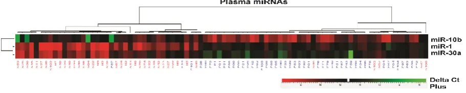
2). Based on the ΔCT values, distances between all
subjects were calculated for hierarchical clustering
(Fig. 3). Accordingly,the correlations between the
expression level of microRNAs and demographic/
clinico-pathologic characteristics of cancer patients
were evaluated. Moreover, the effects of cigarette
smoking on microRNAs expression were analyzed
(Fig. 4).
Fig. 1. MicroRNAs expression in NSCLC patients and non-cancerous group. Expression of miR-10b (a), miR-1 (b) and miR-30a (c) in NSCLC patients and non-cancerous groups was compared (P˂ 0.05).
.
Fig. 2. MicroRNAs expression in different clinical stages of NSCLC. Relative expression (Mean±SE) of miR-10b, miR-1 and miR-30a in four stages of the NSCLC was compared (P˂ 0.05).
.
Fig. 3. Cluster analysis and heat map of microRNAs expression.Differences between all studied subjects and assays were considered for hierarchical clustering and heat map according to the ΔCT values using Pearson’s Correlation. In the present graph, rows represent selected microRNAs, and columns represent all subjects. Furthermore, red indicates an increase with ΔCTs below the middle level, while green represents a decrease with ΔCTs above the middle level (P< 0.05).
MiR-10b was overexpressed in NSCLC
Significant increase in miR-10b expression in
plasma of the NSCLC patients was observed (P
<0.001) (Fig. 1a). Also, miR-10b was found to
show a significant up-regulation in four clinical
stages of NSCLC (P=0.002) (Fig. 2). No significant
correlation between miR-10b overexpression and
demographic/ clinical characteristics of the cancer
group was found.However,we observed a moderate
positive correlation between miR-10b level and
clinical stages of NSCLC patients (P <0.001,
R=+0.54).
MiR-1 was down-regulated in NSCLC
When expression of miR-1 in the NSCLC
group was compared to the cancer- free samples, a
considerable decrease of miR-1 (P<0.001) was
Fig. 4. The effects of cigarette smoking on microRNAs expression in the cancer-free subjects and cancer patients. Significant effects of cigarette smoking on microRNAs expression in the smoker and never smoker individuals of the cancer-free group (a) and lung cancer patients (b) were evaluated (P< 0.05).
Fig. 5. Diagnostic value of microRNAs. MiR-10b produced 0.861 AUC value (%95 CI: 0.785-0.937, P <.001), miR-1 with 0.905 AUC (%95 CI: 0.832-0.978, P <.001) and miR-30a with 0.889 AUC (%95 CI: 0.817-0.961, P <.001).
observed in patients (Fig. 1b). Down-regulation of
miR-1 was also found with significant differences
in four clinical stages of NSCLC (P =0.001) (Fig.
2). Furthermore, we noticed a moderate negative
correlation between miR-1 expression and the
clinical stages of NSCLC (P <0.001, R= -0.596).
MiR-30a was down-regulated in NSCLC
Evaluation of miR-30a expression in NSCLC
cases demonstrated lower levels of this microRNA
in comparison with the cancer-free group (P<0.001)
(Fig. 1c). Down-regulation of miR-30a was also
detected with significant difference in four clinical
stages of NSCLC (P=0.013) (Fig. 2). We also found
a moderate negative correlation between miR-30a
expression and the clinical stages of NSCLC (P
<0.001, R=-0.538).
Evaluation of the effects of smoking on microRNA expression
The effects of smoking on expression of
microRNAs in the cancer-free (Fig. 4a) and cancer
groups (Fig. 4b) was analyzed. Our study found
significant effects of smoking on microRNAs
expression in the smokers in comparison to with
that of never-smoker individuals in the cancer-free
group, including: a considerable elevation of
miR-10b (P =0.004) and significant down-regulation of
miR-1 (P<0.001) and miR-30a (P<0.001) in the
smokers of the control group. Evaluation of the
effects of cigarette smoking on microRNAs level in
lung cancer patients found a significant
up-regulation of miR-10b (P=0.017) in smokers.
ROC curve analysis
ROC anelysis (with %95 CI) of miR-10b and
miR-1/miR-30a indicated a 0.861 AUC
(0.785-0.937, P<0.001) for miR-10b, and higher AUCs of
0.905 (0.832-0.978, P <0.001) and 0.889
(0.817-0.961, P<0.001) for miR-1 and miR-30a,
respectively. Taking the optimum cut-off into
account, miR-10b showed a sensitivity and
specificity of both 78 percent (Fig. 5a),and was
deemed a promising circulating microRNA for
diagnosis of NSCLC. MiR-1 with 95% sensitivity
and 80% specificity (Fig. 5b), and miR-30a with
87% sensitivity and 83% specificity (Fig. 5c) could
also be good (miR-30a) and an excellent (miR-1)
markers for detection of NSCLC, respectively.
Discussion
Current routine diagnostics for lung cancer
include sputum cytology, chest imaging and
bronchoscopy assessments, that can discover only
15-20% of lung cancer cases prior to metastasis.
We suggested a new approach for future clinical
application, not only as a complementary diagnostic
investigation fo rconventional diagnostics, but also
for monitoring of prognosis and response to therapy
through blood sampling with minimal invasiveness,
low cost and time-saving characteristics.
As the evidence points out, circulating
microRNAs are very stable, and resistant to blood
RNase even in severe conditions (22). However,
under pathological conditions, intracellular
microRNAs are released into the circulation
dominantly from tumor cells via tumor-derived
microvesicles/exosomes (31). Despite the few
recent studies, the clinical importance of miR-10b
in the plasma samples of NSCLC has not yet been
clearly explained. Huang et al. and Pan et al. in
their studies found that in NSCLC cells, miR-10b
increased cell proliferation and inhibited apoptosis
by targeting Klotho protein (20, 32). Liu et al.
found that miR-10b acted as an oncomir by
positively targeting KLF4 and consequently promoting proliferation and invasion of A549 cell
line (NSCLC) (16).Furthermore, Huang et al.
showed that in miR-10b-silenced NSCLC cells,
apoptosis-inducing factors like Fas/FasL, Bax and
caspase3 were up-regulated and
apoptosis-inhibiting molecules including Bcl-2 and PCNA
were down-regulated (20). The present study in
agreement with the aforementioned studies was
able to demonstratethe overexpression of miR-10b
in NSCLC. Also, up-regulated miR-10b was
positively associated with the clinical stages of
NSCLC. The present study was also in agreement
with our previous study, which had reported
up-regulation of miR-10b in BAL cell fraction and
sputum cells of NSCLC patients, and a positive
correlation between miR-10b up-regulation and the
clinical stages of cancer (27). These results
complemented the recent study which reported high
miR-10b expression in tissue samples of NSCLC
patients, whichin fact was correlated with advanced
pathological stage of NSCLC patients (32).
However, this study did notfind any correlation
between miR-10b level and demographic/clinic
pathological characteristics of patients. Consistent
with the present findings, an elevation in miR-10b level was discovered in NSCLC patients’ plasma samples, which could be applied as a novel
diagnostic biomarker in the evaluation of lymph
node-positive cases (33). Another research group
found a significantly positive correlation between
miR-10b and the TNM stages. It was suggested that
miR-10b level, as a prognostic biomarker, might be
a notable candidate in development of
additional/alternative therapies for NSCLC (19,
34). Our analysis showed that elevated levels of
miR-10b may be represented as a diagnostic
signature to help with detection of NSCLC patients.
MiR-1 was down-regulated in lung cancer.
Nasser et al. reported that miR-1 by repression of
Mcl-1 function induced apoptosis in lung cancer. Hence, miR-1 affected the tumorigenic potential of
cancer cells by reducing the expression levels of
oncogenes like MET, FoxP1, Pim-1, and HDAC4. Cessationof miR-1 expression facilitates metastasis
(21). MiR-1 regulates tumor growth and metastasis
in lung cancer through down-regulation of MET oncogene(35). Hence, induction of miR-1
expression in A549 cells might potentiate the
sensitivity of these cells to doxorubicin by means of
facilitating the activation of caspase 3/7, PARP-1,
and depletion of Mcl-1 (21). The present study
found that miR-1 level was significantly reduced in
NSCLC patients. Moreover, down-regulated miR-1
expression was negatively correlated with the
advanced clinical stages of NSCLC. These results
were in agreement with our previous study which
had reported a down-regulation of miR-1 in BAL
cell fraction and sputum cells of NSCLC patients,
and found a negative correlation between low levels
of miR-1 and the clinical stages of cancer (27).
Besides, findings of the present study were in agreementwith Hu et al.’sfindings. They found 11 serum microRNAs with more than a 5-fold change
among subjects with longer and shorter survival
rates. According to their findings, miR-1 level was
considerably associated with cancer patients’
survival (22).
More recently, miR-30 down-regulation was
reported in lung cancer (36, 37). Kumarswamy et
al. reported a significant decrease of miR-30a
expression in resected tissue of lung cancer patients
in comparison with the corresponding normal
samples. They found that miR-30a by targeting
Snai1induced suppression of epithelial mesenchymal transition, invasion and metastasis
(24). Consequently, the reduced level of miR-30a in
SCC patients (38) could distinguish SCC patients
from normal in dividuals(39). Boeri et al. in their
study revealed that miR-30a along with another 9
top altered microRNAs were spotted in lung cancer
tissues which had formerly been detected with
computed tomography (CT) (40). Another study
suggested that miR-30a level significantly
decreased in the plasma of NSCLC patients, which
was in agreement with our previous study (27).
Also, the down-regulated miR-30a was negatively
associated with the advanced clinical stages of NSCLC,thatwas in agreement with Cheng et al.’s study. They reported that in the early breast tumors,
miR-30a levels were significantly correlated with
poor clinical characteristics (like advanced
stage/lymph node metastasis) (41).
In the present study, we used a systematic
approach through evaluation of circulating
microRNAs signatures in NSCLC. The developed
plasma-based microRNA yielded a sensitivity of
78% and a specificity of 78% for miR-10b. In 2011,
Roth et al. discovered that elevated levels of
circulating miR-10b with an AUC of 0.899 might
be considered as a novel diagnostic tool with
minimal invasiveness in lung cancer (33). Lu et al.
were able to successfully distinguish normal and
oral cancer plasma samples based on miR-10b
level, which yielded an AUC of 0.87 (42). In total
agreement with the mentioned findings, the present
study found that over-expression of miR-10b in
NSCLC plasma was reasonably accurate for
detection of NSCLC. Furthermore, the present
results indicated that miR-1 and miR-30a with
AUCs of 0.905 and 0.889,respectively, could help
discern NSCLC patients from cancer-free subjects.
These results were partially consistent with our
previous study which had reported that miR-10b
and miR-1 could be excellent and fair candidates to
discriminate NSCLC patients from cancer- free
subjects, respectively (27).
The present study was only designed for
evaluation of microRNAs in patients with NSCLC,
and not in its subtypes. There were 47 patients in
total: 15 adeno, 22 squamous, and 10 large cell
carcinoma. According to sample size and statistical
tests, it was not sensible to divide these subtypes
and draw a ROC curve for each one, separately.
In the present study, evaluation of the effects
of cigarette smoking on microRNAs expression in
both smokers and never-smokers in cancer patients
and cancer-free group showed significant
differences of miR-10b, miR-1 and miR-30a in the
cancer-free group and miR-10b in the cancergroup.
According to previous studies, alteration in
microRNAs levels were correlated with smoking
and human cancer (5, 43, 44). There are several
pathways involved in lung cancer that may affect
the tumor suppressor genes as well as miR-30a and
miR-1. Moreover, the heterogeneity of human
samples, in contrast to the cell lines, could affect
the gene expression. Until now, there has not been
any reported study on miR-1 and miR-30a
expression in NSCLC patients to be implicated in
our selected microRNAs.
Recent clinical studies about NSCLC
mentioned above, have suggested sets of differently
expressed microRNAs as promising circulating
biomarkers for monitoring prognosis and diagnosis
of lung cancer. Because of the high heterogeneity in
human subjects, the data showed a wide range of
distribution especially in small size studies. In
addition, RNase activity of blood might affect the
microRNAs (3-7, 12, 15, 22, 31, 33, 40).
This study suggested that miR-10b, miR-1 and
miR-30a, could be considered as helpful diagnostic
signatures in NSCLC patients. This classifier is
fairly new and could have important molecular
pathologic applications. However, there still
remains an important question concerned with these
signatures, which is concerned with the tissue
origin of the circulating microRNAs. There are
some ambiguous questions about circulating
microRNAs. The first, whether the studied
microRNAs can be emanated from the tumor cells
or maybe from the host reactions to tumorigenesis.
Second, whether the signatures of the evaluated
microRNAs play any role in the lung cancer
development (40). However, according to the
mentioned studies, it was notable that our selected
microRNAs were involved in lung cancer. Also,
another important regard is how to agree on a valid
universal signature. It was surprising gthat results
from different studies often varied. However, there
are complicated reasons for this occurrence, which
may have arisen from the differences in population
intrinsic genetic heterogeneity, pathologic sample
subtypes, different sample size, collection and
processing of samples, technology platforms,
bioinformatic approaches and data analysis,
different experiences and expertise of the
researchers.
In summary, our study showed that the
alteration in microRNAs expression in plasma
could help distinguish the NSCLC patients from
healthy subjects. The small size of the patients was
the main limitation of this study. While our findings
were consistent with the previous studies,
exploration of more samples could be helpful in
improvement of our molecular approach. At the
moment, there is not a comprehensive method for
screening of high risk population or early detection
of lung cancer patients, which can complement the
other existing traditional diagnostic tools.
Considering the noninvasive nature of plasma
sampling and due to its reproducibility and easy
detection of microRNAs, blood-based microRNAs
may be recognized as novel helpful signatures in
lung cancer early detection.
It is notable that the present study is the first
report in Iran about assessment of miR-10b, miR-1
and miR-30a expression profiles in blood samples
of lung cancer patients, and evaluation of the
effects of tobacco smoking on microRNAs level.
Until now, the role of miR-30a has only been
evaluated in other malignancies like breast cancer.
Hence, there are few studies about this particular
type of microRNA, and the combined effects of
these microRNAs in systemic circulation of patients
with lung malignancies.
Acknowledgments
Authors would like to thark the Tuberculosis
and Lung Disease Research Center, Tabriz
University of Medical Sciences, Tabriz, Iran for
financial endorsement of this study as a Ph.D. thesis
(No 93/4–10/3, Grant No 93/5).
Conflict of interest
The authors declare that there is no conflict of
interests regarding the publication of this paper.
References
1. Zhao H, Wang J, Kong X, et al. CD47 Promotes Tumor
Invasion and Metastasis in Non-small Cell Lung Cancer. Sci Rep
2016;6:29719.
2. Bray F, Ferlay J, Soerjomataram I, et al. Global cancer
statistics 2018: GLOBOCAN estimates of incidence and
mortality worldwide for 36 cancers in 185 countries. CA Cancer
J Clin 2018;68:394-424.
3. Heegaard NH, Schetter AJ, Welsh JA, et al. Circulating
micro-RNA expression profiles in early stage nonsmall cell lung
cancer. Int J Cancer 2012;130:1378-86.
4. Wozniak MB, Scelo G, Muller DC, et al. Circulating
MicroRNAs as Non-Invasive Biomarkers for Early Detection of
Non-Small-Cell Lung Cancer. PLoS One 2015;10:e0125026.
5. Huang J, Wu J, Li Y, et al. Deregulation of serum microRNA
expression is associated with cigarette smoking and lung cancer.
Biomed Res Int 2014;2014:364316.
6. Bianchi F, Nicassio F, Marzi M, et al. A serum circulating
miRNA diagnostic test to identify asymptomatic high-risk
individuals with early stage lung cancer. EMBO Mol Med
2011;3:495-503.
7. Cazzoli R, Buttitta F, Di Nicola M, et al. microRNAs derived
from circulating exosomes as noninvasive biomarkers for
screening and diagnosing lung cancer. J Thorac Oncol
2013;8:1156-62.
8. Sozzi G, Boeri M, Rossi M, et al. Clinical utility of a
plasma-based miRNA signature classifier within computed tomography
lung cancer screening: a correlative MILD trial study. J Clin
Oncol 2014;32:768-73.
9. Suzuki K, Yamada H, Nagura A. Association of cigarette
smoking with serum microRNA expression among middle-aged
Japanese adults. Fujita Medical Journal 2016;2:1-5.
10. Sheervalilou R, Shirvaliloo S, Fekri Aval S, et al. A new
insight on reciprocal relationship between microRNA expression
and epigenetic modifications in human lung cancer. Tumour Biol
2017;39:1010428317695032.
11. Kafshdooz L, Pourfathi H, Akbarzadeh A, et al. The role of
microRNAs and nanoparticles in ovarian cancer: a review. Artif
Cells Nanomed Biotechnol 2018;46:241-7.
12. Zhao C, Lu F, Chen H, et al. Clinical significance of
circulating miRNA detection in lung cancer. Med Oncol
2016;33:41.
13. Sheervalilou R, Ansarin K, Fekri Aval S, et al. An update on
sputum MicroRNAs in lung cancer diagnosis. Diagn Cytopathol
2016;44:442-9.
14. Aval SF, Lotfi H, Sheervalilou R, et al. Tuning of major
signaling networks (TGF-beta, Wnt, Notch and Hedgehog) by
miRNAs in human stem cells commitment to different lineages:
Possible clinical application. Biomed Pharmacother
2017;91:849-60.
15. Kroh EM, Parkin RK, Mitchell PS, et al. Analysis of
circulating microRNA biomarkers in plasma and serum using
quantitative reverse transcription-PCR (qRT-PCR). Methods
2010;50:298-301.
16. Liu Y, Li M, Zhang G, et al. MicroRNA-10b overexpression
promotes non-small cell lung cancer cell proliferation and
invasion. Eur J Med Res 2013;18:41.
17. Sasayama T, Nishihara M, Kondoh T, et al. MicroRNA-10b
is overexpressed in malignant glioma and associated with tumor
invasive factors, uPAR and RhoC. Int J Cancer
2009;125:1407-13.
18. Ma L, Teruya-Feldstein J, Weinberg RA. Tumour invasion
and metastasis initiated by microRNA-10b in breast cancer.
Nature 2007;449:682-8.
19. Zhang J, Xu L, Yang Z, et al. MicroRNA-10b indicates a
poor prognosis of non-small cell lung cancer and targets
E-cadherin. Clin Transl Oncol 2015;17:209-14.
20. Pan J-Y, Sun C-C, Li S-J. Role of miR-10b in non-small cell
lung cancer (NSCLC) cells by targeting Klotho. Cancer Cell &
Microenvironment 2015;2:e936.
21. Nasser MW, Datta J, Nuovo G, et al. Down-regulation of
micro-RNA-1 (miR-1) in lung cancer. Suppression of
tumorigenic property of lung cancer cells and their sensitization
to doxorubicin-induced apoptosis by miR-1. J Biol Chem
2008;283:33394-405.
22. Hu Z, Chen X, Zhao Y, et al. Serum microRNA signatures
identified in a genome-wide serum microRNA expression
profiling predict survival of non-small-cell lung cancer. J Clin
Oncol 2010;28:1721-6.
23. Patnaik SK, Kannisto E, Yendamuri S. Overexpression of
microRNA miR-30a or miR-191 in A549 lung cancer or
BEAS-2B normal lung cell lines does not alter phenotype. PLoS One
2010;5:e9219.
24. Kumarswamy R, Mudduluru G, Ceppi P, et al.
MicroRNA-30a inhibits epithelial-to-mesenchymal transition by targeting
Snai1 and is downregulated in non-small cell lung cancer. Int J
Cancer 2012;130:2044-53.
25. Baffa R, Fassan M, Volinia S, et al. MicroRNA expression
profiling of human metastatic cancers identifies cancer gene
targets. J Pathol 2009;219:214-21.
26. Kong W, Yang H, He L, et al. MicroRNA-155 is regulated
by the transforming growth factor beta/Smad pathway and
contributes to epithelial cell plasticity by targeting RhoA. Mol
Cell Biol 2008;28:6773-84.
27. Sheervalilou R, Khamaneh AM, Sharifi A. Using miR-10b,
miR-1 and miR-30a expression profiles of bronchoalveolar
lavage and sputum for early detection of non-small cell lung
cancer. Biomedicine & Pharmacotherapy 2017;88:1173-82.
28. Schmittgen TD, Livak KJ. Analyzing real-time PCR data by
the comparative C(T) method. Nat Protoc 2008;3:1101-8.
29. Xiang M, Zeng Y, Yang R, et al. U6 is not a suitable
endogenous control for the quantification of circulating
microRNAs. Biochem Biophys Res Commun 2014;454:210-4.
30. Israel GD. Determining Sample Size. University of Florida
Cooperative Extension Service, Institute of Food and Agriculture
Sciences, EDIS, Florida1992.
31. Hunter MP, Ismail N, Zhang X, et al. Detection of
microRNA expression in human peripheral blood microvesicles.
PLoS One 2008;3:e3694.
32. Huang J, Sun C, Wang S, et al. microRNA miR-10b
inhibition reduces cell proliferation and promotes apoptosis in
non-small cell lung cancer (NSCLC) cells. Mol Biosyst
2015;11:2051-9.
33. Roth C, Kasimir-Bauer S, Pantel K, et al. Screening for
circulating nucleic acids and caspase activity in the peripheral
blood as potential diagnostic tools in lung cancer. Mol Oncol
2011;5:281-91.
34. Li Y, Li Y, Liu J, et al. Expression levels of microRNA-145
and microRNA-10b are associated with metastasis in non-small
cell lung cancer. Cancer Biol Ther 2016;17:272-9.
35. Datta J, Kutay H, Nasser MW, et al. Methylation mediated
silencing of MicroRNA-1 gene and its role in hepatocellular
carcinogenesis. Cancer Res 2008;68:5049-58.
36. Izzotti A, Calin GA, Arrigo P, et al. Downregulation of
microRNA expression in the lungs of rats exposed to cigarette
smoke. FASEB J 2009;23:806-12.
37. Volinia S, Galasso M, Costinean S, et al. Reprogramming of
miRNA networks in cancer and leukemia. Genome Res
2010;20:589-99.
38. Xi S, Yang M, Tao Y, et al. Cigarette smoke induces
C/EBP-beta-mediated activation of miR-31 in normal human respiratory
epithelia and lung cancer cells. PLoS One 2010;5:e13764.
39. Tan X, Qin W, Zhang L, et al. A 5-microRNA signature for
lung squamous cell carcinoma diagnosis and hsa-miR-31 for
prognosis. Clin Cancer Res 2011;17:6802-11.
40. Boeri M, Verri C, Conte D, et al. MicroRNA signatures in
tissues and plasma predict development and prognosis of
computed tomography detected lung cancer. Proc Natl Acad Sci
U S A 2011;108:3713-8.
41. Cheng CW, Wang HW, Chang CW, et al. MicroRNA-30a
inhibits cell migration and invasion by downregulating vimentin
expression and is a potential prognostic marker in breast cancer.
Breast Cancer Res Treat 2012;134:1081-93.
42. Lu Y-C, Cheng A-J. Profiling of microRNA in oral cancer
cells identifying miR-10b functions in oncogenesis and
up-regulates in plasma of oral cancer patients. Cancer Research
2011;71:162.
43. Vrijens K, Bollati V, Nawrot TS. MicroRNAs as potential
signatures of environmental exposure or effect: a systematic
review. Environ Health Perspect 2015;123:399-411.
44. Wang G, Wang R, Strulovici-Barel Y, et al. Persistence of
smoking-induced dysregulation of miRNA expression in the
small airway epithelium despite smoking cessation. PLoS One
2015;10:e0120824.