© 2020 by the Serbian Biological Society How to cite this article: Vakili S, Zal F, Mostafavipour Z, Savardashtaki A, Jafari 95 Khorchani M, Hassanpour A. Effects of quercetin and vitamin E on
ovariectomy-induced oxidative stress in ratserum and tibia. Arch Biol Sci. 2020;72(1):95-104.
Effects of quercetin and vitamin E on ovariectomy-induced oxidative stress in rat serum
and tibia
Sina Vakili1, Fatemeh Zal1,2,*, Zohreh Mostafavipour1, Amir Savardashtaki3, Majid Jafari Khorchani1 and Ashraf Hassanpour4
1Biochemistry Department, Medical School, Shiraz University of Medical Sciences, Shiraz, Iran
2Traditional Medicine and Medical History Research Centre, Shiraz University of Medical Sciences, Shiraz, Iran
3Biotechnology Department, School of Advanced Medical Sciences and Technologies, Shiraz University of Medical Sciences,
Shiraz, Iran
4Anatomical Department, Medical School, Shiraz University of Medical Sciences, Shiraz, Iran
*Corresponding author: [email protected]
Received: November 15, 2019; Revised: December 15, 2019; Accepted: January 13, 2020; Published online: January 20, 2020
Abstract: Estrogen deficiency after menopause accelerates redox imbalance, leading to oxidative stress (OS) and other postmenopausal complications such as osteoporosis and cardiovascular disease. In the present study, the effects of querce-tin (Q), vitamin E (vitE) and estradiol (E2) on the oxidative status in ovariectomized (OVX) rats were investigated. OVX animals were treated with Q (15 mg/kg/day), vitE (60 mg/kg/day), E2 (10 µg/kg/day) and Q (7.5 mg/kg/day)+vitE (30 mg/ kg/day) for 10 weeks. OS markers were analyzed in the serum and tibia of rats. Data indicated that after ovariectomy, rats exhibited a reduction in serum and tibia antioxidants and elevation of oxidant markers. The activities of antioxidant enzymes (AOEs), glutathione peroxidase (GPx), catalase (CAT) and glutathione reductase were decreased and the glutathione (GSH) content was reduced, whereas the malondialdehyde (MDA) level was increased. Treatment with Q, vitE and E2 markedly reversed these changes and improved OS. In conclusion, prevention by antioxidant agents, including Q and vitE, could be a potential approach in the management of menopause-related complications.
Keywords: menopause; ovariectomy; osteoporosis; oxidative stress; quercetin; vitamin E
INTRODUCTION
Menopause is defined by the final menstrual period and generally occurs when women are between 45 and 55 years of age [1]. Menopause is associated with different short-term consequences such as mood changes, vasomotor symptoms, urogenital difficulties, joint pains and long-term consequences, including osteoporosis, cardiovascular diseases (CVDs), neu-rologic disorders, sexual difficulties and metabolic dysfunction [1,2]. The marked decrease in ovarian steroid secretion, mainly estradiol (E2), is responsible for these symptoms [2]. Estrogen deficiency has been reported to reduce antioxidant defenses and to in-crease the expression of some inflammatory cytokines [3]. Osteoporosis is one of the most common conse-quences of the menopause [2]. Bone homeostasis is
maintained through a dynamic balance between bone-forming osteoblasts and bone-resorbing osteoclasts [4]. Oxidative stress (OS) can disrupt this balance, leading to the advent of osteoporosis [5]. The role of estrogens is clearly indicated in osteoporosis through their protective effects against OS and inflammation in bone [6,7]. Furthermore, the beneficial effects of estrogen, such as those on neurons, endothelial cells and other tissues, are considered to occur through improved defenses against OS [3,8].
endo-metrial hyperplasia [10,11]. Moreover, the beneficial effects of HRT failed to prevent the development of cardiovascular complications according to the Heart and Estrogen/Progestin Replacement Study (HERS) and the Women’s Health Initiative (WHI) [12]. Ac-cordingly, several studies have suggested alternative treatments to HRT with few or no reported side ef-fects, based on the phytoestrogenic, antioxidant and antiinflammatory properties of natural compounds and vitamins [13,14].
Quercetin (3,5,7-trihydroxy-2-(3,4-dihydroxy- phenyl)-4Hchromen-4-one) (Q) [5] is a dietary fla-vonol phytoestrogen found in tomato, onion, black chokeberry, caper and lettuce [15]. Q has several health benefits, including antioxidant [16], antiin-flammatory [17], hepatoprotective , gastroprotective [1], anticancer and antibacterial [18], as well as the ability to attenuate some CVDs [15]. This indicates that the prescription of Q might become a potential alternative treatment for diseases induced by oxidative stress (OS) and inflammation, including menopausal consequences [15].
Vitamin E (vitE), mainly α-tocopherol, is a pow-erful antioxidant with a significant effect in protect-ing bone loss caused by OS [19,20]. There are sev-eral reports on vitE actions in neuroprotection and antiinflammation. Prevention of atherosclerosis and ovariectomy-induced memory deficits are another protective effects of vitE [21,22]. The main idea be-hind the researches on vitE and bone metabolism stems from the concept that OS can affect the bone formation activity of osteoblasts, which in turn can result in osteoporosis [23].
In light of the above, the OS footprint is seen in most menopausal consequences, including osteoporo-sis. Protecting against oxidative stress after menopause seems to be an important approach along with HRT. Thus, the current study was designed to evaluate the antioxidant effect of Q and vitE in serum and tibia of ovariectomized (OVX) rats, as a well-established animal model for postmenopausal pathophysiologi-cal changes [24], compared with synthetic steroidal estrogen as a reference HRT.
MATERIALS AND METHODS Chemicals
Q, GSH, GR, oxidized glutathione (GSSG), nicotin-amide adenine dinucleotide phosphate (NADPH), bovine serum albumin (BSA), 1,1,3,3-tetraethoxypro-pane (TEP), 5,5'-dithiobis-2-nitrobenzoic acid and tert-butyl hydroperoxide (tBuOOH) were purchased from the Sigma Chemical Company (St. Louis, Mis-souri, USA). Hydrogen peroxide (H2O2), trichloro-acetic acid, potassium phosphate, ethylenediamine-tetraacetic acid (EDTA), sodium azide and sodium chloride were obtained from Merck (Darmstadt, Ger-many). VitE (α-tocopherol), E2 and other chemicals were obtained from standard commercial suppliers.
Animals
This study was approved by the ethics committee of the Shiraz University of Medical Sciences. A total of 64 three-month-old female Sprague-Dawley rats in regu-lar estrous cycles were used in the study. The animals were maintained under a 12h/12h dark/light regimen at room temperature, with free access of water and ro-dent standard chow. The rats were randomly divided into eight different groups (8 rats in each group). All treatments were administered by gavage in a maxi-mum volume of 1 mL during 10 weeks. All rats were randomly divided into 8 groups, as follows: Group 1: sham operated (SH); Group 2: ovariectomy (OVX); Group 3: ovariectomy + 3% ethanol as Q vehicle (OVX-ETH); Group 4: ovariectomy + rapeseed oil as vitE vehicle (OVX-Oil); Group 5: ovariectomy + 15 mg/kg/day Q (OVX-Q); Group 6: ovariectomy + 60 mg/kg/day vitE (OVX-vitE); Group 7: ovariectomy + 10 µg/kg/day E2 (OVX-E2); Group 8: ovariectomy + 7.5 mg/kg/day Q + 30 mg/kg/day vitE (OVX-Q-vitE). All treatments were initiated one week after ovariec-tomy. Dosage and duration of Q, E2 and vitE admin-istrations were based on previous works [25-27].
Ovariectomy procedure
region of animals was shaved and cleaned with 70% ethanol. Under sterile conditions, a single 2-cm-long incision was made and both ovaries were removed. The gap was then sutured and tetracycline and lidocaine ointments were applied locally over the incision area.
Sample collection
After 10 weeks of treatment, blood samples were col-lected from the heart and the obtained serum was stored at -80°C for biochemical analysis; both tibias from each killed rat were dissected and cleaned of adherent tissues and stored at -80°C before analysis.
Tibia homogenate preparation
Frozen tibias were placed into liquid nitrogen and completely pulverized with a pestle. Powdered bone was homogenized in 4 mL of normal saline with a ho-mogenizer for 30 s. The homogenate was centrifuged at 10000×g for 45 min at 4°C and the supernatant was used for biochemical analysis.
Serum 17β-estradiol measurement
The 17β-estradiol serum level was measured using a commercially-available ELISA kit according to the manufacturer’s instructions.
Determination of GPx activity
To determine GPx activity, the rate of NADPH oxida-tion was measured by spectrophotometry at 340 nm for 150 s in the presence of GSH, tBuOOH and GR [29]. Enzyme activity was expressed as U/L and U/g of protein for the serum and tibia, respectively.
Determination of GR activity
GR activity was determined by the method of Hafe-man with minor modifications [30]. The tibia homog-enate supernatant (or serum) was added to the mix-ture containing potassium phosphate buffer, EDTA, sodium azide, GSSG and BSA. The reaction was ini-tiated by adding NADPH. GR activity was assayed by reduction of absorbance at 340 nm over a 3 min period and was expressed as U/L and U/g of protein for serum and tibia respectively.
Determination of CAT activity
The method of Aebi was used to measure CAT activ-ity based on some modifications in previous studies [31]. Briefly, a mixture containing the homogenate supernatant, H2O2, potassium phosphate buffer and deionized water was prepared and the reaction was initiated by the addition of H2O2, and release of H2O2 by CAT was monitored by following the reduction in absorbance at 240 nm for 2 min. Enzyme activity was expressed as U/g of protein.
Estimation of the GSH content
The GSH content of serum and tibia homogenate supernatant was determined by the reaction of GSH with 5,5'-dithiobis-2-nitrobenzoic acid. The colored product of the reaction was measured by spectro-photometry at 412 nm [32]. The obtained tibia GSH content was normalized by total protein concentration estimated from each sample by the Bradford method. Data was expressed as nmol/mL and nmol/mg of pro-tein for serum and tibia, respectively.
Estimation of MDA levels
MDA in the serum and tibia was measured as de-scribed [33] with some modifications. Briefly, the serum or tibia homogenate supernatant was mixed with 2 mL of thiobarbituric acid (TBA) reagent and then incubated at 100°C for 15 min. After centrifuga-tion at 1500×g at 4°C for 5 min, the MDA level was spectrophotometrically measured at 532 nm using tet-raethoxypropane (TEP) as standard and expressed as nmol/mg of protein and nmol/mL for tibia and serum, respectively. Tibia MDA values were normalized us-ing total protein concentration, measured by Brad-ford’s method using BSA as standard. A SHIMADZU UV-1700 UV/visible spectrophotometer (Kyoto, Ja-pan) was used for all colorimetric assays.
Statistical analysis
analyses were performed using SPSS software version 23.0 for Windows. A value of P<0.05 was considered as statistically significant.
RESULTS
Changes in serum 17β-estradiol levels
Serum 17β-estradiol levels were significantly decreased in OVX rats compared to the SH group (p<0.05). Treatments did not make any significant change in 17β-estradiol levels of ovariectomized rats (Fig.1).
Effect of quercetin, vitE and estradiol on GPx activity
The serum GPx activity in the OVX rats was signifi-cantly reduced by 42% in com-parison to the SH group (p<0.05). Daily administration of E2, vitE and Q to OVX rats for 10 weeks significantly increased the activity of GPx compared to the untreated OVX group (p<0.05 for all three). Simultaneous administration of Q and vitE had a similar effect but with lower intensity (p<0.05) (Fig. 2A). Tibia GPx activity was reduced by 47% when rats were ovariectomized (p<0.05). Tibia GPx activity was significantly in-creased in OVX-E2, OVX-vitE, OVX-Q and OVX-vitE-Q rats compared to the OVX group (p<0.05) to the extent that they did not differ significantly from the SH group (Fig. 2B).
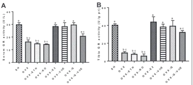
Effect of Q, vitE and E2 on GR activity
The serum GR activity in the OVX rats was significantly reduced by 47% in comparison to SH group (p<0.05). Treating the OVX rats with E2, vitE or Q significantly in-creased serum GR activity (p<0.05). Although the co-administration of
Fig. 2. Changes in (A) serum and (B) tibia GPx activity in OVX rats treated with E2, vitE, Q and Q+vitE. Data are expressed as means±SEM, (n=8). Histograms with different letters are significantly different at P<0.05 (Kruskal-Wallis with post-hoc Mann-Whitney U test). E2 – estradiol, Q – quercetin, vitE – vitamin E, OVX – ovariectomized, ETH – ethanol, RO – rapeseed oil.
Fig. 1. Effects of E2, Q and vitE on serum 17β-estradiol levels. Data are expressed as means±SEM, (n=8). Histograms with dif-ferent letters are significantly difdif-ferent at P<0.05 (Kruskal-Wallis with post-hoc Mann-Whitney U test). E2 – estradiol, Q – quer-cetin, vitE – vitamin E, OVX – ovariectomized, ETH – ethanol, RO – rapeseed oil.
Effects of Q, vitE and E2 on CAT activity
Tibia CAT activity was reduced by 73% when rats were ovariectomized (p<0.001). CAT activity was in-creased significantly in E2, vitE and OVX-Q rats when compared to the OVX group (p<0.05). Co-administration of Q and vitE increased CAT activ-ity in comparison with the OVX group but the differ-ence was not significant (Fig. 4).
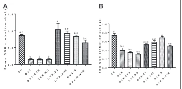
Effects of Q, vitE and E2 on the GSH content
The serum GSH content in OVX rats was significantly decreased by more than 81% when compared to the SH group (P<0.05). E2, vitE and Q treatments resulted in a significant increase in the level of GSH in the serum of OVX rats (p<0.001 for E2, p<0.05 for Q and vitE). The increase in the GSH content in the OVX-Q-vitE group was not significant when com-pared to the OVX group (P<0.05) (Fig. 5A). The tibia GSH content decreased by 47% in rats after ovariectomy (p<0.05). Tibia GSH was increased by 26%, 33%, 42% and 20% in OVX-E2, OVX-vitE, OVX-Q and OVX-vitE-Q groups, respectively, compared to the OVX group (p>0.05 for E2 and Q+vitE, p<0.05 for Q and vitE). There were no significant differences regard-ing the GSH content between SH, OVX-E2, OVX-vitE, OVX-Q and OVX-vitE-Q groups (Fig. 5B).
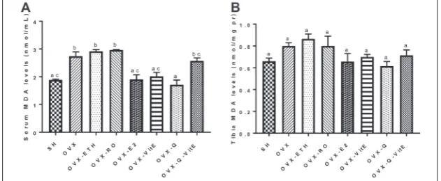
Effects of Q, vitE and E2 on MDA levels
The serum MDA level of OVX rats was significantly higher than the SH group (p<0.05). The MDA lev-els were decreased by 30%, 26% and 37% in E2, vitE and Q treated rats, respectively, when compared to the non-treated OVX group (p<0.05). Although co-administration of Q and vitE reduced the level of MDA, the difference was not significant compared to the OVX group (Fig. 6A). The level of tibia MDA increased by 19% in OVX rats when compared to the SH group (p>0.05). Compared to the OVX group, rats
Fig. 5. Changes in (A) serum and (B) tibia GSH content in OVX rats treated with E2, vitE, Q and Q+vitE. Data are expressed as means±SEM, (n=8). Histograms with different letters are significantly different at P<0.05 (Kruskal-Wallis with post-hoc Mann-Whitney U test). E2 – estradiol, Q – quercetin, vitE – vitamin E, OVX – ovariectomized, ETH – ethanol, RO – rapeseed oil.
Fig. 4. Changes in tibia CAT activity in OVX rats treated with E2, vitE, Q and Q+vitE. Data are expressed as means±SEM, (n=8). Histograms with different letters are significantly different at P<0.05 (Kruskal-Wallis with post-hoc Mann-Whitney U test). E2 – estradiol, Q – quercetin, vitE – vitamin E, OVX – ovariec-tomized, ETH – ethanol, RO – rapeseed oil.
treated with E2, vitE, Q or Q+vitE showed no signifi-cant reductions in the levels of MDA (Fig. 6B).
DISCUSSION
Menopause, a form of reproductive aging, is char-acterized by a complete failure of ovarian tissue to produce estrogen, which leads to an increase in OS, bone loss, CVDs, neurologic and sexual disorders [34]. Ovariectomy is the most common animal model for postmenopausal changes [21]. In the present study, we evaluated the effects of E2, vitE and Q on ovari-ectomy-induced OS in the serum and tibia of female rats. Our data demonstrated that ovariectomy induced an imbalance between ROS production and the an-tioxidant defense system, and the resulting OS was accompanied by a marked decrease in GPx, GR and CAT activities, GSH content and an increase in MDA level. These changes were improved by administration of either E2, vitE or Q in comparison with untreated OVX rats. Co-administration of Q and vitE had a weaker improving effect than when each compound was applied alone. Our findings demonstrated that Q and vitE markedly protected against ovariectomy-induced OS in female rats.
GPx and GR are the first line of defense of anti-oxidant enzymes against OS. We found that ovariec-tomy reduced GPx and GR activities in both serum and tibia of female rats. E2 treatment reversed these changes to the extent that they did not differ signifi-cantly from the SH group, suggesting that estrogen depletion after ovariectomy is likely the main reason
for OS induction. Degradation of hydrogen peroxide is catalyzed by CAT enzyme. Tibia CAT activity was also decreased after ovariec-tomy and improved by E2 admin-istration, emphasizing the role of estrogen on preventing OS. Serum CAT activity was not assayed be-cause of sample volume limitation. It was previously suggested [3] that the increase in hydrogen perox-ide together with the decrease in the superoxide dismutase (SOD), GPx and glutathione S transferase (GST) activities in ovariectomized rats is the main factor behind bone loss in experimental animals.
GSH is a cellular thiol-containing nonenzymatic antioxidant that plays an important role in the re-moval of free radicals [35]. In the current study, the tibia and serum GSH contents in the OVX rats were markedly depleted compared to the SH group and im-proved after E2 treatment. Other studies [1] indicate that ovariectomy induces OS and lipid peroxidation, as demonstrated by the increase in MDA level and decrease in GSH content. Since GR is responsible for GSSG reduction, the decrease in GSH after ovariec-tomy may be due to GR depletion.
MDA is a lipid peroxidation product that increas-es as a rincreas-esult of the oxidant/antioxidant imbalance in tissues [36-38]. We observed that ovariectomy could induce MDA production in the serum and tibia of female rats. The MDA level was decreased after E2 treatment, suggesting that estrogen deficiency and the resulting OS after ovariectomy may be the reason of MDA production. These changes were not significant as regards the tibia, indicating that MDA alterations start earlier in the serum compared to bone tissue. In agreement with current data, several studies showed that MDA levels in tissues increase after ovariectomy because of estrogen depletion [1,3,28,36]. Oxidized lipids can affect osteoclastic and osteoblastic bone cells [39]. As MDA reacts with amino groups, it can inhibit the synthesis of proteins and also deactivate certain enzymes [40]. For this reason, the reduction in tibia and serum antioxidant enzyme activities ob-served after OVX may be due to increased lipid per-Fig. 6. Changes in (A) serum and (B) tibia MDA levels in OVX rats treated with E2, vitE,
oxidation, as demonstrated in other studies [3,36]. OS induction has been suggested to result in bone loss [36,41], to contribute to CVDs [42], neurologic disorders [43,44] and other consequences of meno-pause or ovariectomy. Thus, we assumed that defense against OS could be a potential approach to improve menopause-derived complications.
Q, a potent antioxidant and phytoestrogen, is widely present in human diet. Thus, it is essential to understand the effects of Q on human health [15]. Compared to trolox, a water-soluble analog of vitE, Q could remarkably improve the endogenous antioxi-dant activity of 2,2’-azino-bis(3-ethylbenzothiazoline-6-sulphonic acid (ABTS) [45]. Our results indicate that Q treatment after ovariectomy reduced the levels of MDA and increased the GSH content and antioxi-dant enzyme activities in serum and tibia compared to the OVX group. These ameliorating effects of Q on OS are comparable to the effects of E2 on rats for all measured OS markers in both the serum and the tibia. In agreement with the current study, Dong et al. [46] indicated that treatment with a high dose of Q significantly enhanced the activities of GPx and SOD although it significantly decreased the level of MDA. Coskun et al. [47] demonstrated that the protective effect of Q on β-cell is attributed to its antioxidant nature. Wei Liang et al. [48] found that Q has some beneficial effects on osteopenia induced by diabetes in rats and suggested that Q could be a potential thera-peutic drug for diabetic osteopenia. Further studies indicated that Q is capable of reversing bone resorp-tion, subsequently reducing loss of bone mass in rats [48]. Q also has phytoestrogenic properties as a result of structural similarities to estrogen and binding ca-pabilities to estrogen receptors that stimulate calcium absorption [1]. This may explain the beneficial ef-fects of Q on bone health in addition to its antioxidant properties. Because phytoestrogens have been sug-gested to be estrogen receptor modulators, they may be used in postmenopausal women to improve some health risks associated with estrogen deficiency [13]. Q has both phytoestrogenic and antioxidant prop-erties and could be a potential therapeutic approach for relieving menopausal complications. However, there are inconsistent literature data with regard to the effects of Q on estrogen receptors [1], and more research is needed to prove its beneficial effects as an alternative to HRT.
VitE defends cells against damage from oxygen free radicals and lipid peroxides [49]. We evaluated the antioxidant ability of Q along with vitE as a wide-ly-known antioxidant. Our results showed that there was no significant difference between the antioxidant effects of Q and vitE in both serum and tibia. Similar to Q, vitE reduced lipid peroxidation and increased the GSH content and antioxidant enzyme activities in both serum and tibia to the extent that they did not differ significantly from the SH group. This is compat-ible with previous findings [50] where it was revealed that vitE supplementation protected several tissues against radical damage. It was observed [50] that there is a significant protective effect of vitE in combina-tion with Se on GPx, GR, SOD and CAT of the heart, kidney, liver and testis of sodium azide-treated mice. VitE prevents peroxidation of membrane lipids and is converted into a tocopheroxyl radical by scaveng-ing lipid peroxyl radicals [51]. VitE has been reported to protect from bone loss and damage caused by OS [20,52]. However, no significant association between vitE and bone structural changes or negative results has been reported in human epidemiological studies [53,54], suggesting that amelioration of OS alone may not guarantee bone health.
Q [59], indicating a contrary finding to that reported in the present study. This discrepancy may be due to the fact that the authors evaluated hepatic oxidative markers and the co-administration of three Q, vitE and vitamin C antioxidants, whereas we assessed only Q and vitE in combination in serum and bone tissue.
It is concluded from the present study that meno-pause/ovariectomy-induced OS can be ameliorated by the administration of Q and vitE and this can relieve menopause/ovariectomy-related dysfunctions. With quercetin’s documented safety profile in humans, clin-ical trials into the potential benefit of Q in treating menopause-derived complications in humans should be performed.
Funding: This paper is a part of the PhD thesis of Sina Vakili and was supported by Grant Number 18208 from the Vice-Chancellor for Research Affairs of Shiraz University of Medical Sciences.
Author contributions: Sina Vakili assisted with the design of the study, carried out all the experiments and participated in manu-script preparation. Fatemeh Zal carried out the design and coor-dinated the study, participated in most of the experiments and data analysis and manuscript correction. Zohreh Mostafavipour and Amir Savardashtaki assisted with the design of the study and provided assistance for enzyme assay experiments. Majid Jafari Khorchani and Ashraf Hassanpour provided assistance for the experimental design and animal surgery. All authors have read and approved the content of the manuscript.
Conflict of interest disclosure: The authors declare that there are no conflicts of interest.
REFERENCES
1. El-Fattah AIA, Fathy MM, Ali ZY, El-Garawany AE-RA, Mohamed EK. Enhanced therapeutic benefit of quercetin-loaded phytosome nanoparticles in ovariectomized rats. Chem Biol Interact. 2017;271:30-8.
2. Gompel A. Menopausal Treatment. In: Huhtaniemi I, Mar-tini L, editors. Encyclopedia of Endocrine Diseases. 2nd ed. Oxford: Academic Press; 2019. p. 634-45.
3. Muthusami S, Ramachandran I, Muthusamy B, Vasudevan G, Prabhu V, Subramaniam V, Jagadeesan A, Narasimhan S. Ovariectomy induces oxidative stress and impairs bone anti-oxidant system in adult rats. Clin Chim Acta. 2005;360(1-2):81-6.
4. Vijayan V, Khandelwal M, Manglani K, Singh RR, Gupta S, Surolia A. Homocysteine alters the osteoprotegerin/RANKL system in the osteoblast to promote bone loss: pivotal role of the redox regulator forkhead O1. Free Radic Biol Med. 2013;61:72-84.
5. Huang Q, Shi J, Gao B, Zhang H-Y, Fan J, Li X-J, Fan J-Z, Han Y-H, Zhang J-K, Yang L. Gastrodin: an ancient Chinese herbal
medicine as a source for anti-osteoporosis agents via reducing reactive oxygen species. Bone. 2015;73:132-44.
6. Baek KH, Oh KW, Lee WY, Lee SS, Kim MK, Kwon HS, Rhee EJ, Han JH, Song KH, Cha BY. Association of oxidative stress with postmenopausal osteoporosis and the effects of hydro-gen peroxide on osteoclast formation in human bone marrow cell cultures. Calcif Tissue Int. 2010;87(3):226-35.
7. O’Neill S, Eden J. The pathophysiology of menopausal symp-toms. Obstet Gynaecol Reprod Med. 2012;22(3):63-9. 8. Sawada H, Ibi M, Kihara T, Urushitani M, Honda K,
Nakani-shi M, Akaike A, Shimohama S. Mechanisms of antiapoptotic effects of estrogens in nigral dopaminergic neurons. FASEB J. 2000;14(9):1202-14.
9. Morrone MDS, Schnorr CE, Behr GA, Gasparotto J, Bortolin RC, Moresco KS, Bittencourt L, Zanotto-Filho A, Gelain DP, Moreira JCF. Oral administration of curcumin relieves behav-ioral alterations and oxidative stress in the frontal cortex, hip-pocampus, and striatum of ovariectomized Wistar rats. J Nutr Biochem. 2016;32:181-8.
10. Potter B, Schrager S, Dalby J, Torell E, Hampton A. Meno-pause. Prim Care. 2018;45(4):625-41.
11. Sarri G, Pedder H, Dias S, Guo Y, Lumsden MA. Vasomotor symptoms resulting from natural menopause: a systematic review and network meta‐analysis of treatment effects from the National Institute for Health and Care Excellence guide-line on menopause. BJOG. 2017;124(10):1514-23.
12. Svatikova A, Hayes SN. Menopause and menopausal hormone therapy in women: cardiovascular benefits and risks. Revista Colombiana de Cardiología. 2018;25:30-3.
13. Chen J, Zhang N, Wang Y, Wang J, Ji S, Dang W, Li S, Feng L. Estrogenic effects of flavonoid components in xiaoyao pow-der. Genet Mol Res. 2016;15(1):gmr7500.
14. Borrelli F, Ernst E. Alternative and complementary therapies for the menopause. Maturitas. 2010;66(4):333-43.
15. Wang W, Sun C, Mao L, Ma P, Liu F, Yang J, Gao Y. The bio-logical activities, chemical stability, metabolism and delivery systems of quercetin: A review. Trends Food Sci Technol. 2016;56:21-38.
16. Ekinci Akdemir FN, Gülçin İ, Karagöz B, Soslu R. Quercetin protects rat skeletal muscle from ischemia reperfusion injury. J Enzyme Inhib Med Chem. 2016;31(Sup2):162-6.
17. Li C, Wang T, Zhang C, Xuan J, Su C, Wang Y. Quercetin attenuates cardiomyocyte apoptosis via inhibition of JNK and p38 mitogen-activated protein kinase signaling pathways. Gene. 2016;577(2):275-80.
18. Yang X, Zhang W, Zhao Z, Li N, Mou Z, Sun D, Cai Y, Wang W, Lin Y. Quercetin loading CdSe/ZnS nanoparticles as effi-cient antibacterial and anticancer materials. J Inorg Biochem. 2017;167:36-48.
19. Smith B, Lucas E, Turner R, Evans G, Lerner M, Brackett D, Stoecker B, Arjmandi B. Vitamin E provides protection for bone in mature hindlimb unloaded male rats. Calcif Tissue Int. 2005;76(4):272-9.
21. Monteiro SC, Matté C, Bavaresco CS, Netto CA, Wyse AT. Vitamins E and C pretreatment prevents ovariectomy-induced memory deficits in water maze. Neurobiol Learn Mem. 2005;84(3):192-9.
22. Galli F, Azzi A, Birringer M, Cook-Mills JM, Eggersdorfer M, Frank J, Cruciani G, Lorkowski S, Özer NK. Vitamin E: Emerging aspects and new directions. Free Radic Biol Med. 2017;102:16-36.
23. Guralp O. Effects of vitamin E on bone remodeling in peri-menopausal women: mini review. Maturitas. 2014;79(4):476-80.
24. Feng Z, Zhang J-t. Long-term melatonin or 17β-estradiol supplementation alleviates oxidative stress in ovariectomized adult rats. Free Radic Biol Med. 2005;39(2):195-204. 25. Muhammad N, Luke DA, Shuid AN, Mohamed N, Soelaiman
I-N. Two different isomers of vitamin e prevent bone loss in postmenopausal osteoporosis rat model. Evid Based Comple-ment Alternat Med. 2012;2012:161527.
26. Yang Y, Zheng X, Li B, Jiang S, Jiang L. Increased activity of osteocyte autophagy in ovariectomized rats and its correlation with oxidative stress status and bone loss. Biochem Biophys Res Commun. 2014;451(1):86-92.
27. Mostafavi Pour Z, Vessal M, Zal F, Khoshdel Z, Torabinejad S. Protective effects of vitamin E and/or quercetin co-sup-plementation on the morphology of kidney in cyclosporine A-treated rats. Iranian J Medic Sci. 2009;34(4):271-6. 28. Sankar P, Zachariah B, Vickneshwaran V, Jacob SE, Sridhar
M. Amelioration of oxidative stress and insulin resistance by soy isoflavones (from Glycine max) in ovariectomized Wistar rats fed with high fat diet: the molecular mechanisms. Exp Gerontol. 2015;63:67-75.
29. Zal F, Mahdian Z, Zare R, Soghra B, Mostafavi-Pour Z. Com-bination of vitamin E and folic acid ameliorate oxidative stress and apoptosis in diabetic rat uterus. Int J Vitam Nutr Res. 2014;84(1-2):55-64.
30. Zal F, Taheri R, Khademi F, Keshavarz E, Rajabi S, Mostafavi-Pour Z. The combined effect of furosemide and propranolol on GSH homeostasis in ACHN renal cells. Toxicol Mech Methods. 2014;24(6):412-6.
31. Yarahmadi A, Zal F, Bolouki A. Protective effects of quercetin on nicotine induced oxidative stress in ‘HepG2 cells’. Toxicol Mech Methods. 2017;27(8):609-14.
32. Mashhoody T, Rastegar K, Zal F. Perindopril may improve the hippocampal reduced glutathione content in rats. Adv Pharm Bull. 2014;4(2):155-9.
33. Zal F, Mostafavi-Pour Z, Amini F, Heidari A. Effect of vitamin E and C supplements on lipid peroxidation and GSH-depen-dent antioxidant enzyme status in the blood of women con-suming oral contraceptives. Contraception. 2012;86(1):62-6. 34. Li X, Song Q-S, Wang J-Y, Leng H-j, Chen Z-Q, Liu Z-J, Dang
G-T, Song C-L. Simvastatin induces estrogen receptor-alpha expression in bone, restores bone loss, and decreases ERα expression and uterine wet weight in ovariectomized rats. J Bone Miner Metab. 2011;29(4):396-403.
35. Dickinson DA, Forman HJ. Glutathione in defense and sig-naling. Ann N Y Acad Sci. 2002;973(1):488-504.
36. Arslan A, Orkun S, Aydin G, Keles I, Tosun A, Arslan M, Caglayan O. Effects of ovariectomy and ascorbic acid
supple-ment on oxidative stress parameters and bone mineral density in rats. Libyan J Med. 2011;6(1):5965.
37. Vakili S, Savardashtaki A, Moghaddam MAM, Nowrouzi P, Shirazi MK, Ebrahimi G. The effects of saffron consump-tion on lipid profile, liver enzymes, and oxidative stress in male hamsters with high fat diet. Trends Pharmacol Sci. 2017;3(3):201-8.
38. Tabrizi R, Vakili S, Akbari M, Mirhosseini N, Lankarani KB, Rahimi M, Mobini M, Jafarnejad S, Vahedpoor Z, Asemi Z. The effects of curcumin‐containing supplements on biomark-ers of inflammation and oxidative stress: A systematic review and meta‐analysis of randomized controlled trials. Phytother Res. 2019;33(2):253-62.
39. Manolagas SC. Birth and death of bone cells: basic regulatory mechanisms and implications for the pathogenesis and treat-ment of osteoporosis. Endocr Rev. 2000;21(2):115-37. 40. Bird R, Draper H. Effect of malonaldehyde and
acetalde-hyde on cultured mammalian cells: Growth, morphology, and synthesis of macromolecules. J Toxicol Environ Health. 1980;6(4):811-23.
41. Mada SB, Reddi S, Kumar N, Kumar R, Kapila S, Kapila R, Trivedi R, Karvande A, Ahmad N. Antioxidative peptide from milk exhibits antiosteopenic effects through inhibition of oxidative damage and bone-resorbing cytokines in ovari-ectomized rats. Nutrition. 2017;43:21-31.
42. Lamas AZ, Caliman IF, Dalpiaz PLM, de Melo Jr AF, Abreu GR, Lemos EM, Gouvea SA, Bissoli NS. Comparative effects of estrogen, raloxifene and tamoxifen on endothelial dysfunc-tion, inflammatory markers and oxidative stress in ovariecto-mized rats. Life Sci. 2015;124:101-9.
43. Moreira AC, Silva AM, Branco AF, Baldeiras I, Pereira GC, Seiça R, Santos MS, Sardão VA. Phytoestrogen coumestrol improves mitochondrial activity and decreases oxidative stress in the brain of ovariectomized Wistar-Han rats. J Funct Foods. 2017;34:329-39.
44. Delrobaei F, Fatemi I, Shamsizadeh A, Allahtavakoli M. Ascorbic acid attenuates cognitive impairment and brain oxidative stress in ovariectomized mice. Pharmacol Rep. 2019;71(1):133-8.
45. Arts MJ, Dallinga JS, Voss H-P, Haenen GR, Bast A. A new approach to assess the total antioxidant capacity using the TEAC assay. Food Chem. 2004;88(4):567-70.
46. Dong Y-s, Wang J-l, Feng D-y, Qin H-z, Wen H, Yin Z-m, Gao G-d, Li C. Protective effect of quercetin against oxida-tive stress and brain edema in an experimental rat model of subarachnoid hemorrhage. Int J Med Sci. 2014;11(3):282. 47. Coskun O, Kanter M, Korkmaz A, Oter S. Quercetin, a
fla-vonoid antioxidant, prevents and protects streptozotocin-induced oxidative stress and β-cell damage in rat pancreas. Pharmacol Res. 2005;51(2):117-23.
48. Liang W, Luo Z, Ge S, Li M, Du J, Yang M, Yan M, Ye Z, Luo Z. Oral administration of quercetin inhibits bone loss in rat model of diabetic osteopenia. Eur J Pharmacol. 2011;670(1):317-24.
49. Zingg J-M. Vitamin E: an overview of major research direc-tions. Mol Aspects Med. 2007;28(5-6):400-22.
sodium azide in liver, kidney, testis and heart of male mice. Biomed Pharmacother. 2017;91:602-10.
51. Arita M, Sato Y, Arai H, Inoue K. Binding of α‐tocopheryl-quinone, an oxidized form of α‐tocopherol, to glutathione‐S‐ transferase in the liver cytosol. FEBS Lett. 1998;436(3):424-6. 52. Deng L, Ding Y, Peng Y, Wu Y, Fan J, Li W, Yang R, Yang M,
Fu Q. γ-Tocotrienol protects against ovariectomy-induced bone loss via mevalonate pathway as HMG-CoA reductase inhibitor. Bone. 2014;67:200-7.
53. Macdonald HM, New SA, Golden MH, Campbell MK, Reid DM. Nutritional associations with bone loss during the meno-pausal transition: evidence of a beneficial effect of calcium, alcohol, and fruit and vegetable nutrients and of a detrimental effect of fatty acids. Am J Clin Nutr. 2004;79(1):155-65. 54. Wolf RL, Cauley JA, Pettinger M, Jackson R, Lacroix A, Leboff
MS, Lewis CE, Nevitt MC, Simon JA, Stone KL. Lack of a relation between vitamin and mineral antioxidants and bone mineral density: results from the Women’s Health Initiative. Am J Clin Nutr. 2005;82(3):581-8.
55. Ranilla LGl, Genovese MI, Lajolo FM. Effect of different cooking conditions on phenolic compounds and antioxidant capacity of some selected Brazilian bean (Phaseolus vulgaris L.) cultivars. J Agric Food Chem. 2009;57(13):5734-42. 56. Buchner N, Krumbein A, Rohn S, Kroh LW. Effect of thermal
processing on the flavonols rutin and quercetin. Rapid Com-mun Mass Spectrom. 2006;20(21):3229-35.
57. Pękal A, Biesaga M, Pyrzynska K. Interaction of quercetin with copper ions: complexation, oxidation and reactivity towards radicals. Biometals. 2011;24(1):41-9.
58. Fabre G, Bayach I, Berka K, Paloncýová M, Starok M, Rossi C, Duroux J-L, Otyepka M, Trouillas P. Synergism of antioxidant action of vitamins E, C and quercetin is related to formation of molecular associations in biomembranes. Chem Commun. 2015;51(36):7713-6.