1. Serviço de Otorrinolaringologia. Hospital de Egas Moniz. Lisboa. Portugal. 2. Faculdade de Ciências Médicas. Universidade Nova de Lisboa. Lisboa. Portugal.
Recebido: 07 de Janeiro de 2013 - Aceite: 17 de Março de 2013 | Copyright © Ordem dos Médicos 2013
Pedro ESCADA1,2
Acta Med Port 2013 May-Jun;26(3):200-207
RESUMO
Introdução: A distribuição da mucosa olfactiva humana só pode ser determinada em estudos que avaliem a totalidade da região olfac-tiva. O objectivo deste trabalho é determinar a distribuição da mucosa olfactiva humana a partir do estudo histológico, por microscopia óptica, de peças anatómicas da região olfactiva obtidas do cadáver.
Material e Métodos: Utilizaram-se peças anatómicas da região olfactiva colhidas durante a autópsia de cadáveres recentes. Em cada uma das peças foi determinada a distância entre a lâmina crivosa e o limite inferior da região olfactiva em três localizações diferentes da parede septal e da parede lateral.
Resultados: das 230 peças anatómicas disponíveis, 217 foram excluídas por razões clínicas ou técnicas. Realizaram-se estudos morfométricos em 13 peças num total de 156 medições. O limite inferior da mucosa olfactiva no septo nasal estava a 15,9 ± 3,2 mm, a 15,3 ± 3 mm e a 16 ± 2,8 mm nas porções anterior, média e posterior da região olfactiva. O limite inferior da mucosa olfactiva na parede turbinal estava a 15,3 ± 2,4 mm, a 14,8 ± 2,3 mm e a 12,3 ± 1,9 mm nas mesmas localizações. O valor mínimo observado foi de 12 mm.
Conclusões: A mucosa olfactiva estende-se pelo corneto superior e médio e pelo septo nasal confrontante numa distância que nunca é inferior a 12 mm e que pode ultrapassar os 16 mm. O conhecimento da distribuição exacta da mucosa olfactiva nas fossas nasais pode ser útil para orientar a colheita em seres humanos, com propósitos diagnósticos ou terapêuticos.
Palavras-chave: Antropometria; Cadaver; Colheita de Tecidos e Orgãos; Mucosa Olfactiva/ anatomia e histologia; Neurónios Recep-tores Olfactivos.
ABSTRACT
Introduction: The exact distribution of the human olfactory mucosa can only be determined in studies that evaluate the entire olfactory region. The purpose of this study is to determine the distribution of human olfactory mucosa in the nasal cavities, by performing the histological analysis, by light microscopy, of anatomical specimens of the olfactory region obtained from cadavers.
Material and Methods: The specimens were taken during the autopsy of fresh cadavers. In each of the specimens, the distance be-tween the cribiform plate and the lower limit of the olfactory region was determined in three different locations of the septal and lateral walls.
Results: Of the 230 anatomical specimens available, 217 were excluded for medical or technical reasons. Morphometric studies were performed on 13 specimens (total 156 measurements). The lower limit of the olfactory mucosa in the nasal septum was 15.9 ± 3.2 mm, 15.3 mm ± 3 and 16 ± 2.8 mm in the anterior, middle and posterior olfactory regions. The lower limit of the olfactory mucosa in the tur-binate wall was 15.3 ± 2.4 mm, 14.8 ± 2.3 mm and 12.3 ± 1.9 mm in the equivalent regions. The minimum value observed was 12 mm. Conclusions: The olfactory mucosa extends through the upper and middle turbinates and the confronting nasal septum in a minimum distance of 12 mm and that may exceed 16 mm. Knowing the exact distribution of the olfactory mucosa can guide the collection of this tissues in humans, for diagnostic or therapeutic purposes.
Keywords: Anthropometry; Cadaver; Nasal Septum; Olfactory Mucosa/anatomy and histology; Olfactory Receptor Neurons; Tissue and Organ Harvesting
INTRODUCTION
Olfactory mucosa histology
The human olfactory mucosa is a pseudostratified columnar epithelium with a highly cellular lamina propria (Figure 1).1
This epithelium has four different cell types: ciliated olfactory receptors, supporting cells, microvillar cells and basal cells.2,3
Olfactory receptors are bipolar neurons from which a single dendrite extends to the olfactory neuroepithelium surface and one axon to the olfactory bulb. Bipolar neuron nuclei are located at all levels of the epithelium thickness,
the oldest cells’ nuclei located closer to the epithelium surface. The nuclei variable configuration explains this typical pseudostratified pattern.
The bipolar neuron dendrite has a spindle-shaped tip, known as the olfactory vesicle. The olfactory vesicle extends towards the epithelial surface (Figures 2a and 2b), containing non-motile cilia with membrane receptors to which the odour mollecules attach (this aspect is only observed through electron microscopy).
Each axon of the 6 to 20 million bipolar cells crosses the basement membrane towards the lamina propria, joins
ARTIGO ORIGINAL
Localization and Distribution of Human
Olfactory Mucosa in the Nasal Cavities
together to form fascicles and nerves and passes through the 15 to 20 holes of each cribiform plate, forming a synapse within the olfactory bulb.3
The supporting cells, which are the prevailing cells, surround the bipolar cells contributing to the regulation and maintenance of the ionic environment and in this way
favouring olfactory transduction.4
The microvillar cells, only identifiable by electron microscopy, were described for the first time in 1982 and were considered a second class of morphologically distinct
receptor cells in the olfactory mucosa.5
The basal cells, the only kind of cells that do not extend towards the epithelial surface and lie over the basement membrane, are recognized as a different population of cells from the olfactory epithelium stem cells, with a capacity to continuously regenerate the affected olfactory cells throughout the life of the individual 6-8 They
have auto-renovation and multipotent properties and are experimentally capable of differentiating into other cells, both neural and non-neural.1,9-11
The lamina propria contains fascicles of axons, blood
vessels, connective tissue and Bowman glands.12 The axons
of olfactory neurons are wrapped, in its transition from the olfactory neuroepithelium towards the olfactory bulb, by a unique glial cell lineage: the olfactory ensheathing cells.13
The Bowman’s glands are tubuloalveolar glands, consisting of secretory acini arranged in circles and excreting its products through a duct that crosses towards
the olfactory epithelium (Figures 3a and 3b).14 The products
secreted by these glands towards the mucosal layer of the olfactory are essential for olfactory transduction.
Distribution of the olfactory mucosa in the nasal cavities
Human distribution of olfactory mucosa in nasal cavities
remains to be adequately documented.1,15 The existing
studies about olfactory mucosal structure are based on olfactory mucosa biopsies, are very limited in number and only performed for research purposes, aiming primarily to
establish the correlation between the observed structural and histopathological changes and the nature or degree of
olfactory disfunction.16 More recently, the olfactory mucosa
has attracted researcher’s attention, as it may be used as an early marker for neurodegenerative disorders and as a multipotent neural stem cell source, with application in regenerative medicine.17-21 We should remark, within this
scope, the basic research and clinical research works of Portuguese researcher Carlos Lima regarding the use of olfactory mucosa in the treatment of chronic spinal cord traumatic lesions, in which the author of the present work participated,15,19-26 or the more recent animal research works
in dogs, largely divulged in scientific community and in the media.27
In 1892, von Brunn made measurements in the olfactory region in two post-mortem cases, showing areas of 307 and
238 mm2 (Case 1: 133 mm2 in nasal septum and 174 mm2
in the lateral wall; Case 2: 99 mm2 in nasal septum and 139
mm2 in the lateral wall).28 From the figure drawn by Lang,
based on von Brunn measurements, we understand that the olfactory region extends to a position lower than the plane of the sphenoid sinus ostium and that it is distributed in areas
of the middle turbinate,29 contradicting the assumption
sometimes made that the olfactory mucosa in the lateral wall would only involve the upper turbinate.
Many subsequent studies also confirm that the olfactory mucosa has a lower and more anterior distribution than that which is traditionally considered. In one of these studies, Leopold et al used electro-olfactogram (EOG) and localized biopsies to identify the presence of olfactory mucosa near the anterior attachment of the middle turbinate and in front of this insertion, in the medial wall, as well as in the lateral wall of the nasal cavity.30 Restrepo et al collected olfactory
receptor neurons in biopsies carried out in the nasal septum opposed to the upper portion of the middle turbinate,31
and Féron et al demonstrated the presence of olfactory epithelium distributed in regions of the middle turbinate in more than 50% of 71 biopsy specimens collected from healthy volunteers.32 Similar results have been obtained
Figure 1 -Original optical microscopy image of the olfactory muco-sa (cadaver specimen, Masson’s trichrome, 200X original magnifi-cation). Pseudostratified epithelium. Thin basement membrane and cell-enriched lamina propria.
Figures 2a and 2b - Original optical microscopy image of the ol-factory neuroepithelium (cadaver specimen, Masson’s trichrome, 400X original magnification). The olfactory neuroepithelium surface is characterized by the presence of bipolar neurones with dendritic bulb-shaped projections, called the olfactory vesicles (b = detail of a; OV = olfactory vesicle).
by Rawson et al, who obtained morphologically identifiable olfactory neurons from the nasal septum opposed to the upper portion of the middle turbinate and in the middle turbinate itself.33 The same authors observed that in the
obtained biopsies, they were able to produce, in the most part of the specimens, olfactory neurons responsive to
odorous substances.34 Immunochemical studies carried out
by Nibu et al have also confirmed that the olfactory mucosa is present in the lining epithelium of the lower portion of the middle turbinate.35
Examination of the olfactory mucosa in nasal cavities
Most of the studies carried out to study and to identify the olfactory mucosa in humans were based on biopsies. Interestingly, the authors who developed the olfactory mucosa endoscopic biopsy technique recognized at the same time their insufficiency to achieve studies aiming to obtain mapping of the olfactory mucosa in the nasal cavities.36 According to these authors, the exact distribution
of the olfactory mucosa in nasal cavities could only be obtained in cadaver studies as the only method, by technical and ethical reasons, that allowed for the analysis of larger quantities of tissue or anatomical specimens that included the total olfactory region.
Until now, the only published studies in which more extensive cadaver anatomical specimens were used, were
those by Paik et al and by Nibu et al.35,37 In both studies,
anatomical sampling was carried out through an intracranial approach, upon craniotomy and brain and dura mater removal. The specimen included the cribiform plate, the nasal septum and upper and middle turbinates, removed as a whole using circular saws.
In the first study, 12 specimens were collected but in only three of them the epithelium was kept preserved and could be analysed. The donors were individuals with no past or present rhino-sinus pathology, with 34, 57 and 82 years old, the last one being a female. The technique used to visualize the olfactory mucosa (neuro epithelium) was optical microscopy of silver-stained samples by the Bodian
method. The criteria for identification were the presence of bipolar cell dendrites (olfactory vesicles) and the absence of respiratory cilia. The results were the identification of olfactory mucosa in all three specimens, in a medium vertical extension of 14.3 mm of the olfactory region (from the cribiform plate). The presence of respiratory epithelium areas within the olfactory neuroepithelium was identified, but only in a relevant form in the 82-year old patient.37
In the second study, the two cadaver specimens used were obtained less than 24 hours after death. No demographic or clinical data from donors was mentioned. The identification of the olfactory mucosa was made using immunohistochemical techniques with antibodies that mark the olfactory neuroepithelium. The results showed that the olfactory mucosa extends more inferior than it was previously established, comprising the lower portion of the middle turbinate in its area of distribution.35
Identification of the olfactory mucosa
The different methods that have been used for human olfactory mucosa identification in specimens obtained from living donor biopsies include optical microscopy, transmission electron microscopy and
immunohistochemical techinques.38-40 Nevertheless,
electron microscopy and immunohistochemistry performed in a deceased body are affected by protein and neural tissue olfactory neuroepithelium degeneration occurring in the first hours after death.37,41,42 The study by Nibu et al
using immunological markers used specimens from only two individuals and specimens were obtained less than 24 hours before death, a condition which is difficult to achieve in most of specimen collections from cadaver.
Due to all these reasons and until the present time, conventional optical microscopy is the most appropriate technique for identification of olfactory and respiratory mucosa in cadaveric specimens. The optical microscopy differentiation between olfactory and respiratory mucosa
has been defined by Robert Kern in 2000.43 According
to this author, the criteria to distinguish olfactory from respiratory mucosa are as follows: both mucosa (olfactory and respiratory) consist of a pseudostratified epithelium; the olfactory mucosa has irregular cilia, no caliciform cells, a thin basement membrane and a cellular lamina propria and big and numerous nerve bundles; the respiratory mucosa has regular cilia, numerous caliciform cells, a thick basement membrane and a vascular lamina propria with scarce nerve bundles.
As described by Paik et al, normal olfactory mucosa identification must also be obtained through a positive identification of bipolar cells, which may be achieved by the presence of the olfactory vesicle; respiratory mucosa identification must be obtained by the identification of long, regular and uniformly distributed cilia in the cell epithelial surface.37
Aim of the study
The aim of this work is to evaluate human olfactory
Figures 3a and 3b - Original optical microscopy image of the ol-factory neuroepithelium (cadaver specimen, Masson’s trichrome, 100X original magnification). a) Bowman’s glands are spherical, with cells surrounding a central lumen (alveolar portion) a) and b) the Bowman’s glands cross the olfactory neuroepithelium (tubular portion), opening in a gap located in its surface (arrows = alveolar portion of the Bowman’s glands).
mucosa distribution in nasal cavities. This study was performed through histological examination, with optical microscopy, of anatomical parts from the olfactory region of human cadaver body parts.
MATERIAL AND METHODS Obtaining anatomical specimens
Anatomical specimens of the olfactory region were selected from the human olfactory organs tissue bank at the Unidade Funcional de Neuropatologia - Hospital de Egas Moniz. Histopathological analysis was carried out at the Unidade de Microcirurgia – Centro Hospitalar Lisboa Ocidental.
Specimens were collected during clinical or forensic autopsy performed in recently deceased bodies. Specimen sampling was preceded by all necessary institutional authorizations, in accordance with current legislation and the present study was performed upon approval of the Ethics Committee of Centro Hospitalar de Lisboa Central. Most specimens were collected by Dr. Carlos Lima between 1987 and 1994.
The technique used for collecting anatomical specimens of the olfactory region was the intracranial approach. The upper surface of the anterior cranial fossa was exposed upon craniotomy and the brain and dura mater removed in addition to the olfactory bulb section. The specimens included the base of the skull and the nasal structures included in the cutting planes were obtained with the use of circular saws: on the foreground, sections were performed in front of the anterior limit of the cribiform plate and of the crista galli; on the background, sections were performed immediately behind the posterior limit of the cribiform plate at the anterior limit of the sphenoid planum; laterallly, sections were performed one centimeter outside the lateral limit of the cribiform plate, in order to include, in the specimen, the full insertion of the middle and upper turbinates at the base of the skull.
Specimens were fixed in a 10% tamponated formalin solution and then placed in a decalcifying solution for 24 hours. After dehydration they were stored in paraffin. In addition to the archive of paraffinated blocks with the anatomical specimens, the anatomical specimen bank also keeps a record of donors characteristics, including demographic and clinically available data.
Selection of anatomical specimens
At first, the anatomical specimen donor clinical records were checked, in order to exclude those with any disorder inducing olfactory neuroepithelial pathological changes. Exclusion criteria were as follows: any known olfactory disorder, rhinitis and rhinosinusitis, nasal or skull injuries, nasal tumors or surgeries, neurovegetative disorders and specimens from individuals over 50 years of age 44- 47 The exclusion criteria based on age were established on the assumption and on the established evidence that with ageing, the olfactory mucosa is gradually replaced by respiratory mucosa.1,37,48,49
Processing and observation of sections
The paraffinated blocks were cut in three different sections following the anterior, the middle and posterior portions of the specimen. All 3μm sections were performed following a cross-sectional direction of the block, and the sections followed a Hematoxylin-Eosine (H&E) and Mas-son’s trichrome staining protocol.
The sections were observed using a optical microscope
Olympus®, model CX40, with 40X, 100X, 200X, 400X and
600X magnifications. A Meiji® microscope, model MX5300H
was used for the image record, with a connected digital
camera Del tapix® model Infinity X equipped with the Deltapix
Viewer Pro fessional 1.7 software.
The first observation of the sections, performed in small magnification (40X), was intended to check the correct section orientation and specimen integrity. The quality of the histological technique was also evaluated, if necessary, with higher magnifications. All three sections of each anatomical specimen were observed.
Sections that allowed a positive identification of a bilateral olfactory region with an extension of at least 10 to 15 mm below the cribiform plate were selected. The olfactory region identification was based on the visualization of the most important anatomical features: cribiform plate; nasal septum and upper or middle turbinates. The histological technique quality was evaluated using parameters such as chromatin aggregation, cellular volume reduction, loss of
intercellular contact and loss of microscopic resolution.50
Olfactory region histological and morphometric analysis
Mucosa of both olfactory regions (right and left) in each section was observed at the microscope in all its length and was defined as olfactory or respiratory, according to the above mentioned criteria. The prevailing criteria in the differentiation between olfactory and respiratory mucosa were: the presence of the irregular cilia or of the dendritic ending (olfactory vesicle) of bipolar cells considered the specimen as olfactory mucosa while the uniform distribution cilia at cell’s epithelial surface was indicative of the presence of respiratory mucosa.
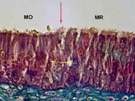
In each observation, the distance between the cribiform plate and the lower limit of the olfactory mucosa (i.e. the transition from olfactory to respiratory mucosa) was determined and recorded (Figure 4) in both walls: middle (nasal septum) and lateral (middle or upper turbinate). The measurements were obtained with the help of a millimeter scale inserted in the slide and visualized in low magnification.
It must be mentioned that the different steps for tissue histological processing, in particular the paraffination process, reduces the size of structures when compared to
their in vivo dimension.50-52 In what concerns the olfactory
region processing, this reduction has already been
estimated at approximately 30% by other authors.37 As
such a possible error in our study would occur were we to estimate the olfactory region by default, which may be safer
Figura 4 - Imagem original de microscopia óptica da mucosa da re-gião olfactiva (amostra de cadáver, Tricrómico de Masson, amplia-ção original de 400X). Observa-se neste caso uma transiamplia-ção nítida entre a mucosa olfactiva e a mucosa respiratória (seta vermelha). O epitélio olfactivo ém é identificável pela sua característica distin-tiva mais importante, a presença de cílios irregulares e vesículas dendríticas, contrastando com o epitélio respiratório, que apresen-ta maior densidade de cílios uniformes e paralelos. Outro aspecto muito característico na diferenciação entre a mucosa olfactiva e a mucosa respiratória é a membrana basal, muito espessa na mu-cosa respiratória (barra amarela) e fina na mumu-cosa olfactiva (seta amarela) (MO = mucosa olfactiva; MR = mucosa respiratória).
as the results of the study are aimed to predict the most correct location for obtaining olfactory tissue in the living individual.
Used images
All images used in this work have been specifically obtained and prepared for the particular purpose of the present work and obtained from the existing specimens kept in the abovementioned human anatomical tissue bank.
RESULTS
Selection of anatomical specimens and positive identification of the olfactory region
Following the criteria described, 52 out of a total of 230 surveyed specimens were selected for further study. The exclusion criteria applied to the remaining 178 specimens are presented in Table 1. The excluded specimens presented in the age exclusion criteria row were specimens which were not excluded due to any other criteria.
From the 52 selected, sectioned and stained anatomical specimens, 39 further specimens were excluded after low magnification observation of the section and section repetition for reconfirmation of the exclusion criteria. The results of this phase of the study are presented in Table 1. In the end, only 13 anatomical specimens were selected, considered suitable for olfactory mucosa mapping in serial histological sections.
Histological/morphometric analysis of the olfactory region
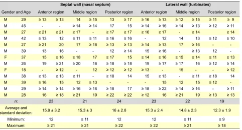
Table 2 summarizes all observations and measurements, indicating gender and age of the individual corresponding to the evaluated specimen. The measurements carried out in the middle and lateral walls were recorded separately. Two measurements were made for each section in each wall, as the same section included the olfactory regions of both nasal cavities.
In 59% of the performed measurements in the septal wall and in 62% of those performed in the lateral wall of the olfactory region, the limits of the observable specimen in the section consisted of olfactory mucosa and therefore did not include the transition from the olfactory to the respiratory mucosa. The maximum limit of the olfactory mucosa included in the specimen was recorded in these cases and these measurements were presented by its value preceded by the sign ≥.
Changes in the mucosa, particularly in the epithelium, were observed in 13% of the performed measurements in the septal wall and in 18% of those performed in the lateral wall of the olfactory region, preventing a conclusive identification of the lining mucosa. These cases are presented in the table with an empty cell (missing value). The real measurements of the histological transition between olfactory and respiratory mucosa have been used to calculate the average values of olfactory mucosa distribution which was calculated using the observable limit of the specimen that included olfactory mucosa in the remaining cases.
The lower limit of the olfactory mucosa in the nasal septum, determined by histology from the position of the cribiform plate, has been found, on average, at 15.9 ± 3.2 mm in the most anterior portion of the olfactory region, at 15.3 ± 3 mm in the middle portion and at 16 ± 2.8 mm in the most posterior portion. The minimum value of the distance between the base of the skull and the olfactory to respiratory mucosa transition (12 mm) has only been recorded in four observations (5% of the measurements).
The lower limit of the olfactory mucosa in the upper and middle turbinates, determined by histology from the cribiform plate position, was found on average at 15.3 ± 2.4 mm in the most anterior portion of the olfactory region, at 14.8 ± 2.3 mm in the middle portion and at 12.3 ± 1.9 mm in the most posterior portion.
Table 1 - Specimens excluded for clinical and technical reasons.
Absence of demographic or clinical data 18
Generalized CNS Infections (including HIV) 86
Neurodegenerative disorders 40
Over 50 years of age 21
Rhinitis or sinusitis 13
Insufficient size specimen 18
Incorrect section 16
Incorrect histological technique 5
Total 217
DISCUSSION
Comprehensive summary of the results
The histological and morphometric studies performed demonstrate the following extension of olfactory mucosa in the olfactory region of nasal cavities: in the septal wall, the lower limit of the olfactory mucosa at the nasal septum was found at 15.9 ± 3.2 mm, 15.3 ± 3 mm and 16 ± 2.8 mm from the base of the skull and in the lateral wall, this limit was found at 15.3 ± 2.4 mm, 14.8 ± 2.3 mm and 12.3 ± 1.9 mm from the base of the skull, respectively at the anterior, middle and posterior portions of the olfactory region. These values show that the olfactory mucosa extends to a lower level than the plane defined by the position of the sphenoid ostium, as this structure is located at 10.3 ± 4.3
mm from the base of the skull.53
Limitations of the study
Despite the study design using cadaver anatomical specimens, aimed to overpass the limitations already
mentioned by other authors,36 some difficulties have arisen.
A relevant percentage of the anatomical specimens could not be used, either due to the existence of pathologies producing changes in the olfactory mucosa, or due to technical reasons: difficulty in obtaining sections with an anatomical orientation adjusted to the objective of the study and poor histological quality in some of the preparations. In some cases, specimen collection did not preserve the lower portion of the olfactory region. This was due to the collection technique, which did not include the transnasal section of the base of the septum, a step which was already proposed by other authors for a human whole olfactory
organ removal.35,54 Nevertheless, we should remark that
the establishment of an anatomical specimens tissue bank preceded the publication of the abovementioned works.
Finally, the results of our study are still undervalued due to the fact that we admittedly did not apply the correction factor resulting from the need to compensate the effects of tissue histological processing when we performed
morphometric studies.37
Comparison of the obtained results with those from other studies
As we already mentioned, pioneer studies performed more than 100 years ago and several publications in particular over the last decades, described the olfactory mucosa area as extending lower than the sphenoid ostium plane and with a distribution to lower and more anterior regions than it was previously established, including lower regions of the middle turbinate, areas of the confronting nasal septum and even areas of the nasal septum located in front of the anterior insertion of the middle turbinate.3,16,18,28,30-35,37,55-60 The results
that we present confirm these findings.
Clinical application of the study results
Nasal cavity biopsy has been increasingly used as a mean to obtain olfactory tissue for diagnostic purposes or neural multipotent stem cell collection. The correct knowledge of the exact distribution of the olfactory mucosa in the nasal cavities will facilitate clinical application of those
techniques that require biopsy and collection of this tissue.1
CONCLUSION
The results of this study confirm the presence of olfactory mucosa in a well identified area of the olfactory region, which may also be recognized through measurements or using endoscopic identifiable surgical references, as the sphenoid sinus ostium.
Table 2 - Distance between the cribiform plate and the lower limit of the olfactory mucosa.
Septal wall (nasal septum) Lateral wall (turbinates)
Gender and Age Anterior region Middle region Posterior region Anterior region Middle region Posterior region
M 29 ≥ 13 ≥ 13 14 ≥ 15 13 ≥ 17 ≥ 16 ≥ 13 ≥ 12 ≥ 15 ≥ 11 ≥ 9
M 45 - - ≥ 14 ≥ 14 17 15 ≥ 14 ≥ 16 ≥ 14 ≥ 13 ≥ 12 ≥ 11
M 27 ≥ 21 ≥ 21 ≥ 17 - ≥ 17 ≥ 17 ≥ 16 ≥ 17 - ≥ 14 ≥ 14
M 42 ≥ 13 12 ≥ 11 ≥ 11 ≥ 16 ≥ 16 - 12 14 13 ≥ 12 ≥ 10
M 27 ≥ 21 20 17 ≥ 18 ≥ 13 ≥ 13 ≥ 14 ≥ 13 17 ≥ 16 -
-M 39 13 16 - - 12 ≥ 14 15 ≥ 16 - ≥ 13 12
-F 37 15 ≥ 16 ≥ 18 17 ≥ 17 15 ≥ 14 ≥ 16 ≥ 15 ≥ 14 ≥ 11 ≥ 13
M 26 19 ≥ 21 ≥ 20 16 ≥ 18 ≥ 18 19 ≥ 17 ≥ 17 16 ≥ 12 ≥ 14
F 18 - ≥ 12 - 12 ≥ 12 ≥ 12 ≥ 13 - 15 - - ≥ 12
M 38 ≥ 13 ≥ 13 ≥ 11 - ≥ 18 14 15 ≥ 13 - ≥ 11 ≥ 18 14
M 39 ≥ 16 15 12 ≥ 13 - - - 15 12 15 ≥ 12
-M 29 ≥ 14 ≥ 14 ≥ 16 ≥ 16 ≥ 18 17 ≥ 18 ≥ 22 ≥ 14 ≥ 16 - ≥ 11
M 28 16 ≥ 18 ≥ 21 19 ≥ 22 ≥ 22 ≥ 12 16 ≥ 21 19 ≥ 13 ≥ 13
n: 23 21 24 23 22 19
Average and
standard deviation: 15.9 ± 3.2 15.3 ± 3 16 ± 2.8 15.3 ± 2.4 14.8 ± 2.3 12.3 ± 1.9
Minimum: 12 ≥ 11 12 12 ≥ 11 ≥ 9
Maximum: ≥ 21 ≥ 21 ≥ 22 ≥ 22 ≥ 21 ≥ 18
ACKNOWLEDGEMENTS
I wish to acknowledge Carlos Lima (1955-2012) the facilities granted for the use of the human anatomical specimens from the Unidade Funcional de Neuropatologia, Hospital de Egas Moniz, Centro Hospitalar Lisboa Ocidental, as well as the general supervision of the histological examinations.
I wish to acknowledge José Francisco Madeira da Silva and João Goiry O’Neill for their critical revision of the original manuscript.
CONFLICT OF INTERESTS
The author declares there is no conflict of interests regarding the writing of this manuscript.
FINANTIAL SOURCES
There were no external financial sources for writing this manuscript.
REFERENCES
1. Escada PA, Lima C, Madeira da Silva J. The human olfactory mucosa. Eur Arch Otorhinolaryngol. 2009;266:1675-80.
2. Morrison EE, Costanzo RM. Morphology of the human olfactory epithe-lium. J Comp Neurol. 1990;297:1-13.
3. Moran DT, Rowley JC 3rd, Jafek BW, Lovell MA. The fine structure of the olfactory mucosa in man. J Neurocytol. 1982;11:721-46.
4. Nickell WT. Basic anatomy and phisiology of olfaction. In: Seiden AM, editor. Taste and smell disorders. New York: Thieme; 1997. p.20-37. 5. Moran DT, Rowley JC 3rd, Jafek BW. Electron microscopy of human
olfactory epithelium reveals a new cell type: the microvillar cell. Brain Res. 1982;253:39-46.
6. Calof AL, Chikaraishi DM. Analysis of neurogenesis in a mammalian neuroepithelium: proliferation and differentiation of an olfactory neuron precursor in vitro. Neuron. 1989;3:115-27.
7. Hahn CG, Han LY, Rawson NE, Mirza N, Borgmann-Winter K, Lenox RH, et al. In vivo and in vitro neurogenesis in human olfactory epithe-lium. J Comp Neurol. 2005;483:154-63.
8. Murrell W, Feron F, Wetzig A, Cameron N, Splatt K, Bellette B, et al. Mul-tipotent stem cells from adult olfactory mucosa. Dev Dyn. 2005;233:496-515.
9. Chen X, Fang H, Schwob JE. Multipotency of purified, transplanted glo-bose basal cells in olfactory epithelium. J Comp Neurol. 2004;469:457-74.
10. Roisen FJ, Klueber KM, Lu CL, Hatcher LM, Dozier A, Shields CB, et al. Adult human olfactory stem cells. Brain Res. 2001;890:11-22. 11. Carter LA, MacDonald JL, Roskams AJ. Olfactory horizontal basal cells
demonstrate a conserved multipotent progenitor phenotype. J Neurosci. 2004;24:5670-83.
12. Menco BP, Morrison EE. Morphology of the mammalian olfactory epi-thelium: form, fine structure, function ans pathology. In: Doty RL, editor. Handbook of olfaction and gustation. New York: Marcel Dekker; 2003. p.17-49.
13. Bartolomei JC, Greer CA. Olfactory ensheathing cells: bridging the gap in spinal cord injury. Neurosurgery. 2000;47:1057-69.
14. Moulton DG, Beidler LM. Structure and function in the peripheral olfac-tory system. Physiol Rev. 1967;47:1-52.
15. Escada P, Madeira da Silva J. Anatomia endoscópica e relevância cirúr-gica da região olfactiva. Clin Invest Otorrinolaringol. 2008;2:256-67. 16. Jafek BW, Murrow B, Michaels R, Restrepo D, Linschoten M. Biopsies of
human olfactory epithelium. Chem Senses. 2002;27:623-8.
17. Lima C, Escada P, Pratas-Vital J, Branco C, Arcangeli CA, Lazzeri G, et al. Olfactory mucosal autografts and rehabilitation for chronic traumatic spinal cord injury. Neurorehabil Neural Repair. 2010;24:10-22. 18. Lima C, Pratas-Vital J, Escada P, Hasse-Ferreira A, Capucho C,
Pe-duzzi JD. Olfactory mucosa autografts in human spinal cord injury: a pilot clinical study. J Spinal Cord Med. 2006;29:191-203.
19. Lima C, Vital JP, Escada P, Ferreira AH, Capucho C, Peduzzi J. Cellular therapies for human spinal cord injury: The olfactory mucosa autograft experience “ongoing clinical trial”. Eur J Neurol. 2005;12(Suppl. 2): 34. 20. Escada P, Capucho C, Lima C, Pratas Vital J, Madeira da Silva J. Trans-plantes autólogos de mucosa olfactiva em doentes traumatizados da medula espinal. Clin Invest Otorrinolaringol. 2007;1:128-38.
21. Carvalhal AV, Lima C, Basto V, Cunha C, Escada P, Cruz H, et al. Adult human neural stem/progenitor cells from the olfactory epithelium and ol-factory lamina propria, isolation method, proliferation and differentiation in serum free culture medium and utilization for transplantation. Patente WO/020611, 2007.
22. Escada P. Autotransplantação de células estaminais olfactivas no trata-mento das lesões traumáticas crónicas da medula espinal. Estudos da
região olfactiva e da sua mucosa: Departamento de Otorrinolaringo-logia. Lisboa: Faculdade de Ciências Médicas, Universidade Nova de Lisboa; 2010.
23. Lima C, Pratas-Vital J, Escada P, Hasse-Ferreira A, Peduzzi JD. Olfac-tory mucosa autografts in human spinal cord injury: from rats to humans. 2004 International Campaign for cures of spinal cord injury paralysis: spinal cord injury clinical trials workshop. Vancouver, Canadá; 2004. 24. Escada PA, Lima C, da Silva JM. The human olfactory mucosa. Eur Arch
Otorhinolaryngol. 2009;266:1675-80.
25. Lima C, Escada P, Pratas-Vital J, Branco C, Arcangeli CA, Lazzeri G, et al. Olfactory mucosal autografts and rehabilitation for chronic traumatic spinal cord injury. Neurorehabilitation Neural Repair. 2010;24:10-22. 26. Lima C, Pratas-Vital J, Escada P, Hasse-Ferreira A, Capucho C,
Pe-duzzi JD. Olfactory mucosa autografts in human spinal cord injury: a pilot clinical study. J Spinal Cord Med. 2006;29:191-203.
27. Granger N, Blamires H, Franklin RJ, Jeffery ND. Autologous olfactory mucosal cell transplants in clinical spinal cord injury: a randomized dou-ble-blinded trial in a canine translational model. Brain. 2012;135:3227-37.
28. von Brunn A. Beiträge zur mikroskopischen Anatomie der menschlichen Nasenhöhle. Arch Mikr Anat. 1892;39:632-51.
29. Lang J. Clinical anatomy of the nose, nasal cavity and paranasal si-nuses. New York: Thieme Medical Publishers;1989.
30. Leopold DA, Hummel T, Schwob JE, Hong SC, Knecht M, Kobal G. Anterior distribution of human olfactory epithelium. Laryngoscope. 2000;110:417-21.
31. Restrepo D, Okada Y, Teeter JH, Lowry LD, Cowart B, Brand JG. Human olfactory neurons respond to odor stimuli with an increase in cytoplas-mic Ca2+. Biophys J. 1993;64:1961-6.
32. Feron F, Perry C, McGrath JJ, Mackay-Sim A. New techniques for bi-opsy and culture of human olfactory epithelial neurons. Arch Otolaryngol Head Neck Surg. 1998;124:861-6.
33. Rawson NE, Brand JG, Cowart BJ, Lowry LD, Pribitkin EA, Rao VM, et al. Functionally mature olfactory neurons from two anosmic patients with Kallmann syndrome. Brain Res. 1995;681:58-64.
34. Rawson NE, Gomez G, Cowart B, Brand JG, Lowry LD, Pribitkin EA, et al. Selectivity and response characteristics of human olfactory neurons. J Neurophysiol. 1997;77:1606-13.
35. Nibu K, Li G, Zhang X, Rawson NE, Restrepo D, Kaga K, et al. Olfac-tory neuron-specific expression of NeuroD in mouse and human nasal mucosa. Cell Tissue Res. 1999;298:405-14.
36. Lanza DC, Deems DA, Doty RL, Moran D, Crawford D, Rowley JC, 3rd, et al. The effect of human olfactory biopsy on olfaction: a preliminary report. Laryngoscope. 1994;104:837-40.
37. Paik SI, Lehman MN, Seiden AM, Duncan HJ, Smith DV. Human olfac-tory biopsy. The influence of age and receptor distribution. Arch Otolar-yngol Head Neck Surg. 1992;118:731-8.
38. Jafek BW, Johnson EW, Eller PM, Murrow B. Olfactory mucosal biopsy and related histology. In: Seiden AM, editors. Taste and smell disorders. New York: Thieme; 1997.p.107-27.
39. Hempstead JL, Morgan JI. A panel of monoclonal antibodies to the rat olfactory epithelium. J Neurosci. 1985;5:438-49.
40. Nakashima T, Kimmelman CP, Snow JB, Jr. Olfactory marker protein in the human olfactory pathway. Arch Otolaryngol. 1985;111:294-7. 41. Jafek BW. Ultrastructure of human nasal mucosa. Laryngoscope.
1983;93:1576-99.
42. Graziadei PP. Cell dynamics in the olfactory mucosa. Tissue Cell. 1973;5:113-31.
43. Kern RC. Chronic sinusitis and anosmia: pathologic changes in the
factory mucosa. Laryngoscope. 2000;110:1071-7.
44. Doty RL, Mishra A. Olfaction and its alteration by nasal obstruction, rhi-nitis, and rhinosinusitis. Laryngoscope. 2001;111:409-23.
45. Doty RL. Olfactory dysfunction in neurodegenerative disorders. In: Getchell TV, Doty RL, Bartoshuk LM, Snow JB, editors. Smell and taste in Health and disease. New York: Raven Press; 1991. p.735-51. 46. Seiden AM. Olfactory loss secondary to nasal and sinus pathology.
In: Seiden AM, editor. Taste and smell disorders. New York: Thieme; 1997.p.52-71.
47. Jafek BW, Eller PM, Jonhson EW, Linschoten MR, Sheikali S. Hystopa-thology of olfactory mucosa. In: McCaffrey TV, editor. Rhinologic diagno-sis and treatment. New York: Thieme; 1997.p.1-28.
48. Robinson AM, Conley DB, Shinners MJ, Kern RC. Apoptosis in the aging olfactory epithelium. Laryngoscope. 2002;112:1431-5.
49. Doty RL, Shaman P, Applebaum SL, Giberson R, Siksorski L, Rosen-berg L. Smell identification ability: changes with age. Science. 1984;226:1441-3.
50. Uraih LC, Maronpot RR. Normal histology of the nasal cavity and ap-plication of special techniques. Environ Health Perspect. 1990;85:187-208.
51. McLean M, Prothero JW. Three-dimensional reconstruction from serial sections. V. Calibration of dimensional changes incurred during tissue preparation and data processing. Anal Quant Cytol Histol. 1991;13:269-78.
52. Denef JF, Cordier AC, Mesquita M, Haumont S. The influence of fixation
procedure, embedding medium and section thickness on morphometric data in thyroid gland. Histochemistry. 1979;63:163-71.
53. Kim HU, Kim SS, Kang SS, Chung IH, Lee JG, Yoon JH. Surgical anatomy of the natural ostium of the sphenoid sinus. Laryngoscope. 2001;111:1599-602.
54. Nibu K, Sasaki T, Kawahara N, Sugasawa M, Nakatsuka T, Yamada A. Complications of craniofacial surgery for tumors involving the anterior cranial base. Neurosurgery. 1998;42:455-61.
55. Thurauf N, Gjuric M, Kobal G, Hatt H. Cyclic nucleotide-gated chan-nels in identified human olfactory receptor neurons. Eur J Neurosci. 1996;8:2080-9.
56. Gomez G, Rawson NE, Hahn CG, Michaels R, Restrepo D. Character-istics of odorant elicited calcium changes in cultured human olfactory neurons. J Neurosci Res. 2000;62:737-49.
57. Bianco JI, Perry C, Harkin DG, Mackay-Sim A, Feron F. Neurotrophin 3 promotes purification and proliferation of olfactory ensheathing cells from human nose. Glia. 2004;45:111-23.
58. Choi D, Li D, Law S, Powell M, Raisman G. A prospective observational study of the yield of olfactory ensheathing cells cultured from biopsies of septal nasal mucosa. Neurosurgery. 2008;62:1140-4.
59. Rawson NE, Gomez G. Cell and molecular biology of human olfaction. Microsc Res Tech. 2002;58:142-51.
60. Lane AP, Gomez G, Dankulich T, Wang H, Bolger WE, Rawson NE. The superior turbinate as a source of functional human olfactory receptor neurons. Laryngoscope. 2002;112:1183-9.