Ceramic abutments and ceramic
oral implants. An update
R
A L F-J. K
O H A L, W
A E LA
T T, M
A R I AB
A¨ C H L E& F
R A N KB
U T ZA focus of interest in implant dentistry is the appli-cation of ceramic materials for the fabriappli-cation of implant abutments as well as for dental implants. The ceramic materials of choice are currently alu-mina and zirconia. The present review discusses the available literature on the use of these ceramic materials in implant dentistry.
Ceramic abutments
Dental implants are considered an essential treat-ment modality. Published data have demonstrated high success rates for implants placed in partially edentulous arches for the replacement of both single teeth (42, 66, 88) and multiple teeth (87, 117, 121, 163, 200). However, the use of implants to replace missing teeth in the aesthetic zone is challenging (64, 127, 211). The restorations are subjected, especially in patients with a gummy smile or a high lip line, to direct visual comparison with the adjacent natural teeth (81, 118). Perfect three-dimensional implant positioning and well-designed superstructures are therefore essential to mimic the appearance of a natural tooth and to achieve an optimal aesthetic outcome (118, 197, 209). Dental implants and abut-ments are usually fabricated out of commercially pure titanium, primarily because of its well-docu-mented biocompatibility and mechanical properties (2). However, despite numerous modifications to the fabrication and design of metal abutments, there is still the disadvantage of metallic components show-ing through when such abutments are used (81, 83, 85, 126). The resultant dull grayish background may give the soft tissue an unnatural bluish appearance (64, 118, 209). The presence of a gray gingival discoloration may be attributed to a thin gingival
biotype that is incapable of blocking reflective light from the metallic abutment surface (64, 209). Gingi-val biotype switching has been suggested when using a metal abutment to increase the thickness of the gingiva; this thicker gingiva will block the reflective light from the abutmentÕs surface from showing through and thus improve the aesthetic outcome (91, 104, 105, 192). Biotype switching, however, requires an additional surgical procedure, which is unpleasant for most patients (105). Recent years have shown a consistent trend toward aesthetic improvements in implant restorative materials and in treatment out-come. To achieve optimal mucogingival aesthetics, ceramic abutments were developed (Figs 1 and 2).
Development of ceramic
abutments
The first ceramic abutment ÔCeramic CoreÕ was introduced in 1993 in small and large diameters (not commercially available) (156, 157). The abutment was a prototype of alumina ceramic with resistance to shearing forces that reached values up to those of the metal–ceramic crowns (130). Compared to metal abutments, these new abutments offered optically favorable characteristics, low corrosion potential, high biocompatibility, and low thermal conductivity (156). On the other hand, restorations made out of such ceramic cores were weaker when compared to metal–ceramic restorations (98). Such controversies led to further investigations into new designs and materials for ceramic abutments. Custom-made ceramic abutments were fabricated using Alumina blocks (InCeram, Vita, Bad Sa¨ckingen, Germany) and milled on a coping milling machine (Celay, Mikrona, Spreitenbach, Switzerland) (198). The
Printed in Singapore. All rights reserved Journal compilation2008 Blackwell Munksgaard
abutments showed improved values for resistance to fracture but they were still weaker than the CeraOne abutments (Nobel Biocare, Go¨teborg, Sweden) (36, 198). Another step toward perfecting the overall aesthetic outcome was taken with the development of the customizable CerAdapt abutment (Nobel Biocare). The abutment was made of pure, highly sintered aluminum oxide and demonstrated signifi-cantly improved resistance compared to previous abutments (13, 158). It was indicated for the fabri-cation of implant-supported single crowns and short-span fixed partial dentures in both anterior and premolar regions. Technically, an impression is taken at the implant level. Then, the ceramic abutment can be prepared in the dental laboratory like a die on the master cast with water-cooled rotary instruments. After a try-in of the abutment, the fabrication pro-cedures of the restoration can be resumed, which will
depend on the type and material used, following the same technique as for tooth-supported restorations. Finally, the abutment is fixed onto the implant in-traorally using a CeraOne gold screw and a torque regulator, and the final restoration can be either conventionally or adhesively cemented. Clinical studies have demonstrated high success rates of the CerAdapt abutment (12–14). Additional attempts were made to enhance the resistance of the abut-mentÕs material by combining alumina with zirconia (136, 164). Taking advantage of computer-aided design⁄computer-aided manufacturing technolo-gies, the zirconia abutment, which will be discussed later in this paper, is a recent addition to the range of ceramic abutments (209). Further design improve-ments led to the application of a concept in which metals were used to reinforce the ceramic abutment (24, 27). This design was intended to provide an im-plant abutment that presented a metal reinforcement at the implant–abutment interface and thus provided improved aesthetics combined with increased resis-tance to fracture (ZiReal abutment, 3i, Palm Beach Gardens, FL, USA) (27).
Contemporary ceramic abutments
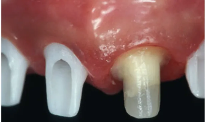
Today, the majority of implant manufacturers offer ceramic abutments. The abutments are available in pre-fabricated or customizable forms and can be prepared in the dental laboratory either by the technician or by utilizing computer-aided design⁄computer-aided manufacturing techniques. The materials of preference are densely sintered high-purity alumina (Al2O3) ceramic and yttria (Y2O3) -stabilized tetragonal zirconia polycrystal ceramics. These high-strength ceramics have improved mechanical properties (15, 38, 125, 180). Alumina ceramic has a flexural strength of 400 MPa, a fracture toughness value between 5 and 6 MPa⁄m0.5, and a modulus of elasticity of 350 GPa (148). The yttria stabilized zirconia ceramic has twice the flexural strength of alumina ceramic (900–1400 MPa), a fracture toughness of up to 10 MPa⁄m0.5, and a modulus of elasticity value of 210 GPa (38, 148). Compared to alumina ceramic, the enhanced strength of zirconia (ZrO2) can be explained by microstructural differences, such as higher density, smaller particle size, and polymorphic mechanism against flaw propagation (38, 67). The main reason for the superior resistance of zirconia lies in the sta-bilizing effect of yttria, which allows the processing of zirconia in the metastable tetragonal crystallineFig. 1. Ceramic abutments (zirconia) attached to the im-plants intraorally. Aesthetically pleasing appearance of the soft tissue.
Fig. 2. The same situation as in Fig. 1 with the all-ceramic crowns cemented on to the ceramic abutments.
structure at room temperature (18C–23C). The tetragonal phase at room temperature allows for transformation to the monoclinic phase under stress and represents an efficient mechanism against flaw propagation. The transformation results in a com-pressive stress as the result of volume expansion and slows down further crack propagation, resulting in improvement of the mechanical properties (i.e. transformation toughening) (34, 38, 62).
Alumina abutments are composed of 99.5% pure alumina ceramic (15). These abutments provide cer-tain aesthetic advantages when compared to the more whitish zirconia abutments (75, 210). In addi-tion, the alumina ceramic is easier to prepare; this saves time during definitive preparation, which is usually performed intraorally (75, 210). The problems presented by alumina abutments include their radioopalescence at the time of radiographic exami-nation and their weak resistance to fracture (13, 36, 198). In this context, it is commonly agreed that ceramic abutments should show proper resistance against the masticatory forces raised during chewing or swallowing. Several studies reported a mean loading force of approximately 206 N and maximum biting forces of up to 290 N in the aesthetic zone (71, 94). For a successful restoration, the abutment should present resistance to fracture values greater than such forces and, to guarantee long-term success, maintain this resistance for at least 5 years of clinical function. A study performedin vitrocompared tita-nium-reinforced zirconia and pure alumina abut-ments for their outcome after chewing simulation and static loading. After fixation of the abutments and adhesive cementing of metal crowns, the speci-mens were exposed to 1.2 million cycles in a chewing simulator to simulate 5 years of clinical service. The median fracture loads were 294 N, 239 N, and 324 N for the zirconia, alumina, and titanium abutment groups, respectively. The authors concluded that titanium-reinforced zirconia abutments perform in a similar way to metal abutments, and can therefore be recommended as an aesthetic alternative for the restoration of single implants in the anterior region. Ceramic abutments made of alumina showed less favorable properties (30). However, clinical studies using alumina abutments demonstrated excellent aesthetic outcomes and favorable survival rates when accepted treatment concepts were followed and documented components were used (76). Recent studies have shown that alumina implant abutments, when used for the fabrication of implant-supported, short-span, fixed partial dentures, had a cumulative survival rate of 98.1%after an observation period of
5 years (12), whereas alumina abutments used for the fabrication of implant-supported, single crowns had cumulative survival rates between 93% and 100%
after observation periods between 1 and 3 years (14, 76). Clinical studies on zirconia abutments confirmed that these abutments had a cumulative survival rate of 100% after observation periods between 4 and 6 years (64, 65).
Ceramic abutments can be restored using all-ceramic crown systems. The majority of clinical studies and case reports applied glass–ceramic crowns on alumina or zirconia abutments (65, 80, 145, 160, 209). In a study in vitro, the fracture resistance of such restorations was evaluated (210). Alumina and zirconia abutments were prepared and restored with glass–ceramic crowns and placed on Bra˚nemark implants (Nobel Biocare, Go¨teborg, Sweden). No artificial aging was applied to the test specimens. The statistical analysis showed significant differences between both groups, with mean fracture load values of 280.1 N for the group with alumina abutments and 737.6 N for the group with zirconia abutments. The fracture resistance in the zirconia abutment group was more than twice that in the alumina abutment group (210). Recent developments in computer-aided design⁄computer-aided manu-facturing techniques made it possible to use high-strength ceramics to fabricate implant-supported all-ceramic restorations (68, 75, 143, 145, 159). The combination of a high-strength ceramic abutment and a high-strength all-ceramic superstructure sys-tem would enhance the overall resistance of the restoration. Unfortunately, no clinical data on the success of such restorations are available. In two studies conductedin vitroby our group, the fracture resistance of different implant-supported, all-cera-mic restorations was evaluated and compared after chewing simulation and static loading. Ninety-six implants with an internal connection design (Replace, Nobel Biocare) were divided into one control group and two test groups of 32 specimens each. Implants in the control group received tita-nium abutments whereas the implants of the test groups received Procera alumina abutments and Procera zirconia abutments (both Nobel Biocare, Go¨teborg, Sweden; Fig. 3). The abutments were pre-pared to receive standardized maxillary central inci-sor all-ceramic crowns (analog tooth 21); half of each group received Procera alumina crowns while the other half received Procera zirconia crowns. The specimens were exposed to 1.2 million cycles in a chewing simulator to simulate 5 years of clinical service. The median fracture loads after aging were
1454 N, 1251 N, 423 N, 241 N, 444 N, and 457 N for titanium abutment⁄alumina crown, titanium abut-ment⁄ zirconia crown, alumina abutment⁄alumina crown, alumina abutment⁄zirconia crown, zirconia abutment⁄alumina crown, and zirconia abut-ment⁄ zirconia crown combinations, respectively. The highest fracture resistance value was found with the titanium abutment⁄alumina crown combina-tion, whereas the smallest fracture resistance was found with the alumina abutment⁄zirconia crown combination. We concluded that all the abut-ment⁄ crown combinations tested have the potential to withstand physiological occlusal forces in the anterior region (19, 20). Compared with a previous
in vitrostudy (210), it was obvious that the restora-tions in our study had significantly smaller values. The differences in the resistance to fracture, espe-cially in the zirconia abutment groups, could be ex-plained by the aging effect through environmental stresses on the abutments (abutment grinding, chewing simulator including low temperature hydrothermal degradation) (43, 48, 106, 107, 115).
Temperature peaks could alter the metastable tetragonal crystalline phase of the zirconia ceramic and there is controversy over whether this would lead to a reduction in the fracture resistance of the material (35, 48, 67, 122). Like other ceramics, zirco-nia is sensitive to changes in humidity and temper-ature, which is a particularly important issue when prosthetic applications are considered. Long-term exposure of zirconia ceramics to humidity and thermal cycling leads to a slow, low-temperature degradation of the material that might not become significant before several years have passed (128, 182, 193).
Conclusion and future
developments
The diversity of contemporary materials and of methods available for the fabrication of implant-supported, all-ceramic restorations makes it difficult to select the most appropriate treatment modality. New products are constantly being added to the wide range of existing products. For example, efforts are being made to produce low-temperature, degrada-tion-free zirconia–alumina composites for dental implant abutments (96, 97). The first short-term results are encouraging (95).
In addition to correct diagnosis and treatment planning, recognizing the properties, long-term behavior, indications, and contraindications of each material used are essential to guarantee the long-term clinical success of the restoration. Bothin vitro
and in vivo studies show that the indication for ceramic abutments is restricted to the fabrication of single-tooth, implant-supported all-ceramic restora-tions. Enhancing the resistance of the abutment will expand its application to implant-supported, all-ceramic, fixed partial dentures and restorations in the posterior region. Zirconia ceramics and abutments are being intensively investigated and are gaining in popularity. Future improvements in the ceramic will focus on its color and long-term stability. Attempts are being made to add coloring oxides to zirconia ceramic before the sintering process; this would change its whitish color and enhance the aesthetic outcome. It remains important, however, to verify the effectiveness of a suggested method before its rec-ommendation.
Advances in computer-aided design⁄ computer-aided manufacturing technologies, which have made the fabrication procedures of ceramic abutments and implant-supported, all-ceramic restorations faster, easier, and more efficient, are playing a major role in the growing use of ceramic abutments. Future developments will make it possible to produce more resistant abutments and restorations with higher quality and lower fabrication time and costs. Here, it should be noted that the lifetime of any dental ceramic material may be limited because of accu-mulating damage from the oral function (90).
Ceramic implants
Forty years ago, oral implants appeared on the mar-ket for clinical use. A variety of forms, materials, and
Fig. 3. Zirconia (left) and alumina (right) dental abutment with a metal base.
surface treatments evolved, some more and some less for the benefit of the patients. Today, the endos-seous-cylinder⁄screw surface textured type of implant, made from commercially pure titanium, is considered the gold standard for the fabrication of oral implants. Titanium is applied in many fields of dentistry because of its biocompatibility, high corrosion resistance (92), and good mechanical characteristics. Commercially pure titanium has been used as an implant substrate (1, 10), as well as material for implant abutments, for many years. Numerous investigations have demonstrated the reliability of this material for both mid- and long-term use (1, 28, 77, 84, 86, 116, 200). There are some concerns that titanium might provoke unwelcome host reactions, but little evidence is available (199). Tschernitschek et al. (199) found that titaniumÔ…can also cause chemical–biological interactions. Tissue discoloration and allergic reactions in patients who have come in contact with titanium have been re-ported.Õ A small number of investigations showed increased titanium concentrations close to titanium implants (23) and in regional lymph nodes (205). An investigation reported on titanium implant failures of which more than half were, according to the authors, the result of toxic metal ions released from the superstructure (207). In an investigation evaluating tissues from patients who underwent a revision of their hip-joint replacements, Lalor et al. (113) sug-gested a sensitization to titanium because monoclo-nal antibody labeling showed macrophages and T lymphocytes in the presence of titanium particles. However, it should be mentioned that the clinical relevance of these findings is not clear. One fact, however, is that patients – perhaps because of more or less scientific reports in the lay press, which considers metals to be harmful to the body – rely on natural health and holistic medicine where a freedom of metal contamination is part of the philosophy. With implant components produced from ceramic mate-rials, it will be possible to render the option of metal-free treatment to patients asking for such an option.
Ceramic materials are frequently used in dentistry. The application ranges from veneering material for metal substructures, through all-ceramic posts and cores towards frameworks for crowns and bridge-work. Ceramics are highly biocompatible and may improve the aesthetic appearance of dental recon-structions. Furthermore, it is well-known that cera-mic materials are less prone to plaque accumulation than metal substrates (47, 162, 169).
Ceramic implants in dentistry are not new. Sand-haus (166) was one of the first to report on aluminum
oxide ceramic implants (CBS = the Crystalline Bone Screw). However, the Crystalline Bone Screw showed a success rate of only 25%after an average observa-tion period of 5 years (190). In 1987, Sandhaus (167) introduced the Cerasand (Incermed, Lausanne, Switzerland) ceramic implant but unfortunately there are no long-term data on its clinical behavior. To the knowledge of the authors, neither of these ceramic implants is available on the market any more. In 1976, Schulte & Heimke (176) introduced the alumi-num oxide Tu¨ bingen implant (Frialit I; Friadent, Mannheim, Germany) for immediate implant place-ment in the anterior area. Besides clinical reports on this implant system (29, 60, 142, 170–175, 177, 178, 204), there are also scientific data available on the long-term behavior of this system (44, 49–51, 212, 213). Investigations showed that this material inte-grated well into bone and soft tissue (16, 29, 212, 213). DÕHoedt (49) reported on 924 Tu¨ bingen implants in 631 patients after approximately 10 years. A success rate of 92.5%was presented since the routine phase of 1982. There was no report in that investigation on the entire success rate from 1975 until 1985. In a subsequent comparative investigation, dÕHoedt & Schulte (51) reported the results from 1982 to 1987 and showed that only two of the 448 rehabilitations were not successful; 396 rehabilitations were suc-cessful with a 95%confidence limit of 85–92%for the success rate. No information was provided for the remaining 50 samples. No clear information could be drawn on implant success rate from that investiga-tion. However, De Wijs et al. (45) presented a clinical evaluation over a 10-year period for 127 Tu¨ bingen implants. In 101 patients, implants were placed in the maxillary incisor, canine, and premolar regions. The authors found an overall survival rate in their inves-tigation of 87%with a mean follow-up of 4.5 years.
Since the Frialit polycrystalline aluminum oxide implant system did not meet the expectations in terms of long-term stability, it was withdrawn from the market and eventually replaced by the titanium Frialit-II system (Friadent, Mannheim, Germany).
Another aluminum oxide implant system was the Bionit implant, which was developed by Mu¨ ller, Piesold, and Glien (139, 151, 153). However, there is only limited information from laboratory investiga-tions (150, 152, 153) and no information from clinical investigations regarding the (long-term) behavior of this implant system. The authors do not know whether this implant system is still available on the market.
Much more scientific support exists for single-crystal alumina (sapphire) oral implants. Numerous
single-crystal alumina oral implants (131, 132, 134, 135) have been placed over the last 20 years in animal (7, 8, 55, 72, 131, 133, 188) and clinical (54, 56, 57, 108–111, 185–187) investigations. This material proved to be biocompatible (214). One commercially produced single-crystal oral implant was the Bioce-ram implant by Kyocera (Kyoto, Japan). Koth et al. (111) and Steflik et al. (187) reported 5- and 10-year results of the single-crystal sapphire (alumina) threaded cylindrical endosteal oral Bioceram im-plants. Twenty-eight implants were placed in the lower jaws of 17 patients. Six weeks after implant insertion, 23 implants were included into fixed prostheses as distal abutments. Twenty-one of the 23 implants could be recalled after 10 years. The authors reported a success rate of 81%. It is noteworthy that these two investigations were included in a system-atic review on the survival and complication of tooth– implant-supported fixed partial dentures (114). The estimated survivals in that review were 75.7% and 64.7% after 5 and 10 years, respectively, demon-strating survival rates lower than those of titanium implants in the same review. A further report on the long-term behavior of these sapphire implants was presented by Fartash & Arvidson (54). They reported on the treatment of total or partial edentulism with fixed prosthodontics supported by Bioceram sapphire implants. The implant system performed well in the lower jaw and in partial edentulism. This investiga-tion was also included in the above-meninvestiga-tioned sys-tematic review (114); the 10-year results showed an estimated survival of 100% for combined tooth– implant reconstructions. The same investigation by Fartash & Avidson (154) was included in a second systematic review by the same group that included only implant-supported fixed partial dentures. The estimated survival for the Bioceram implants was 96.6%. The results were similar (implant-supported) or better (implant–tooth-supported) when compared to those of titanium implants.
Another report on sapphire implants (Bioceram) used to support mandibular overdentures was pre-sented by Berge & Gronningsaeter (22). They showed the results from 30 patients who had been treated between 1984 and 1991 and reported a cumulative survival rate for the implants of 69% over a mean observation time of 11 years. The authors concluded that the long-term results with this implant system for the use of mandibular overdenture support were inferior to those with other systems.
Except for the investigation by Fartash & Avidson (54), the single-crystal alumina implant showed inferior results to titanium implants, which might be
why this implant system was not as accepted as titanium implants. According to the manufacturer, the sapphire oral Bioceram implant is no longer produced.
Although it might be common sense that alumina is prone to fracture because of its brittleness, low fracture strength, and long-term aging, fracture was only mentioned once as the reason for the loss of a maxillary posterior implant in the long-term investi-gations mentioned above (54). Therefore, the reason for the withdrawal of some of the alumina implant systems from the market remains unclear. It can be assumed, though, that the anxieties raised by dentists that ceramic implants are prone to fracture might have played a role. There was therefore a search for tougher ceramic materials for use as an oral implant substrate instead of alumina. The ceramic of choice – it has already been in use in orthopedics since the 1990s (31, 37, 39) – seems to be zirconia. Zirconia was introduced into dentistry in the 1990s and is currently being used as the material for posts for tooth build up, for frameworks for crown and bridgework, and for implant abutments (3, 4, 9, 17, 18, 26, 27, 53, 63, 64, 78, 79, 93, 100, 112, 123, 124, 137, 147, 155, 165, 189, 191, 193–196, 202, 203, 209, 210).
Cell culture testing of zirconia
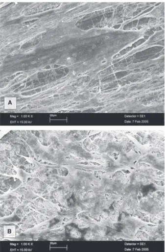
Since zirconia is of certain interest in orthopedics and implant dentistry, its biocompatibility towards bone and soft connective and epithelial tissue is essential. To determine the biocompatibility or interactions at the biomaterial–tissue interface stud-iesin vitrousing cell cultures provide an important tool (Fig. 4A, B) (138). Advantages of such studies include the isolated, homogeneous nature of the culture system, a defined temporal course of events, relatively limited expense, the reproducible growth of multiple cultures, and reduced animal morbidity and mortality (40).
While early in vitro studies were performed with zirconia in powder form, co-cultured mostly with fi-broblasts or lymphocytes – well-summarized in the reviews by Piconi et al. (148, 149) – newer experi-ments focused on osteoblasts and compact zirconia substrates (Table 1). Josset et al. (89) tested prolifer-ation, and total protein and DNA content of human osteoblasts on zirconia and alumina disks with ref-erence to control cells cultured on glass coverslips (89). They found that neither material altered the cell ploidy or the cell growth rate, which was in accor-dance with the absence of any inducing effect on DNA synthesis or proliferation.In vitrostudies using
human osteoblasts are considered to be of particular interest because these cells are involved in the tissue reaction at the implant site in patients (129, 161). The effect of zirconia, alumina, and titanium on the expression and secretion of the matrix metallopro-teinases MMP-2, MMP-9 and their natural tissue inhibitors TIMP-1 and TIMP-2 by primary human osteoblasts was studied by OumÕhamed et al. (146). Matrix metalloproteinases (MMPs) are a family of proteolytic enzymes that are capable of degrading all the major components of the extracellular matrix (140). Their activities can be regulated by natural specific inhibitors (tissue inhibitors of metallopro-teinases, TIMPs). A balance between MMPs and TIMPs is necessary for many physiological processes and an imbalance can result in a number of patho-logical events (32, 46, 141). MMPs play an important role in the development of osteolysis and implant loosening (120, 141). Zirconia did not have a signifi-cant effect on MMP-2 messenger RNA expression and
even decreased MMP-9 expression. Whereas TIMP-2 levels were very low in this experiment, TIMP-1 in-creased to a maximum level at 48–72 hours. In their biomolecular study Carinci et al. (33) determined the genetic effect of zirconia on an osteoblast-like cell line (MG63) using microarray slides with 19,200 dif-ferent oligonucleotides. Several genes were identified of which the expression was either up- or down-regulated. It was shown that zirconia was able to modulate immunity, vesicular transport, and cell cycle regulation. The authors suggested that the regulation of immunity may positively affect the foreign body reaction in vivo, allowing for a close apposition of bone, as found in histological studies (5, 6, 52, 82, 168, 179). Furthermore, it was concluded that the regulation of vesicular transport and cell cycle mechanisms might produce modifications in the turnover of extracellular matrix and in the pro-liferation of osteoblasts. Using another approach, the effect of zirconia surface property modification on human fetal osteoblasts was analyzed (69, 70). Car-bon dioxide laser treatment was applied on magnesia partially stabilized zirconia (69) and yttria partially stabilized zirconia (70). The surface of the magnesia partially stabilized zirconia was roughened and the polar component of the surface energy was enhanced by the laser treatment, resulting in an increased number of cells attaching and spreading compared to the untreated control. In contrast, the surface of yttria partially stabilized zirconia was smoothed by carbon dioxide laser irradiation. Nevertheless, cell attach-ment was also increased, probably as a result of surface energy enhancement, i.e. alterations in the wettability characteristics. A recent investigation evaluated differently roughened yttrium-stabilized tetragonal zirconia polycrystal samples using the CAL72 osteoblast-like cell line (21). This investigation showed that there was no difference in cell prolifer-ation between the different materials and surfaces later in the culture. From their results the authors concluded that the yttrium-stabilized tetragonal zir-conia polycrystal material used might be an appro-priate substrate for the proliferation and spreading of osteoblastic cells.
Further studiesin vitroare required to understand the behavior of osteoblasts on zirconia substrates more comprehensively. So far, little is known about the effects of zirconia on gene⁄extracellular matrix protein expression patterns (183) and healing-spe-cific enzyme kinetics. Since surface topography has proven to be one of the key factors in bone–titanium integration (208), this may be another field that needs to be addressed in future studies.
A
B
Fig. 4. Scanning electron microscopy images of primary human osteoblasts cocultured with zirconia; (A) machined substrate surface, (B) rough, air-borne particle abraded substrate surface.
Zirconia oral implants
Zirconia was applied relatively early as a coating material for oral implants in animal investigations. In 1975, Cranin et al. (41) used zirconia flame-spray-deposition-coated Vitallium implants in beagle dogs. They showed that five of nine zirconia-coated im-plants were surrounded by connective tissue and that the results were not satisfactory. In a histological interface analysis of titanium and zirconium bone implants, Albrektsson et al. (11) observed a fibrous, tissue-free zone with a 20- to 40-nm thick proteo-glycan layer at the titanium implants. At a distance of approximately 100 nm, collagen bundles were de-tected. The proteoglycan zone at the zirconia-coated implants showed a thickness of 30–50 nm and the collagen fibers were further apart from zirconia compared to titanium. From these two earlier inves-tigations, it could be concluded that zirconia might not be an adequate alternative to titanium for
im-plant fabrication. Further investigations in the early 1990s (73, 74) compared the biocompatibility of alumina, zirconia, and stainless steel in dogs. In several experiments it was shown that the affinity of bone towards the different materials was not differ-ent. However, the authors reported on thin inter-vening fibrous membranes between the bone and the implants. There was no detailed description of the kind of soft tissue that was found.
In a histomorphometric study in rats, Fini et al. (58) evaluated implants of different materials placed in the femur (hydroxyapatite, titanium alloy, yttrium partially stabilized zirconia, alumina, and two bio-logical glasses). In non-ovariectomized rats there were no significant differences regarding the affinity index of bone towards the different implant materials (hydroxyapatite 77.0%, zirconium 58.2%, titanium alloy 61.2%).
Summarizing the results of later investigations, it seems that the bone integration of zirconia has
im-Table 1. Summary of cell culture studies involving compact zirconia substrates
Reference Material Cell type Test Main findings
Josset et al. (89) Zirconia Alumina Glass
Primary human osteoblasts
Cell proliferation Total protein synthesis Cell morphology Evidence of osteoblastic
proteins Carcinogenicity (DNA image cytometry
Ag-NORs quantification) No adverse response OumÕhamed et al. (146) Zirconia Alumina Titanium Primary human osteoblasts Expression analysis of MMP-2, MMP-9, TIMP-1, TIMP-2 No effect on MMP-2, decrease of MMP-9, increase of TIMP-1 Carinci et al. (33) Zirconia Osteoblast-like
MG63 cells Gene expression analysis (DNA microarray) Modulation of immunity, vesicular transport, and cell cycle regulation
Hao et al. (69) Zirconia* (MgO-PSZ) Human fetal osteoblast cells (hFOB 1.19) Surface analysis Cell proliferation Cell adhesion Increase of cell attachment and spreading by surface treatment
Hao et al. (70) Zirconia* (Y-PSZ) Human fetal osteoblast cells (hFOB 1.19) Surface analysis Cell adhesion Increase of cell attachment by surface treatment
Ba¨chle et al. (21) Zirconia (Y-TZP) CAL72 osteoblast-like cells Cell proliferation Cell morphology Cell-covered surface area No adverse response Similar cell proliferation
of different substrates
*Carbon dioxide laser irradiation.
Ag-NOR, argyrophilic nuclear organizer region; MgO-PSZ, magnesia partially stabilized zirconia; MMP, matrix metalloproteinase; TIMP, tissue inhibitor of metalloproteinases; Y-PSZ, yttria partially stabilized zirconia; Y-TZP, yttria partially stabilized zirconia.
proved and is no longer different to that of titanium (59, 119, 168, 179). This might be because of refine-ments made in the preparation and production of zirconia materials.
Akagawa et al. (6) were the first to report the use of oral implants made of zirconia in beagle dogs. In
their experiment they evaluated loaded (1 week after implant placement) and unloaded zirconia dental implants. Histology was performed 3 months after implant placement. The authors reported that no implant was mobile and no fracture occurred during the experiment. Direct bone apposition to the implant could be observed in both treatment groups. The bone-to-implant contact ratio was 81.9%for the
Fig. 5. En-face photograph of a patient participating in the clinical investigation on zirconia implants.
Fig. 6. Overview (retracted lips). Tooth 12 (FDI nomen-clature) is restored with an insufficient crown which had to be re-cemented several times.
Fig. 7. Close-up of Fig. 6. Marginal gingival inflammation is visible. The papillae towards the neighboring teeth are present.
Fig. 8. A periapical radiograph showing an apical radio-lucency (result of an apicectomy) as well as a transdental fixation. The crown is insufficiently attached to the tooth with an endodontic post.
nonloaded implants and 69.8%for the loaded group and the loaded group showed more loss of marginal bone than the nonloaded group. Unfortunately, no
titanium control group was included for comparison. In a second investigation, Akagawa et al. (5) com-pared entirely implant-borne reconstructions with implant–tooth-connected restorations in monkeys. Again, implants fabricated from partially stabilized zirconia were inserted into the mandibles of eight monkeys and 3 months later three different types of superstructures were provided (single crowns on
Fig. 9. The tooth was extracted with caution so as not to damage the soft and hard tissues. After tooth extraction, there was an alveolar defect of the buccal bone, which was treated according to the guided bone regeneration prin-ciple and with a soft connective tissue graft in the same session (treatment not shown).
Fig. 10. Initial drilling with a 2-mm twist drill and a direction aid.
Fig. 11. Subsequent enlargement of the implant osteoto-my with the respective drills.
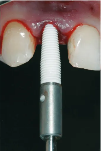
Fig. 12. Insertion of the ceramic implant.
Fig. 13. Placement of the implant to the correct depth so that only the implant head is visible.
implants, implant-borne connected restorations, or implant–tooth-supported reconstructions). Clini-cally, there were no differences between the groups. Histology was performed after 12 and 24 months. Direct bone apposition was observed in all groups. Histomorphometry showed bone-to-implant contact between 66 and 81%. In general, there was no sig-nificant difference between the different treatment groups.
Our group was the first to compare loaded titanium implants with loaded zirconia implants in the same model (103). Twelve custom-made titanium and 12 zirconia implants were inserted in the upper anterior jaw of six monkeys. Six months after implant instal-lation single crowns were fabricated and inserted on all implants. Five months after crown installation (i.e. the loading time) the implants, with their soft and hard tissues, were harvested and evaluated micro-scopically. No implant was lost over the observation period. There were no statistically significant differ-ences in the soft tissues around the titanium and zirconia implants and no differences in respect to the
bone-to-implant contact between the two materials (titanium 72.9%; zirconia 67.4%). The surface treat-ments used to change the surface topography for the titanium implants included air-borne particle abra-sion and acid etching. The zirconia implants, how-ever, were only air-borne particle abraded because acid etching had no effect on zirconia (unpublished data). So far, air-borne particle abrasion has been the
Fig. 14. Adaptation of the temporary to the clinical situ-ation and subsequent relining.
Fig. 15. The provisional is cemented with temporary cement at the end of session.
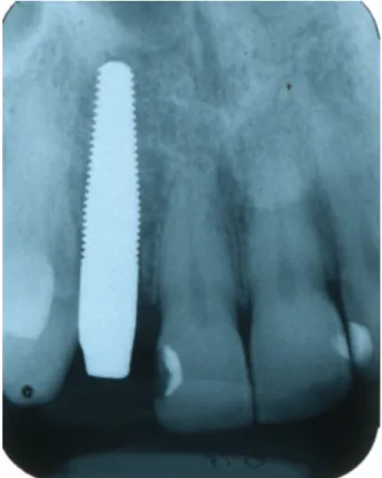
Fig. 16. Standardized periapical radiograph after implant placement showing the implant position in the mesio-distal and apicocoronal direction.
Fig. 17. Clinical appearance 13 weeks after installation of implant. Marginal soft tissue overgrowth made it neces-sary for a minor gingivectomy to be performed at the peri-implant mucosal margin.
method of choice for increasing the surface rough-ness of zirconia. However, Sennerby et al. (181) developed a technique that was different to air-borne particle abrasion to achieve a porous surface. They coated zirconia implants with slurry containing zir-conia powder and a pore-former. While sintering the coating the pore-former was burned and a porous surface was the result. Their topographic analyses showed that the zirconia implant surface modified in this way resembled the well-known TiUnitesurface (Nobel Biocare, Goteborg, Sweden). The authors installed threaded zirconia implants with either a machined surface or one of two surface modifica-tions into the tibia and femur of rabbits. Oxidized titanium implants served as controls. Removal torque tests showed higher values for the surface-modified zirconia implants than for the titanium implants. The lowest values were found for the machined zirconia implants. However, no significant differences regarding bone-to-implant contact and bone area
filling could be observed between the different treatment groups. Another two animal investigations showed that zirconia implants undergo osseointe-gration and that a rough surface is of benefit (61, 184).
Biomechanical investigations on zirconia oral implants are rare in the literature. In a three-dimensional finite element analysis, the stress dis-tribution patterns of implants made of commercially pure titanium and yttrium partially stabilized zir-conia were analyzed. Two three-dimensional finite element analysis models of a maxillary incisor with Re-Implant (Hagen, Germany) implants (99) were made, surrounded by cortical and cancellous bone. A porcelain-fused-to-metal crown for the commer-cially pure titanium implant and a ceramic crown for the yttrium partially stabilized zirconia implant were modeled and the stress levels were calculated according to von Mises criteria. Yttrium partially stabilized zirconia implants had similar stress dis-tribution to commercially pure titanium implants (102). In another investigation by our group on biomechanical behavior, two groups of two-piece zirconia implants were tested in the artificial mouth (101). The implants restored with Procera crowns seemed possibly to fulfill the biomechanical
Fig. 19. Situation with retracted lips. The dental and soft tissue symmetry is not perfect when tooth 12 is compared to tooth 22 but the overall result is satisfactory.
Fig. 20. Standardized periapical radiograph after crown placement.
Fig. 18. Close-up of the final restoration with the clinical appearance of the red and white aesthetics. Slight reces-sion of the interdental papillae can be observed.
requirements for anterior teeth, showing mean fracture loads of the crowns of 575.7 N when not artificially loaded and 555.5 N when aged in a chewing simulator for 1.2 million cycles. Further testing in vitro (i.e. artificial mouth) mimicking long-term clinical service would surely be of benefit to obtain additional information on the long-term fatigue behavior of zirconia implants.
Although there have been reports of problems with zirconia (e.g. increased fracture rate of hip prosthe-sis), which occurred some years ago in orthopedics (34, 206), the zirconia production process for medical applications is currently one of the best-controlled fabrication processes in industry. High reproducibil-ity and constant production chains seem to ensure a high quality of zirconia products for medical applications so zirconia appears to be the ceramic material of choice for metal-free reconstructions in implant dentistry.
However, regarding the clinical use of zirconia oral implants, scientific information is lacking. To the authorsÕ knowledge there are only two clinical patient case reports using zirconia implants and
two retrospective observational case series pub-lished in the international literature. The first report on the application of a zirconia implant – crown system in a patient dated from 2004 (100). The authors used a custom-made two-piece dental implant to replace a left upper central incisor. After an unloaded healing period of 6 months, abutment connection was performed using a pre-fabricated zirconia abutment, which was cemented to the implant with Panavia (Kuraray, Osaka, Japan). The implant was subsequently restored with a zirconia-based single crown. A second report presented a patient case in which eight, one-piece zirconia implants had been placed. After implant placement the patient had to wear a protective splint over a 6-month period for 24 hours a day. After that time the implants were restored with zirconia crowns (201). There was, however, no follow-up presented on either of the two reports.
Two clinical retrospective case series were reported by the developers of two different zirconia ceramic implant systems (25, 144). However, because of bias the scientific value of these reports is questionable. Furthermore, the authors did not clearly state the aim of their investigation nor the inclusion or exclusion criteria used. No additional parameters (radiographic bone remodelling, soft tissue health, mobility) were evaluated. The only result that was reported was the ÔsurvivalÕ rate in per cent. The level of evidence in these two clinical reports might therefore be regarded as very low.
There are currently five zirconia implant systems commercially available. Sandhaus, again, was the first to develop a zirconia implant system in 1987: the SIGMA implant system (Incermed, Lausanne, Swit-zerland). Further zirconia implant systems are the Ceraroot system (Ceraroot, Barcelona, Spain), the White Sky system (Bredent Medical, Senden, Ger-many), the z-systems implant system (z-systems, Konstanz, Germany), and the zit-z ceramic implant system (Ziterion GmbH, Uffenheim, Germany). However, there are no data on the histological and biomechanical behavior of these different implant systems in the international literature. Furthermore, no scientific short-, middle- or long-term clinical data on the above-mentioned zirconia implant sys-tems can be found.
The only prospective ongoing clinical evaluation on zirconia implants of which the authors are aware is on a one-piece yttria-stabilized tetragonal zirconia polycrystal reinforced with alumina implant with an implant surface modification similar to that pre-sented by Sennerby et al. (181). This investigation is
being performed at the Department of Prosthodon-tics at the University of Freiburg, Germany. So far, 92 patients have received 119 implants: 65 patients received one implant (Figs 5–21) and 27 patients received two implants each for a three-unit bridge. All implants were immediately temporized with composite temporary crowns or bridges. After an observation time of up to 1 year four implants have so far been lost, giving a cumulative survival rate of 96.6%. Besides clinical measurements, aesthetic measurements are also being performed, and bone modeling⁄remodeling will be evaluated using stan-dardized radiographs.
Since the clinical use of zirconia implants lacks scientific support, the authors do not currently rec-ommend their use. Prospective clinical investigations are needed before these implant systems can be recommended for clinical use.
References
1. Adell R, Eriksson B, Lekholm U, Bra˚nemark P-I, Jemt T. A long-term follow-up study of osseointegrated implants in the treatment of totally edentulous jaws.Int J Oral Max-illofac Implants1990:5: 347–359.
2. Adell R, Lekholm U, Rockler B, Bra˚nemark PI. A 15-year study of osseointegrated implants in the treatment of the edentulous jaw.Int J Oral Surg1981:10: 387–416. 3. Ahmad I. Yttrium-partially stabilized zirconium dioxide
posts: an approach to restoring coronally compromised nonvital teeth.Int J Periodontics Restorative Dent1998:18: 454–465.
4. Ahmad I. Zirconium oxide post and core system for the restoration of an endodontically treated incisor. Pract Periodontics Aesthet Dent1999:11: 197–204.
5. Akagawa Y, Hosokawa R, Sato Y, Kamayama K. Compar-ison between freestanding and tooth-connected partially stabilized zirconia implants after two yearsÕfunction in monkeys: a clinical and histologic study. J Prosthet Dent 1998:80: 551–558.
6. Akagawa Y, Ichikawa Y, Nikai H, Tsuru H. Interface his-tology of unloaded and early loaded partially stabilized zirconia endosseous implant in initial bone healing. J Prosthet Dent1993:69: 599–604.
7. Akagawa Y, Matsumoto T, Hashimoto M, Tsuru H. Clini-cal evaluation of the gingiva around single-crystal sap-phire endosseous implant after experimental ligature-induced plaque accumulation in monkeys.J Prosthet Dent 1992:68: 111–115.
8. Akagawa Y, Matsumoto T, Kawamura M, Tsuru H. Changes of subgingival microflora around single-crystal sapphire endosseous implants after experimental liga-ture-induced plaque accumulation in monkeys.J Prosthet Dent1993:69: 594–598.
9. Akkayan B, Gulmez T. Resistance to fracture of end-odontically treated teeth restored with different post systems.J Prosthet Dent2002:87: 431–437.
10. Albrektsson T, Dahl E, Enbom L, Engevall S, Engquist B, Eriksson AR, Feldmann G, Freiberg N, Glantz P-O, Kjellman O, Kristersson L, Kvint S, Ko¨ndell P-A˚ , Palm-quist J, Werndahl L, A˚ strand P. Osseointegrated oral implants. A Swedish multicenter study of 8139 consec-utively inserted Nobelpharma implants. J Periodontol 1988:59: 287–296.
11. Albrektsson T, Hansson HA, Ivarsson B. Interface analysis of titanium and zirconium bone implants.Biomaterials 1985:6: 97–101.
12. Andersson B, Glauser R, Maglione M, Taylor A. Ceramic implant abutments for short-span FPDs: a prospective 5-year multicenter study.Int J Prosthodont2003:16: 640– 646.
13. Andersson B, Schaerer P, Simion M, Bergstrom C. Cera-mic implant abutments used for short-span fixed partial dentures: a prospective 2-year multicenter study. Int J Prosthodont1999:12: 318–324.
14. Andersson B, Taylor A, Lang BR, Scheller H, Schaerer P, Sorensen JA, Tarnow D. Alumina ceramic implant abutments used for single-tooth replacement: a pro-spective 1- to 3-year multicenter study.Int J Prosthodont 2001:14: 432–438.
15. Andersson M, Oden A. A new all-ceramic crown. A dense-sintered, high-purity alumina coping with porcelain.Acta Odontol Scand1993:51: 59–64.
16. Anneroth G, Ericsson AR, Zetterqvist L. Tissue integration of A12O3-ceramic dental implants (Frialit) – a case report. Swed Dent J1990:14: 63–70.
17. Ardlin BI. Transformation-toughened zirconia for dental inlays, crowns and bridges: chemical stability and effect of low-temperature aging on flexural strength and surface structure.Dent Mater2002:18: 590–595.
18. Asmussen E, Peutzfeldt A, Heitmann T. Stiffness, elastic limit, and strength of newer types of endodontic posts. J Dent1999:27: 275–278.
19. Att W, Kurun S, Gerds T, Strub JR. Fracture resistance of single-tooth implant-supported all-ceramic restorations after exposure to the artificial mouth.J Oral Rehabil2006: 33: 380–386.
20. Att W, Kurun S, Gerds T, Strub JR. Fracture resistance of single-tooth implant-supported all-ceramic restorations: An in vitro study.J Prosthet Dent2006:95: 111–116. 21. Ba¨chle M, Butz F, Hu¨ bner U, Bakalinis E, Kohal RJ.
Behavior of CAL72 osteoblast-like cells cultured on zir-conia ceramics with different surface topographies.Clin Oral Implants Res2007:18: 53–59.
22. Berge TI, Gronningsaeter AG. Survival of single crystal sapphire implants supporting mandibular overdentures. Clin Oral Implants Res2000:11: 154–162.
23. Bianco PD, Ducheyne P, Cuckler JM. Local accumulation of titanium released from a titanium implant in the ab-sence of wear.J Biomed Mater Res1996:31: 227–234. 24. Binon PP. Implants and components: entering the new
millennium.Int J Oral Maxillofac Implants2000:15: 76–94. 25. Blaschke C, Volz U. Soft and hard tissue response to zirconium dioxide implants – a clinical study in man. Neuroendocrinol Lett2006:27: 69–72.
26. Bornemann G, Rinke S, Huels A. Prospective clinical trial with conventionally luted zirconia-based fixed partial dentures – 18 months results.J Dent Res2003:82Spec Iss B: 117 (abstract no 842).
27. Brodbeck U. The ZiReal Post: A new ceramic implant abutment.J Esthet Restor Dent2003:15: 10–23; discussion 24.
28. Buser D, Mericske-Stern R, Bernard JP, Behneke A, Be-hneke N, Hirt HP, Belser UC, Lang NP. Long-term evaluation of non-submerged ITI implants. Part I: 8-year life table analysis of a prospective multi-center study with 2359 implants. Clin Oral Implants Res1997: 8: 161–172.
29. Bu¨ sing CM, dÕHoedt B. Human bone attachment to the Tu¨ bingen implant.Dtsch Zahna¨rztl Z1981:36: 563–566. 30. Butz F, Heydecke G, Okutan M, Strub JR. Survival rate,
fracture strength and failure mode of ceramic implant abutments after chewing simulation.J Oral Rehabil2005: 32: 838–843.
31. Cales B, Stefani Y, Lilley E. Long-term in vivo and in vitro aging of a zirconia ceramic used in orthopaedy.J Biomed Mater Res1994:28: 619–624.
32. Cambray GJ, Murphy G, Page-Thomas DP, Reynolds JJ. The production in culture of metalloproteinases and an inhibitor by joint tissues from normal rabbits, and from rabbits with a model arthritis. I. Synovium.Rheumatol Int 1981:1: 11–16.
33. Carinci F, Pezzetti F, Volinia S, Francioso F, Arcelli D, Farina E, Piattelli A. Zirconium oxide: analysis of MG63 osteoblast-like cell response by means of a microarray technology.Biomaterials2004:25: 215–228.
34. Chevalier J. What future for zirconia as a biomaterial? Biomaterials2006:27: 535–543.
35. Chevalier J, Deville S, Munch E, Jullian R, Lair F. Critical effect of cubic phase on aging in 3mol%yttria-stabilized zirconia ceramics for hip replacement prosthesis. Biomaterials2004:25: 5539–5545.
36. Cho HW, Dong JK, Jin TH, Oh SC, Lee HH, Lee JW. A study on the fracture strength of implant-supported restora-tions using milled ceramic abutments and all-ceramic crowns.Int J Prosthodont2002:15: 9–13.
37. Christel P, Meunier A, Dorlot JM, Crolet JM, Witvoet J, Sedel L, Boutin P. Biomechanical compatibility and de-sign of ceramic implants for orthopedic surgery.Ann N Y Acad Sci1988:523: 234–256.
38. Christel P, Meunier A, Heller M, Torre JP, Peille CN. Mechanical properties and short-term in-vivo evaluation of yttrium-oxide-partially-stabilized zirconia. J Biomed Mater Res1989:23: 45–61.
39. Christel PS. Zirconia: the second generation of ceramics for total hip replacement. Bull Hosp Jt Dis Orthop Inst 1989:49: 170–177.
40. Cooper LF, Masuda T, Yliheikkila PK, Felton DA. Gener-alizations regarding the process and phenomenon of osseointegration. Part II. In vitro studies. Int J Oral Maxillofac Implants1998:13: 163–174.
41. Cranin AN, Schnitman PA, Rabkin SM, Onesto EJ. Alumina and zirconia coated vitallium oral endosteal implants in beagles. J Biomed Mater Res 1975: 9: 257– 262.
42. Creugers NH, Kreulen CM, Snoek PA, de Kanter RJ. A systematic review of single-tooth restorations supported by implants.J Dent2000:28: 209–217.
43. De Boever JA, McCall WD Jr, Holden S, Ash MM Jr. Functional occlusal forces: an investigation by telemetry. J Prosthet Dent1978:40: 326–333.
44. De Wijs FL, De Putter C, Cune MS. Front tooth replace-ment with Tu¨ bingen (Frialit) implants: a radiographical evaluation.J Oral Rehabil1996:23: 97–100.
45. De Wijs FL, Van Dongen RC, De Lange GL, De Putter C. Front tooth replacement with Tu¨ bingen (Frialit) implants. J Oral Rehabil1994:21: 11–26.
46. Dean DD, Martel-Pelletier J, Pelletier JP, Howell DS, Woessner JF Jr. Evidence for metalloproteinase and metalloproteinase inhibitor imbalance in human osteo-arthritic cartilage.J Clin Invest1989:84: 678–685. 47. Degidi M, Artese L, Scarano A, Perrotti V, Gehrke P,
Piattelli A. Inflammatory infiltrate, microvessel density, nitric oxide synthase expression, vascular endothelial growth factor expression, and proliferative activity in peri-implant soft tissues around titanium and zirconium oxide healing caps.J Periodontol2006:77: 73–80.
48. Deville S, Gremillard L, Chevalier J, Fantozzi G. A crit-ical comparison of methods for the determination of the aging sensitivity in biomedical grade yttria-stabilized zirconia.J Biomed Mater Res B Appl Biomater2005:72: 239–245.
49. dÕHoedt B. 10 Jahre Tu¨ binger Implantat aus Frialit. Eine Zwischenauswertung der Implantatdatei. Z Zahna¨rztl Implantol1986:2: 6–10.
50. dÕHoedt B, Lukas D, Schulte W. Das Tu¨ binger Implantat als Sofort- und Spa¨timplantat, ein statistischer Vergleich. Dtsch Zahna¨rztl Z1986:41: 1068–1072.
51. dÕHoedt B, Schulte W. A comparative study of results with various endosseous implant systems.Int J Oral Maxillofac Implants1989:4: 95–105.
52. Dubruille´ JH, Viguier E, Le Naour G, Dubruille MT, Auriol M, Le Charpentier Y. Evaluation of combinations of tita-nium, zirconia, and alumina implants with 2 bone fillers in the dog.Int J Oral Maxillofac Implants1999:14: 271– 277.
53. Edelhoff D, Sorensen JA. Retention of selected core materials to zirconia posts.Oper Dent2002:27: 455–461. 54. Fartash B, Arvidson K. Long-term evaluation of single crystal sapphire implants as abutments in fixed prostho-dontics.Clin Oral Implants Res1997:8: 58–67.
55. Fartash B, Arvidson K, Ericsson I. Histology of tissues surrounding single crystal sapphire endosseous dental implants: an experimental study in the beagle dog. Clin Oral Implants Res1990:1: 13–21.
56. Fartash B, Eliasson S, Arvidson K. Mandibular single crystal sapphire implants: changes in crestal bone levels over three years.Clin Oral Implants Res1995:6: 181–188. 57. Fartash B, Tangerud T, Silness J, Arvidson K. Rehabilita-tion of mandibular edentulism by single crystal sapphire implants and overdentures: 3–12 year results in 86 patients. A dual center international study. Clin Oral Implants Res1996:7: 220–229.
58. Fini M, Giavaresi G, Aldini NN, Torricelli P, Morrone G, Guzzardella GA, Giardino R, Krajewski A, Ravaglioli A, Belmonte MM, Benedittis AD, Biagini G. The effect of osteopenia on the osteointegration of different biomate-rials: histomorphometric study in rats.J Mater Sci Mater Med2000:11: 579–585.
59. Franchi M, Orsini E, Trire A, Quaranta M, Martini D, Piccari GG, Ruggeri A, Ottani V. Osteogenesis and mor-phology of the peri-implant bone facing dental implants. Scientific World Journal2004:4: 1083–1095.
60. Fritzemeier CU, Lentrodt J, Holtje W, Osborn JF. Prior experience with the Tu¨ bingen immediate implant made of aluminum oxide ceramic.Dtsch Zahnarztl Z1981:36: 579–584.
61. Gahlert M, Gudehus T, Eichhorn S, Steinhauser E, Kniha H, Erhardt W. Biomechanical and histomorphometric comparison between zirconia implants with varying sur-face textures and a titanium implant in the maxilla of miniature pigs.Clin Oral Implants Res2007:18: 662–668. 62. Garvie RC, Hannink RH, Pascoe RT. Ceramic steel?Nature
1975:258: 703–704.
63. Gernhardt CR, Bekes K, Schaller HG. Short-term retentive values of zirconium oxide posts cemented with glass io-nomer and resin cement: an in vitro study and a case report.Quintessence Int2005:36: 593–601.
64. Glauser R, Sailer I, Wohlwend A, Studer S, Schibli M, Scha¨rer P. Experimental zirconia abutments for implant-supported single-tooth restorations in esthetically demanding regions: 4-year results of a prospective clinical study.Int J Prosthodont2004:17: 285–290.
65. Glauser R, Wohlwend A, Studer S. Application of zirconia abutments on single-tooth implants in the maxillary es-thetic zone. A 6-year clinical and radiographic follow-up report.Applied Osseointegration Research2004:4: 41–45. 66. Gotfredsen K. A 5-year prospective study of single-tooth
replacements supported by the Astra Tech implant: a pilot study.Clin Implant Dent Relat Res2004:6: 1–8.
67. Guazzato M, Quach L, Albakry M, Swain MV. Influence of surface and heat treatments on the flexural strength of Y-TZP dental ceramic.J Dent2005:33: 9–18.
68. Haag P, Andersson M, Steyern PV, Oden A. 15 years of clinical experience with Procera alumina. A review.Appl Osseointegration Res2004:4: 7–12.
69. Hao L, Lawrence J, Chian KS. Effects of CO2laser irradi-ation on the surface properties of magnesia-partially stabilised zirconia (MgO-PSZ) bioceramic and the sub-sequent improvements in human osteoblast cell adhe-sion.J Biomater Appl2004:19: 81–105.
70. Hao L, Lawrence J, Chian KS. Osteoblast cell adhesion on a laser modified zirconia based bioceramic. J Mater Sci Mater Med2005:16: 719–726.
71. Haraldson T, Carlsson GE, Ingervall B. Functional state, bite force and postural muscle activity in patients with osseointegrated oral implant bridges.Acta Odontol Scand 1979:37: 195–206.
72. Hashimoto M, Akagawa Y, Nikai H, Tsuru H. Single-crystal sapphire endosseous dental implant loaded with functional stress – clinical and histological evaluation of peri-implant tissues.J Oral Rehabil1988:15: 65–76. 73. Hayashi K, Inadome T, Tsumura H, Mashima T, Sugioka
Y. Bone-implant interface mechanics of in vivo bio-inert ceramics.Biomaterials1993:14: 1173–1179.
74. Hayashi K, Matsuguchi N, Uenoyama K, Sugioka Y. Re-evaluation of the biocompatibility of bioinert ceramics in vivo.Biomaterials1992:13: 195–200.
75. Hegenbarth EA. Esthetic and prosthetic considerations of Procera development on implants and teeth – a technical analysis.Appl Osseointegration Res2004:4: 22–26. 76. Henriksson K, Jemt T. Evaluation of custom-made
pro-cera ceramic abutments for single-implant tooth replacement: a prospective 1-year follow-up study. Int J Prosthodont2003:16: 626–630.
77. Henry PJ, Laney WR, Jemt T, Harris D, Krogh PH, Polizzi G, Zarb GA, Herrmann I. Osseointegrated implants for single-tooth replacement: a prospective 5-year multicenter study.Int J Oral Maxillofac Implants1996:11: 450–455. 78. Heydecke G, Butz F, Hussein A, Strub JR. Fracture
strength after dynamic loading of endodontically treated teeth restored with different post-and-core systems. J Prosthet Dent2002:87: 438–445.
79. Heydecke G, Butz F, Strub JR. Fracture strength and sur-vival rate of endodontically treated maxillary incisors with approximal cavities after restoration with different post and core systems: an in-vitro study.J Dent2001:29: 427– 433.
80. Heydecke G, Sierraalta M, Razzoog ME. Evolution and use of aluminum oxide single-tooth implant abutments: a short review and presentation of two cases.Int J Prosth-odont2002:15: 488–493.
81. Holst S, Blatz MB, Hegenbarth E, Wichmann M, Eitner S. Prosthodontic considerations for predictable single-im-plant esthetics in the anterior maxilla.J Oral Maxillofac Surg2005:63: 89–96.
82. Ichikawa Y, Akagawa Y, Nikai H, Tsuru H. Tissue com-patibility and stability of a new zirconia ceramic in vivo. J Prosthet Dent1992:68: 322–326.
83. Jemt T. Modified single and short-span restorations sup-ported by osseointegrated fixtures in the partially eden-tulous jaw.J Prosthet Dent1986:55: 243–247.
84. Jemt T. Fixed implant-supported prostheses in the eden-tulous maxilla. A five-year follow-up report. Clin Oral Implants Res1994:5: 142–147.
85. Jemt T. Customized titanium single-implant abutments: 2-year follow-up pilot study.Int J Prosthodont 1998:11: 312–316.
86. Jemt T, Chai J, Harnett J, Heath MR, Hutton JE, Johns RB, McKenna S, McNamara DC, van Steenberghe D, Taylor R, Watson RM, Herrmann I. A 5-year prospective multicen-ter follow-up report on overdentures supported by os-seointegrated implants. Int J Oral Maxillofac Implants 1996:11: 291–298.
87. Jemt T, Lekholm U. Oral implant treatment in posterior partially edentulous jaws: a 5-year follow-up report.Int J Oral Maxillofac Implants1993:8: 635–640.
88. Jemt T, Lekholm U, Gro¨ndahl K. 3-year follow up study of early single implant restorations ad modum Bra˚nemark. Int J Periodontics Restorative Dent1990:10: 340–349. 89. Josset Y, OumÕHamed Z, Zarrinpour A, Lorenzato M,
Adnet JJ, Laurent-Maquin D. In vitro reactions of human osteoblasts in culture with zirconia and alumina ceramics. J Biomed Mater Res1999:47: 481–493.
90. Jung YG, Peterson IM, Kim DK, Lawn BR. Lifetime-limit-ing strength degradation from contact fatigue in dental ceramics.J Dent Res2000:79: 722–731.
91. Kan JY, Rungcharassaeng K, Umezu K, Kois JC. Dimen-sions of peri-implant mucosa: an evaluation of maxillary anterior single implants in humans. J Periodontol2003: 74: 557–562.
92. Kasemo B, Lausmaa J. Biomaterial and implant surfaces: a surface science approach.Int J Oral Maxillofac Implants 1988:3: 247–259.
93. Kern M, Simon MHP, Strub JR. Erste klinische Erfahrun-gen mit Wurzelstiften aus Zirkonoxidkeramik. Dtsch Zahna¨rztl Z1998:53: 266–268.
94. Kiliaridis S, Kjellberg H, Wenneberg B, Engstrom C. The relationship between maximal bite force, bite force endurance, and facial morphology during growth. A cross-sectional study.Acta Odontol Scand1993:51: 323– 331.
95. Kim DJ, Han JS, Lee SH, Yang JH, Lee DY. Zirconia⁄ alu-mina composite dental implant abutments.Key Eng Mater 2004:254-256: 699–702.
96. Kim DJ, Lee MH, Lee DY, Han JS. Mechanical properties, phase stability, and biocompatibility of (Y, Nb)-TZP⁄ Al(2)O(3) composite abutments for dental implant. J Biomed Mater Res2000:53: 438–443.
97. Kim DJ, Park IS, Lee KY, Chevalier J, Attaoui HE, Han JS. Subcritical crack growth resistance of low temperature degradation-free zirconia⁄alumina composites for med-ical applications. Key Eng Mater 2005: 284-286: 1007– 1010.
98. Knode H, Sorensen JA. Fracture strength of ceramic single tooth implant restorations. J Dent Res 1992: 71(Special Issue): 248 (Abstract No. 1137).
99. Kohal RJ, Hu¨ rzeler MB, Mota LF, Klaus G, Caffesse RG, Strub JR. Custom-made root analogue titanium implants placed into extraction sockets. An experimental study in monkeys.Clin Oral Implants Res1997:8: 386–392. 100. Kohal RJ, Klaus G. A zirconia implant-crown system: a
case report.Int J Periodontics Restorative Dent2004:24: 147–153.
101. Kohal RJ, Klaus G, Strub JR. Zirconia-implant-supported all-ceramic crowns withstand long-term load: a pilot investigation.Clin Oral Implants Res2006:17: 565–571. 102. Kohal RJ, Papavasiliou G, Kamposiora P, Tripodakis A,
Strub JR. Three-dimensional computerized stress analysis of commercially pure titanium and yttrium-partially sta-bilized zirconia implants.Int J Prosthodont2002:15: 189– 194.
103. Kohal RJ, Weng D, Ba¨chle M, Strub JR. Loaded custom-made zirconia and titanium implants show similar osseointegration: an animal experiment. J Periodontol 2004:75: 1260–1266.
104. Kois JC. Predictable single tooth peri-implant esthetics: five diagnostic keys.Compend Contin Educ Dent2001:22: 199–206; quiz 208.
105. Kois JC. Predictable single-tooth peri-implant esthetics: five diagnostic keys.Compend Contin Educ Dent2004:25: 895–896, 898, 900 passim; quiz 906-897.
106. Kosmac T, Oblak C, Jevnikar P, Funduk N, Marion L. The effect of surface grinding and sandblasting on flexural strength and reliability of Y-TZP zirconia ceramic.Dent Mater1999:15: 426–433.
107. Kosmac T, Oblak C, Jevnikar P, Funduk N, Marion L. Strength and reliability of surface treated Y-TZP dental ceramics.J Biomed Mater Res2000:53: 304–313. 108. Koth DL, McKinney RV Jr, Davis QB. The single-crystal
sapphire endosteal dental implant. A longitudinal human study: one-year results.J Prosthet Dent1983:50: 72–80.
109. Koth DL, McKinney RV Jr, Steflik DE. The single-crystal sapphire endosseous dental implant. III. Preliminary human clinical trials.J Oral Implantol1983:11: 10–24. 110. Koth DL, McKinney RV, Steflik DE, Davis QB. The single
crystal Al2O3 implant: the results of three years of human clinical trials.Implantologist1986:4: 47–53.
111. Koth DL, McKinney RV, Steflik DE, Davis QB. Clinical and statistical analyses of human clinical trials with the single crystal aluminum oxide endosteal dental implant: five-year results.J Prosthet Dent1988:60: 226–234.
112. Koutayas SO, Kern M. All-ceramic posts and cores: the state of the art.Quintessence Int1999:30: 383–392. 113. Lalor PA, Revell PA, Gray AB, Wright S, Railton GT,
Freeman MA. Sensitivity to titanium. A cause of implant failure?J Bone Joint Surg Br1991:73: 25–28.
114. Lang NP, Pjetursson BE, Tan K, Bragger U, Egger M, Zwahlen M. A systematic review of the survival and complication rates of fixed partial dentures (FPDs) after an observation period of at least 5 years. II. Combined tooth–implant-supported FPDs. Clin Oral Implants Res 2004:15: 643–653.
115. Lawson S. Environmental degradation of zirconia ceramics.J Eur Ceram Soc1995:15: 485–502.
116. Lazzara R, Siddiqui AA, Binon P, Feldman SA, Weiner R, Phillips R, Gonshor A. Retrospective multicenter analysis of 3i endosseous dental implants placed over a five-year period.Clin Oral Implants Res1996:7: 73–83.
117. Lekholm U, Gunne J, Henry P, Higuchi K, Linde´n U, Bergstrom C, van Steenberghe D. Survival of the Bra˚ne-mark implant in partially edentulous jaws: a 10-year prospective multicenter study. Int J Oral Maxillofac Implants1999:14: 639–645.
118. Levin L, Pathael S, Dolev E, Schwartz-Arad D. Aesthetic versus surgical success of single dental implants: 1- to 9-year follow-up. Pract Proced Aesthet Dent 2005: 17: 533–538; quiz 540, 566.
119. Li J. Bone–implant interface and remaining tissues on the implant surface after push-out test: an SEM observation. Biomed Mater Eng1997:7: 379–385.
120. Liacini A, Sylvester J, Li WQ, Zafarullah M. Inhibition of interleukin-1-stimulated MAP kinases, activating protein-1 (AP-protein-1) and nuclear factor kappa B (NF-kappa B) tran-scription factors down-regulates matrix metalloproteinase gene expression in articular chondrocytes. Matrix Biol 2002:21: 251–262.
121. Lindh T, Gunne J, Tillberg A, Molin M. A meta-analysis of implants in partial edentulism. Clin Oral Implants Res 1998:9: 80–90.
122. Luthardt RG, Holzhuter MS, Rudolph H, Herold V, Walter MH. CAD⁄CAM-machining effects on Y-TZP zirconia. Dent Mater2004:20: 655–662.
123. Luthardt RG, Sandkuhl O, Reitz B. Zirconia-TZP and alumina – advanced technologies for the manufacturing of single crowns.Eur J Prosthodont Restor Dent1999:7: 113–119.
124. Lu¨ thy H, Filser F, Loeffel O, Schumacher M, Gauckler LJ, Ha¨mmerle CH. Strength and reliability of four-unit all-ceramic posterior bridges.Dent Mater2005:21: 930–937. 125. Mante FK, Brantley WA, Dhuru VB, Ziebert GJ. Fracture toughness of high alumina core dental ceramics: the ef-fect of water and artificial saliva.Int J Prosthodont1993:6: 546–552.
126. Marchack CB, Yamashita T. A procedure for a modified cylindric titanium abutment. J Prosthet Dent 1997: 77: 546–549.
127. Marinello CP, Meyenberg KH, Zitzmann N, Lu¨ thy H, Soom U, Imoberdorf M. Single-tooth replacement: some clinical aspects.J Esthet Dent1997:9: 169–178.