Address for correspondence Dr. Aisha Ghias
Department of Dermatology, King Edward Medical University/ Mayo Hospital, Lahore
Email: [email protected]
Review Article
Skin biopsy- some key concepts and guidelines
Aisha Ghias
Department of Dermatology, King Edward Medical University/ Mayo Hospital, Lahore
Abstract Skin biopsy is an easy, fundamental and outpatient method aiding timely and correct diagnosis in various confusing skin disorders. In addition to diagnosis, it is valuable regarding monitoring purpose, evaluation of therapy and can be curative for certain small skin lesions. Before embarking on the procedure, one must be very specific and clear in mind regarding anticipated benefits, avoiding unnecessary burden on diagnostic centers with limited resources. There are various types of skin biopsies and the choice lies in site involved, suspected disease, specific anatomy and of course size of the lesion.
This article explains pre-biopsy evaluation of patients, site selection, and types of skin biopsies with their respective indications, area preparation, and choice of anesthesia and possible complications that can bother an internist.
Key words
Biopsy, punch biopsy, shave biopsy, excision biopsy.
Introduction
Skin biopsy plays a pivotal role in diagnostic armamentarium of a dermatologist and is a common procedure to be practised. Living in an era of continuous improvement towards practicing evidence-based medicine, patient awareness and legislation, it provides us sound basis for diagnosis of serious, chronic diseases and starting definite management.
Diagnostic yield of the biopsy depends on many factors e.g. clinical insight into the disease in question, selection of proper site for biopsy, activity of disease, accuracy of biopsy technique, processing and interpretation by a qualified dermatohistopathologist.
Before embarking on any surgical procedure we need to take a proper history of presenting
dermatosis including its evolution in time, symptoms, primary site and other areas involved and examination like site, size, colour, shape, nature of lesion etc.
In addition certain important systemic evaluations also need to be carried out as shown in Table 1.1
Site-selection for skin biopsy
Site of biopsy is selected keeping in mind some patient-specific and lesion-specific factors. Patient-specific factors are age, sex, skin type and prior treatments. Whenever possible the operator must take multiple specimens from several representative sites as some of the results might be inconclusive or incomplete; secondly healing is slow at acral areas.
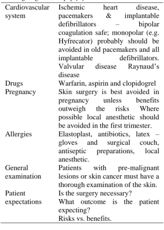
Table 1 Systemic evaluation required in a patient undergoing skin biopsy [1].
Cardiovascular system
Ischemic heart disease, pacemakers & implantable defibrillators – bipolar coagulation safe; monopolar (e.g. Hyfrecator) probably should be avoided in old pacemakers and all implantable defibrillators. Valvular disease Raynaud’s disease
Drugs Warfarin, aspirin and clopidogrel Pregnancy Skin surgery is best avoided in pregnancy unless benefits outweigh the risks Where possible local anesthetic should be avoided in the first trimester. Allergies Elastoplast, antibiotics, latex –
gloves and surgical couch, antiseptic preparations, local anesthetic.
General examination
Patients with pre-malignant lesions or skin cancer must have a thorough examination of the skin. Patient
expectations
Is the surgery necessary?
What outcome is the patient expecting?
Risks vs. benefits.
erythemas, lupus erythematosus and
porokeratosis is preferably taken from the active edge. Infiltrative lesions are to be sampled at their deepest part and vesicles are to be taken intact on a cushion of subcutaneous tissue.2
Another crucial factor is anatomical location of the lesion in skin or subcutaneous tissue. For example lesions which are located in epidermis and papillary dermis e.g. melanocytic nevi, warts, seborrheic keratosis, Paget’s disease, superficial basal cell carcinoma, mycosis fungoides, actinic keratosis, psoriasis, lichen planus, lupus erythematosus etc., biopsy has to be taken at the active edge taking piece of epidermis till reticular dermis at least. However, a lump, which is felt more than seen, is sampled up till subcutaneous tissue.
If the lesion involves deep reticular dermis and subcutaneous tissue, biopsy has to be deeper e.g.
blue nevus, dermatofibroma, melanoma,
panniculitis, sarcoidosis, rheumatoid nodule, polyarteritis nodosa.3
Pigmented lesions must be evaluated by clinical examination followed by dermoscopy and then biopsied. For multiple pigmentary lesions, most suspicious lesion is preferred. Common biopsy techniques used are shave, incisional and excisional biopsies. Current guidelines by National Institute of Health are in favour of taking an excisional biopsy for a suspected lesion of melanoma. This method not only provides proper diagnosis but also aids in staging, future treatments and prognosis.4
The American Academy of Dermatology has recently issued a position statement for the management of melanoma. It says that a narrow excisional biopsy with 1mm to 3mm margins is required to clear the subclinical component of most atypical melanocytic lesions. It is, therefore, preferable in almost all clinical scenarios.5
Regarding pigmented lesion on the lip, shave biopsy is acceptable for a lesion that has not crossed the vermilion border. If the pigmented lesion has crossed vermilion border, a punch biopsy is preferred aligned vertically and the margins must be painted with gentian violet to avoid misalignment while closing the defect.6
Table 2 Type, optimum timing and site of biopsy for a particular dermatological disorder [2,6,14].
Disease Time Site selection Type of biopsy
Alopecia areata Active border Two 4mm punches for
vertical and horizontal sections
Scarring alopecia Active Erythematous follicular plug, inflamed follicle
Punch
Atopic/ contact dermatitis Acute/ chronic Erythematous vesicle/ lichenoid area
Punch
Exanthema Active Lesion Punch
Erythema annulare centrifugum Well-developed lesion Active border punch
Erythema nodosum Active lesion, 1st week Center of lesion Deep incision till sub cutis, send culture
Erythema multiforme Targetoid lesion One from dusky center, other from erythematous border
Punch
Granuloma annulare Active Raised border Punch
Larva migrans Migrating erythema Normal skin 2mm above the erythematous line
Punch
Lichen planus Anytime Violaceous papule Punch
Discoid lupus erythematosus Active lesion Scaly plaque with plugged follicles
punch
Lupus tumidus Active lesion anywhere punch
Sub acute cutaneous LE Active lesion Anywhere punch
Systemic lupus erythematosus Active Anywhere punch
LE profundus Depressed center and adjacent area
center Deep incision or excision
Mycosis fungoides Patch- non-treated Plaque-non-ulcerated Nodule-non-ulcerated
Center
Most infiltrated area Indurated area
Two or more punches One or more punches Punch
Morphea Early
late
Lilac ring center
Punch
Necrobiosis lipoidica Anytime Atrophic center Punch
Nephrogenic fibrosing dermopathy Sclerotic plaque Indurated area of plaque punch Pityriasis lichenoides chronica 2-3 weeks old Papulosquamous area punch Pityriasis lichenoides et
varioliformis acuta
2-3 week old Necrotic papule Punch
Guttate psoriasis Late stage Lesional 4mm punch
Plaque psoriasis Scaly anywhere punch
Pyoderma gangrenosum and ulcerative colitis
Small early, non-ulcerated lesion
Entire lesion Excision with tissue culture
Scleromyxedema Sclerotic skin Beaded papules Punch
Urticaria and urticarial vasculitis Active 3 days lesional punch
Table 3 Types and respective indications of skin biopsy [7,8]. Type of biopsy Indications
Punch biopsy Most superficial inflammatory diseases (erythema multiforme major), papulosquamous disorders (psoriasis), connective-tissue disorders (systemic lupus erythematosus), superficial blistering disorders (pemphigus vulgaris), benign tumors, granulomatous disorders
(sarcoidosis), non-melanotic malignant tumors (infiltrating squamous cell carcinoma) Shave biopsy Raised lesions (skin tags), lesions that separate out easily from underlying tissues (seborrheic
keratosis), dome shaped nevi and benign tumors, non-melanotic malignant tumors (superficial basal cell carcinoma)
Excisional biopsy Subcutaneous or deep dermal tumors, deep inflammatory diseases (erythema nodosum), malignant melanoma, atypical pigmented lesions
Incisional biopsy Inflammatory dermatosis (porokeratosis)
Fine needle aspiration Lymph node, thyroid gland, benign or malignant salivary gland tumors, dermoid cyst, cystic lesions in neck
Saucerization biopsy Vesicobullous disorders, seborrheic keratosis
Wedge biopsy Subcutaneous mycosis, margin of squamous cell carcinoma, tuberculosis verrucosa cutis etc.
Supplies and instruments needed for skin biopsy
Following are minimum necessary instruments including other supplies that we need to have in our outpatient room or theatre to suffice a skin biopsy.
Isopropyl alcohol, povidone-iodine or chlorhexidine for skin preparation
Sterile gauzes
Cotton/ plastic drapes- preferably fenestrated
Disposable syringes 1 and 3 ml
Needles 30G for injections and 22G for drawing solution
Lignocaine 1% or 2% with/without adrenaline
Disposable surgical blade # 15
Double edge razor blade cut in halves
Disposable punches 4, 6 and 8mm
Toothed and non-toothed small tissue forceps
Small tissue scissors
Needle holder
20% aluminum chloride
Packed alcohol swabs
Antibiotic ointment
Band-Aid (spot/ rectangular)
Non-adherent dressings
10% formalin specimen bottle
Surgical marking pen
Pathology request forms
Patients instructions
Preparation of biopsy site and use of anesthesia, some practical considerations
Usually 1 or 2 % lignocaine, isopropyl alcohol, povidone-iodine or chlorhexidine gluconate are used to prepare the biopsy site. Marking with surgical marker or gentian violet can be extremely useful in incisional and excisional biopsies.
Regarding local anesthesia, usually lidocaine with adrenaline is used, however, adrenaline is best avoided and plain lidocaine is to be used in case of fingers, toes and penis; patients suffering from Raynaud’s phenomenon and within six
months of major cardiac events. Major contraindications to use of lidocaine are pregnancy and hypersensitivity. Up to 20ml of 1% and 10ml of 2% plain lignocaine are maximum doses while 50ml of 1% lignocaine with 1: 200000 adrenaline and 25ml of 2% lidocaine with 1:200000 adrenaline are safe.1,9 Make sure to inject in subcutis rather than in dermis because it may cause artefacts in histology specimen.
Infiltrative anesthesia is to be avoided in case of mast cell disorders because of risk of degranulation. Regarding scalp biopsy it is important to consider trichoglyphics and punch should be directed parallel to direction of emerging hair.10
Techniques of important types of skin biopsy procedures in practice
Incisional biopsy It is attempted when we need
to evaluate certain inflammatory dermatoses and the chances of malignancy are less likely. Usually a part of normal skin is removed along with. If the lesion is small enough to be removed in toto, we call it excisional biopsy.
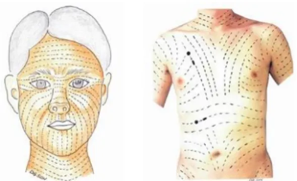
For incisional biopsy the direction of the ellipse of skin, which includes pathological, as well as, normal skin has to be parallel to skin tension or Langer lines (Figure 1) in order to have better healing and scar.11 It holds true for all types of biopsies on face as well.
Figure 2 Marking an ellipse of skin to be removed parallel to skin tension lines
After checking for adequate anesthesia, determine skin tension lines and with the help of surgical pen draw an ellipse of skin to be removed parallel to these lines keeping 30 degree angles at each apex, 2-5mm margin of surrounding skin and length 3 times the width (Figure 2).9,12
Holding the blade perpendicular to skin surface, start incising using tip and gradually coming to belly of blade towards middle and going again to tip of blade for other edge. Carefully lifting the edge with forceps, completely undermine the
tissue at level of subcutaneous fat. Apply pressure in advance of closure.12
Shave biopsy12,13 It is a simple, quick and
sutureless technique. There are two kinds of shave biopsy; superficial and deep also called saucerization biopsy. A double-edge razor blade held in a manner with concavity facing up is used on an elevated lesion in sweeping strokes rather than sawing motion. Near the end of biopsy stabilize the lesion with index finger to avoid unnecessary tears. The blade should be parallel to the skin surface to avoid uneven depths. Superficial shave biopsy removes epidermis and only superficial part of dermis and can also be performed with the help of surgical blade.
Deep or saucerization biopsy removes epidermis and papillary, as well as, upper part of reticular dermis.
Intraoperative complications; their prevention
and management [10]
Table 4 enlists the common complications
encountered during skin biopsy. Other
uncommon or rare complications could be failure to close the wound properly, wound
Table 4 Intraoperative Complications; their prevention & management10
Complications Prevention management
Hypersensitivity Prior intradermal/patch test Manage as anaphylaxis with hydrocortisone, adrenaline, pheniramine etc.
Pain Alkalinize the anesthetic, small-guage needle, slow injection, avoid over inflation, divert patients attention Bleeding Avoid vascular areas, adrenaline where
possible, chalazion clamp
Pressure application, swab soaked in hydrogen peroxide/ aluminum chloride 20-40%, suture, cauterization
Scaring Avoid patients with tendency of scarring, prevent infection
Intralesional injections, surgical excision of scar and suturing
infection Preparation of biopsy site, use sub cutis deep incisions, topical and systemic antibiotics
dehiscence, overgranulation, incomplete excision rate and distortion of local anatomy.1 Proper techniques and protocols of each type of skin biopsy however, are prudent in addition to what has been mentioned above and being beyond the scope of this article, shall be emphasized separately.
Conclusion
Skin biopsy if performed with complete awareness of all the pre- and post-procedure protocols by trained dermatosurgeon is an unmatched, definitive diagnostic and curative
step towards management of long-term
confusing dermatoses. One should not hesitate taking biopsies where definitely required and avoid unnecessary procedures at the same time. Teaching the postgraduate students about every aspect is prudent to continue safe surgical practices
References
1. Cunliffe TP, Chou C. Primary Care Dermatology Society - Skin Surgery Guidelines. 2007. Available from: https://www.scribd.com/document/3200458 80/Skin-Surgery-Guidelines
2. Montani MA, Giors GV. Site selection. Fernando S, Ed. Skin Biopsy. InTech, 2013. DOI: 10.5772/55638. Available from: https://www.intechopen.com/books /skin- biopsy-diagnosis-and-treatment/site-selection
3. Llamas-Velasco M, Paredes E. Basic Concepts in skin biopsy. Part I. Actas Dermosifiliográficas. 2012;103:12-20. 4. Tadiparthi S, Panchani S, Iqbal A. Biopsy
for malignant melanoma – are we following
the guidelines? Ann R Coll Surg Engl. 2008;94:322-5.
5. Bichakjian C, Halpern A, Johnson TM, Foote Hood A, Grichnik JM, Swetter SM et al. Guidelines of care for the management of primary cutaneous melanoma. J Am Acad Dermatol. 2011;65:1032-47.
6. Sina B, Kao G, Deng AC, Gaspari AA. Skin biopsy for inflammatory and common neoplastic skin diseases: optimum time, best location and preferred techniques. A critical review. J Cutan Pathol. 2009;36:505-10. 7. Zuber TJ. Skin biopsy techniques: when and
how to perform punch biopsy. Consultant. 1994;34:1467-70.
8. Skin biopsy. [cited online 2017 march 25]
available from: URL:
https://en.wikipedia.org/wiki/Skin_biopsy 9. Patrick CA, Mathes BM. Skin biopsy
techniques for the internist. J Gen Intern Med. 1998;13:46-54.
10. Nischal U, Nischal KC, Khopkar U. Techniques of skin biopsy and practical considerations. J Cutan Aesthet Surg. 2008;1:107-11.
11. Son D, Harijan A. Overview of surgical scar prevention and management. J Korean Med. 2014;29:751-7.
12. Manocha D, Bansal N, Farah RS. types and selection criteria for various skin biopsy procedures. In: Khopkar U, editor. Skin Biopsy – Perspectives. InTech: 2011,
Available from:
http://www.intechopen.com/books/skin- biopsy-perspectives/types-and-selection-criteria-forvarious-skin-biopsy-procedures 13. Pickett H, O'Callaghan M. Shave and punch
biopsy for skin lesions. Am Fam Physician. 2011;84:995-1002.
![Table 2 Type, optimum timing and site of biopsy for a particular dermatological disorder [2,6,14]](https://thumb-us.123doks.com/thumbv2/123dok_us/7873006.2098970/3.918.107.832.123.770/table-type-optimum-timing-biopsy-particular-dermatological-disorder.webp)