Open Access
Research
Possible involvement of integrin-mediated signalling in oocyte
activation: evidence that a cyclic RGD-containing peptide can
stimulate protein kinase C and cortical granule exocytosis in mouse
oocytes
Carla Tatone* and Maria Cristina Carbone
Address: Department of Biomedical Sciences and Technologies, University of L'Aquila, Via Vetoio, 67100 L'Aquila, Italy Email: Carla Tatone* - [email protected]; Maria Cristina Carbone - [email protected]
* Corresponding author
Abstract
Background: Mammalian sperm-oocyte interaction at fertilization involves several combined interactions between integrins on the oocyte and integrin ligands (disintegrins) on the sperm. Recent research has indicated the ability of peptides containing the RGD sequence that characterized several sperm disintegrins, to induce intracellular Ca2+ transients and to initiate parthenogenetic development in amphibian and bovine oocytes. In the present study, we investigate the hypothesis that an integrin-associated signalling may participate in oocyte activation signalling by determining the ability of a cyclic RGD-containing peptide to stimulate the activation of protein kinase C (PKC) and the exocytosis of cortical granules in mouse oocytes.
Methods: An In-Vitro-Fertilization assay (IVF) was carried in order to test the condition under which a peptide containing the RGD sequence, cyclo(Arg-Gly-Asp-D-Phe-Val), was able to inhibit sperm fusion with zona-free mouse oocytes at metaphase II stage. PKC activity was determined by means of an assay based on the ability of cell lysates to phosphorylate MARKS peptide, a specific PKC substrate. Loss of cortical granules was evaluated by measuring density in the oocyte cortex of cortical granules stained with LCA-biotin/Texas red-streptavidin. In all the experiments, effects of a control peptide containing a non RGD sequence, cyclo(Arg-Ala-Asp-D-Phe-Val), were evaluated.
Results: The IVF assay revealed that the fusion rate declined significantly when insemination was carried out in the presence of cyclic RGD peptide at concentrations > or = 250 microM (P < 0.05, Student-Newman-Keuls Method). When the peptide was applied to the oocytes at these concentrations, a dose-dependent increase of PKC activity was observed, in association with a loss of cortical granules ranging from 38+/-2.5 % to 52+/-5.4 %. Evaluation of meiotic status revealed that cyclic RGD peptide was ineffective in inducing meiosis resumption under conditions used in the present study.
Conclusion: The presents results provide evidence that a cyclic RGD peptide highly effective in inhibiting sperm-oocyte interaction stimulates in mouse oocytes the activation of PKC and the exocytosis of cortical granules. These data support the view that RGD-binding receptors may function as signalling receptors giving rise integrated signalling not sufficient for a full oocyte activation response. This study may contribute to the understanding of possible negative effects of skipping gamete interaction in IVF techniques.
Published: 25 September 2006
Reproductive Biology and Endocrinology 2006, 4:48 doi:10.1186/1477-7827-4-48
Received: 27 July 2006 Accepted: 25 September 2006
This article is available from: http://www.rbej.com/content/4/1/48
© 2006 Tatone and Carbone; licensee BioMed Central Ltd.
Background
At fertilisation the oocyte undergoes a series of rapid changes responsible for the onset of the embryonic devel-opment and the blockage of polyspermy. These changes, collectively known as "oocyte activation", are under the regulation of cytoplasmic signalling events activated in the oocyte following a multi-step interaction with the fer-tilising sperm [1-3]. It is well established that upon fusion sperm releases into the oocyte a sperm-specific phosphol-ipase C-zeta (PLCζ) which induces a rise in intracellular Ca2+ capable of releasing metaphase arrest and driving the
zygote through the embryonic cell cycle [4]. Although the Ca2+-mediated signal transduction pathway at
fertiliza-tion is not fully resolved, it seems to involve specific kinases such as protein kinase C (PKC) known to be acti-vated in many cell types through enzyme- or G-protein-coupled receptors localised on plasma membrane [5,6]. However, due to the effectiveness of intracytoplasmic sperm injection in most mammalian species [7], the hypothesis that receptor-mediated pathways may partici-pate in the oocyte activation process has been poorly investigated.
It is well established that binding of sperm ligands to spe-cific oolemma receptors is a prerequisite step in sperm-oocyte interaction leading to fertilisation [8,9]. Candidate molecules involved in gamete interactions include integrins, transmembrane glycoproteins with het-erodimeric structure (alpha-chain and beta-chain) that act as co-receptors in many cell-cell interaction [10]. Individ-ual integrins can bind to more than one ligand and about half of them recognize the tripeptide sequence Arg-Gly-Asp (RGD) present in the extracellular matrix proteins such as fibronectin and vitronectin [11]. Integrins expressed on the surface of mouse oocytes can be divided into two groups: β 1 integrins (α2 β1, α3β1, α5 β1, α6 β1 and α9β1) and αv integrins (αv β1, αvβ3, αv β5; [12,13]). Integrin recognition sequences known to play a role in fer-tilization are the RGD sequence and other tripeptide sequences such as TDE, QDE and FEE included in the active site of fertilin beta, a component of the first mole-cule identified as a sperm surface protein required for sperm-oocyte fusion [14-18]. Recently it has been sug-gested the hypothesis that sperm-oocyte binding and fusion involve combined interactions between RGD-sen-sitive integrins such as αv β1 and RGD-insensitive integrins such as α6 β1 integrins on the oocyte [19].
In order to clarify the role of integrins at fertilization, it is important to consider that these molecules can serve not only as structural receptors that participate in cell-cell and cell-matrix interaction, but also as signalling receptors that regulate intracellular pH [20], intracellular free Ca2+
[21], inositol lipid turnover [22] and protein phosphor-ylation [23]. Recent research has indicated the ability of
peptides containing a RGD sequence to induce intracellu-lar Ca2+ transients and to initiate parthenogenetic
devel-opment in amphibian and bovine oocytes [24-26], indicating that RGD-binding receptors may function as signalling receptors in oocytes as it occurs in other cell types. Multiple intracellular signalling molecules are stim-ulated following integrin-dependent adhesion. These include members of mitogen-activated protein kinase (MAPK) signalling pathways, Rho family GTPases, non-receptor tyrosine kinases such as focal adhesion kinase (FAK) and Src, and members of the lipid signalling path-ways such as phosphatidyl-inositol 3-kinase (PI 3-K), and protein kinase C [27-29]. PKC signalling is considered a major regulator of oocyte activation acting both depend-ently and independdepend-ently from the fertilization calcium signal [30,31]. Although its role is not clearly established, it has been proposed that this kinase provides integrated signals aimed to modulate the kinetics and the extent of activation events such as Ca2+ spiking and cortical granule
exocytosis [32].
Based on the above observations, we put forward the hypothesis that integrins may participate in the activa-tion-associated signalling in mouse oocytes. To this end, in the present study we investigated the ability of a cyclic RGD peptide to activate a pathway leading to the stimula-tion of protein kinase C and cortical granule exocytosis.
Methods
Reagents
All reagents were purchased from Sigma Chemical Com-pany (St. Louis, MO) unless otherwise stated.
Peptides
The peptide containing the RGD sequence cyclo(Arg-Gly-Asp-D-Phe-Val) [33] and the control peptide containing a non RGD sequence cyclo(Arg-Ala-Asp-D-Phe-Val) [34], were purchased from Peptide International, Inc. (Louis-ville, Kentucky). As demonstrated by the manufacturer when tested by thin layer chromatography both the pep-tides showed a single spot. Lyophilised peppep-tides were resuspended in T6 medium [35] at 500 μM concentration as indicated in the datasheet, aliquotted and stored at -20°C and used within 3 weeks.
Oocyte and sperm isolation
cul-tured at 37°C, 5% CO2 in M16 medium for 1 hr before their use in the experimental groups.
Spermatozoa were obtained by excising the caudae epidi-dymides from two adult CD1 males (3–5 months old, Charles River, Como, Italy) as previously described [31].
In vitro fertilisation and sperm fusion assay
The sperm suspension was diluted to obtain 50 μl insem-ination drops containing 1–5 × 10⁴ sperm/ml covered
with mineral oil. About 25 oocytes preincubated for 30 min in T6 containing peptides at different concentrations or in T6 alone were incubated in each drop and main-tained in the incubator at 37°C in 5% CO2 for 10–30 min. Inseminated oocytes were freed from loosely associated sperm before processing for the evaluation of the desig-nated parameters. Further development of fertilised oocytes was carried out in 50 μl drops of M16 medium.
To visualise sperm-oocyte fusion after fertilisation we used the dye transfer technique as previously described [37]. In this assay, upon fusion with oocytes preloaded with the DNA-staining dye Hoechst 33342, sperm nuclei become brightly fluorescent as the dye gains access and binds sperm DNA. Oocytes were loaded with Hoechst 33342 (0.1 μg/ml) by a 15 min incubation, rinsed thor-oughly in T6 medium and immediately incubated with sperm. After 10 min of insemination, oocytes were col-lected and washed in M2 before being fixed by a 15 min incubation in 3.7% paraformaldehyde in PBS. Fixed oocytes were mounted on slides and scored for the pres-ence of fused sperm under a microscope fitted for epiflu-orescence (Leitz Dialux, Leitz, Wien, Austria).
Protein kinase C assay
PKC activity was assayed using the protocol of Gallicano et al. [30]. For each reaction, groups of 10 oocytes, col-lected at different times of insemination, were washed in collection buffer (phosphate buffered saline (PBS) con-taining 1 mg/ml polyvinyl alcohol, 5 mM EDTA, 10 mM Na3VO4, 10 mM NaF), transferred to a centrifuge tube in 2 μl of collection buffer and immediately submerged in liquid N2 to flash-freeze the oocytes, followed by storage at -80°C until the kinase assay was performed. The frozen oocytes were thawed in 10 μl PKC buffer which contained
β-glycerophosphate (54 mM), para-nitrophenylphos-phate (14.5 mM), MOPS (24 mM), MgCl2 (14.5 mM),
EGTA (14.5 mM), EDTA (0.12 mM), DTT (1 mM), leu-peptin (1μg/ml), aprotinin (1 μg/ml), ML-9 (10 μM), genestein (75 μM), chimostatin (1 μg/ml), tripsin-chimo-tripsin inhibitor (1 μg/ml), PKI (2.4 μM), 50 μCi/ml ν -[32P]ATP(Amersham Pharmacia Biotech, Italy) and
MARKS (2.5 mg/ml, BIOMOL, Plymouth Meeting, PA) as a specific substrate. After 30 min at 37°C, assays were stopped by adding tricine sample buffer (1:1) (Bio-Rad
Laboratories, Hercules, CA). Samples were electro-phoresed and the gel was subjected to autoradiography as previously described [38]. To inhibit PKC activity, BIM was added to the assay buffer at a final concentration of 10
μM (control). Enzyme activity was semiquantified densit-ometrically using a Bio-rad GS-670 computerised imaging densitometer and Molecular Analyst software (Bio-rad Laboratories, Hercules, CA). Intensity of bands were quantified after background subtractions. For each autora-diogram, 3 replicates were performed with 10 oocytes per time-point per replicate and ratios of mean band density in the experimental groups to that of BIM treated samples (control) were evaluated. PKC activity at different time points was expressed as mean ± SEM of ratios obtained from at least 3 autoradiograms.
Staining and quantification of cortical granules
Oocytes were fixed in a 3.7% (w/v) paraformaldeyde and, after permeabilization with Triton X-100, were incubated with LCA (Lens Culinaris Agglutinin)-coupled to biotin and then with Texas red-streptavidin as previously described [38]. Briefly, the oocytes were mounted on slide in 50% w/v glycerol and CGs in the cortex were visualised by a fluorescence microscope equipped with a 100x objec-tive and oil immersion. The CG density for each oocyte was computed by image analysis based on the same prin-ciples as manual counting described previously. The images on flat optical fields of cortex resulting from par-tial compression of the oocyte by the coverslip, were cap-tured by a Vario Cam monochrome CCD and then transferred to a PC with image analysis software (KS300, Kontron Elektronik Gmbh, Germany) [38]. The density of CGs per 100 μm2 for each oocyte was computed by image
analysis as the mean of the counts from tree equal areas of cortex containing cortical granules. For each group, the percentage loss of CGs from the cortex was calculated by comparing the mean density of CGs of the treated group with the mean density of CGs of the untreated control group, according to the following equation: %CG loss = 1-[density of CGs in treated group/density of CGs in untreated group] × 100.
Evaluation of oocyte activation
Oocytes fixed in paraformaldehyde were stained with 3 mg/ml Hoechst 33342 for 10 min, mounted on slides and monitored under an epifluorecence microscope for the presence of chromosomes at anaphase and telophase or pronuclei. As a positive control for oocyte activation, oocytes were activated with 7% ethanol [37].
Statistical analysis
Jandel Scientific Software Corporation, San Rafael, CA). Proportions were compared using z-test. Differences asso-ciated with a P value lower than 0.05 were considered sta-tistically significant.
Results
Effect of a cyclic RGD containing peptide on sperm-oocyte interaction
As in the mouse RGD containing peptides are known to interfere with sperm-oocyte interaction [16,17], we tested the conditions under which a cyclic RGD peptide interacts effectively with the oocyte by monitoring its ability to inhibit fertilization. To this end we performed an IVF assay where successful gamete interaction was assessed by monitoring sperm fusion. As shown in Figure 1, the IVF assay revealed that fusion rate declined significantly when insemination was carried out in the presence of cyclic RGD peptide at 250 μM (P < 0.05). A further increase to 500 μM was responsible for a further reduction of fusion rate to about 20%, a value 3-fold lower than that observed in the presence of the same concentration of the control peptide. Since the control cyclic peptide failed to affect fer-tilisation at any concentration tested (Figure 1), present results were taken an indirect evidence of the peptide interaction with integrin receptors on mouse oocytes.
PKC activity in mouse oocytes exposed to a cyclic RGD containing peptide
In further experiments we applied the peptide to the oocytes at the concentration of 500 μM and then
sub-jected them to a PKC assay. Results from these assays showed that the peptide exposure resulted in a significant stimulation of the enzyme. This effect was seen as early as 5 min post-treatment, and after 60 min the PKC activity reached a level about three-fold higher than that observed in untreated oocytes (time 0; Figure 2A). As shown in Fig-ure 2B, a significant increase in PKC activity could be observed when RGD concentration was lowered to 250
μM although the level of activity was reduced as compared with that observed at 500 μM. When oocytes were exposed to the non RGD-peptide PKC activity did not sig-nificantly increase as compared with that monitored in untreated MII oocytes (Figure 2C). As shown in the same figure, no change was seen when the treatment with the cyclic RGD peptide was carried out in a Mg2+ and Ca2+-free
medium, a condition that prevent ligand-integrin interac-tion [17,39].
Cortical granule exocytosis and meiotic status in mouse oocytes exposed to a cyclic RGD containing peptide
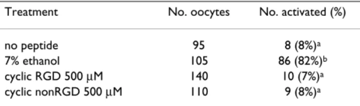
To establish whether the cyclic RGD peptide could induce cortical granule exocytosis, mouse oocytes were incubated in the presence of different concentrations of cyclic RGD-and nonRGD peptides for 1 hr RGD-and processed for the eval-uation of loss of cortical granules 2 hr later. As shown Fig-ure 3, following the exposFig-ure to the cyclic RGD peptide, the oocytes underwent CG exocytosis in a dose-dependent manner. The loss of cortical granules ranged from 38 ± 2.5 % at 250 μM to 52 ± 5.4 % at 500 μM. Both these value were significantly higher than that monitored in oocytes exposed to the nonRGD peptide. As shown in the repre-sentative micrographs in Figure 3b and 3d, evaluation of meiotic status revealed that cyclic RGD peptide was inef-fective in inducing meiosis resumption under conditions used in the present study. To confirm this, in further experiments we monitored the presence of oocytes at ana-phase, telophase or pronuclei stage (activated oocytes) following 8 hr from the exposure to 500 μM cyclic RGD-or nonRGD peptides and to 7% ethanol, as a positive con-trol. As shown in Table 1, the percentage of oocytes treated with the cyclic RGD peptide and showing meiosis resumption was statistically no different from than those exposed to the nonRGD peptide or untreated.
Discussion
In this study, we provide evidence that exposure of mouse oocytes to a cyclic RGD peptide can inhibit fertilization and induce an activation-like response which includes the activation of PKC signalling and exocytosis of cortical granules. To our knowledge, this is the first report identi-fying a potential role of integrins and their ligands in the signalling events underlying mouse oocyte activation.
It is well known that in the mouse RGD-containing pep-tides do not have a substantial inhibitory effect on sperm-Effects of a cyclic RGD peptide on sperm-oocyte fusion
Figure 1
Effects of a cyclic RGD peptide on sperm-oocyte fusion. ZP-free mouse oocyte were incubated in the pres-ence of different concentrations of cyclic RGD or nonRGD peptides for 30 min prior to fertilization. Oocytes were then inseminated with 1–5 × 10⁴ sperm/ml in the presence of
oocyte interaction as it occurs in other species, but cause a partial inhibition of fertilization, an observation taken as an evidence that sperm-oocyte fusion would utilize multi-ple molecules and/or multimulti-ple sites on molecules [16,17]. Although in our IVF assay a complete inhibition of sperm fusion was not achieved, the cyclic RGD peptide we employed reveals a high biological activity being about 50% inhibition observed at 250 μM, a concentration ten-fold lower than that required with linear RGD pep-tides[17]. Nevertheless, we are not able to establish whether higher concentrations of the peptide would have been more effective since in this study the lyophilized compound was dissolved at the maximal concentration allowed according to manufacturer's instructions. How-Cortical granule exocytosis in oocytes exposed to a cyclic RGD peptide
Figure 3
Cortical granule exocytosis in oocytes exposed to a cyclic RGD peptide. ZP-free mouse oocytes were incu-bated in the presence of different concentrations of cyclic RGD or nonRGD peptides for 60 min and stained with LCA-biotin/Texas red-streptavidin 2 hr later. A) Representative photomicrographs of cortical granules localized in the cortex and corresponding chromosomes of a MII oocyte (a, c) and an oocyte exposed to the RGD peptide (b, d). Final magnifi-cation, ×1250. B) Histogram showing CG loss in oocytes exposed to cyclic RGD or nonRGD peptides. The results here represent the average of 3 experiments per peptide concentration ± S.E.M. and a total of 60–80 inseminated oocytes per experimental point. Bar = 10 μm. Means with different letters are statistically different (P < 0.05).
Effects of a cyclic RGD peptide on PKC activity Figure 2
Effects of a cyclic RGD peptide on PKC activity. PKC activity in ZP-free mouse oocytes incubated in the presence of cyclic RGD or nonRGD peptides. A, oocytes incubated in the presence of 500 μM RGD for 5 min or 60 min and proc-essed for PKC assay. B, oocytes incubated in the presence of different concentrations of cyclic RGD peptide for 60 min. C, oocytes exposed for 60 min to 500 μM RGD peptide in the presence or in the absence of Ca2+ and Mg2+, and to 500 μM
ever, given that the RGD peptide inhibited sperm interac-tion whereas the nonRGD did not, we have taken these results as an indirect evidence of peptide binding to integrin receptors.
In studies of integrin functions in gametes and somatic cells, synthetic peptides containing the RGD (Arg-Gly-Asp) motif have been extensively used as the inhibitors of integrin-ligand interactions. Although the inhibitory activity of disintegrins depends mainly from their primary structure, structural and functional studies suggest that the receptor binding ability of these proteins lies in subtle positional requirements of the tripeptide RGD that is har-boured in a defined hairpin loop (the disintegrin loop) projecting from the disintegrin core. This has led to the study of small, chemically synthesised, cyclic-RGD pep-tides, which exert more potency than linear RGD in integrin binding assay [40]. Thus it is likely that the cyclic RGD peptide used in the present study mimics the physi-ological action of RGD-containing proteins, supporting the view that, along with proteins with other tripeptide sequence such as fertilin [14], RGD-containing polypep-tides located on sperm membrane, such as vitronectin [41,42], play an important role in gamete interactions leading to fertilization.
The analysis of PKC activity in oocytes exposed to the cyclic RGD peptide at concentrations effective in inhibit-ing sperm fusion revealed a significant increase in the activity of this enzyme. This finding supports the hypoth-esis that under this condition an oocyte-integrin signal-ling cascade is activated to switch on an oocyte phospholipase C leading to increased production of inosi-tol 1,4,5-triphosphate (IP3) and diacylglycerol (DAG) [43]. DAG activates PKC and IP3 triggers Ca2+ release from
intracellular stores. The observation that oocytes exposed to the RGD peptide undergo a significant loss of cortical granules further suggests that under, our experimental conditions, a RGD-sensitive receptor on the oolemma has activated signalling pathways similar to those triggered by sperm at fertilization. Moreover, the occurrence of cortical granule exocytosis in association with an increased PKC activity supports the role for this kinase in the regulation
of this event. On the other hand, being the loss of cortical granules a Ca2+-dependent event [44], present results
might be an indirect evidence that oocytes exposed to the RGD peptide had undergone an increase of intracellular Ca2+ as suggested by a previous study [45]. There is still the
possibility that PKC activity in response to the RGD pep-tide represents that observed at fertilization in the absence of a Ca2+ signal and probably supported by Ca2+
-inde-pendent PKC isotypes [31]. In this respect, previous results based on the use of PKC agonists and antagonists, suggested that exocytosis can be triggered independently either by Ca2+ rise and PKC [46,47]. Oocyte exposure to a
RGD peptide seems to be responsible for a reorganization of actin network similar to that induced by sperm [48]. Moreover, as discussed by Tsaadon et al. [49], PKC may regulate the cytoskeletal dynamic underlying exocytosis enabling the process of vesicle fusion with plasma mem-brane.
A further observation associated with present results is that RGD-associated signalling leads to PKC activation and cortical granule exocytosis but is not able to stimulate meiosis resumption. This supports the hypothesis that, in contrast to PKC activation achieved by pharmacological agonists, the activation of a PKC signalling through a receptor-mediated mechanism, is not the sufficient trigger for the activation of the anaphase-promoting complex/ cyclosome (APC/C) pathway leading to meiosis resump-tion [32,50]. In contrast to our results in the mouse, in bovine oocytes a release of meiotic arrest is observed after exposure to RGD peptides [25], it is likely that RGD-sen-sitive receptors might be capable of activating additional pathways.
Conclusion
Although further investigation will be needed, our results suggest that, at fertilization, a sperm membrane protein containing a RGD sequence may interact with a RGD-sen-sitive receptor on the oolemma activating a cascade of sig-nalling pathways involved in oocyte activation. Given that in the mouse and bovine sperm injection can induce an abnormal Ca2+ response with developmental
conse-quences [51,52], it can be speculated that a possible role of integrin-mediated pathways may be that to cooperate with those activated in the cytosol by other sperm mole-cules [4] in order to correctly orchestrate oocyte activation events. Although studies on animal models must be inter-preted with caution, this hypothesis raises the need to bet-ter investigate the consequences of skipping gamete interaction at surface level in a number of assisted repro-ductive technologies.
Competing interests
The author(s) declare that they have no competing inter-ests.
Table 1: Analysis of mouse oocyte activation at 8 hr after the exposure to a cyclic RGD peptide
Treatment No. oocytes No. activated (%)
no peptide 95 8 (8%)a
7% ethanol 105 86 (82%)b
cyclic RGD 500 μM 140 10 (7%)a
cyclic nonRGD 500 μM 110 9 (8%)a
Authors' contributions
CT conceived of the study and experimental design, con-tributed to the acquisition of data and wrote the manu-script.
MCC have made substantial contribution to experimental design, acquisition of data and manuscript drafting.
Acknowledgements
This work was supported by a PRIN grant from Ministero dell'Università e della Ricerca. The authors wish to thank Rosella Colonna for supervision of the research group and Francesco Fusi for his useful advice.
References
1. Jones KT: Mammalian egg activation: from Ca2+spiking to cell cycle progression. Reproduction 2005, 130:813-823.
2. Wassarman PM: Contribution of mouse oocyte zona pellucida glycoproteins to gamete recognition during fertilization. J Cell Physiol 2005, 204:388-391.
3. Gardner AJ, Evans JP: Mammalian membrane block to polyspermy: new insights into how mammalian eggs prevent fertilisation by multiple sperm. Reprod Fertil Dev 2006, 18:53-61. 4. Saunders CM, Larman MG, Parrington J, Cox LJ, Royse J, Blayney LM, Swann K, Lai FA: PLC zeta: a sperm-specific trigger of Ca(2+) oscillations in eggs and embryo development. Development
2002, 129:3533-3544.
5. Williams CJ: Signalling mechanisms of mammalian oocyte activation. Hum Reprod Update 2002, 8:313-321.
6. Talmor-Cohen A, Tomashov-Matar R, Eliyahu E, Shapiro R, Shalgi R:
Are Src family kinases involved in cell cycle resumption in rat eggs? Reproduction 2004, 127:455-463.
7. Yanagimachi R: Fertilization and developmental initiation of oocytes by injection of spermatozoa and pre-spermatozoal cells. Ital J Anat Embryol 2005, 110(Suppl 1):145-150.
8. Evans JP: The molecular basis of sperm-oocyte membrane interactions during mammalian fertilization. Hum Reprod Update 2002, 8:297-311.
9. Kaji K, Kudo A: The mechanism of sperm-oocyte fusion in mammals. Reproduction 2004, 127:423-429.
10. Hynes RO: Integrins: versatility, modulation, and signaling in cell adhesion. Cell 1992, 69:11-25.
11. Ruoslahti E, Pierschbacher MD: Arg-Gly-Asp: a versatile cell rec-ognition signal. Cell 1986, 44:517-518.
12. Evans JP, Schultz RM, Kopf GS: Identification and localization of integrin subunits in oocytes and eggs of the mouse. Mol Reprod Dev 1995, 40:211-220.
13. He ZY, Brakebusch C, Fassler R, Kreidberg JA, Primakoff P, Myles DG: None of the integrins known to be present on the mouse egg or to be ADAM receptors are essential for sperm-egg binding and fusion. Dev Biol 2003, 254:226-237.
14. Primakoff P, Hyatt H, Tredick-Kline J: Identification and purifica-tion of a sperm surface protein with a potential role in sperm-egg membrane fusion. J Cell Biol 1987, 104:141-149. 15. Bronson RA, Fusi F: Evidence that an Arg-Gly-Asp adhesion
sequence plays a role in mammalian fertilization. Biol Reprod
1990, 43:1019-25.
16. Almeida EA, Huovila AP, Sutherland AE, Stephens LE, Calarco PG, Shaw LM, Mercurio AM, Sonnenberg A, Primakoff P, Myles DG, White JM: Mouse egg integrin alpha 6 beta 1 functions as a sperm receptor. Cell 1995, 81:1095-1104.
17. Evans JP, Schultz RM, Kopf GS: Mouse sperm-egg plasma mem-brane interactions: analysis of roles of egg integrins and the mouse sperm homologue of PH-30 (fertilin) beta. J Cell Sci
1995, 108:3267-3278.
18. Bigler D, Takahashi Y, Chen MS, Almeida EA, Osbourne L, White JM:
Sequence-specific interaction between the disintegrin domain of mouse ADAM 2 (fertilin beta) and murine eggs. Role of the alpha(6) integrin subunit. J Biol Chem 2000,
275:11576-11584.
19. Ziyyat A, Naud-Barriant N, Barraud-Lange V, Chevalier F, Kulski O, Lemkecher T, Bomsel M, Wolf JP: Cyclic FEE peptide increases
human gamete fusion and potentiates its RGD-induced inhi-bition. Hum Reprod 2005, 20:3452-3458.
20. Schwartz MA, Ingber DE, Lawrence M, Springer TA, Lechene C: Mul-tiple integrins share the ability to induce elevation of intrac-ellular pH. Exp Cell Res 1991, 195:533-535.
21. Schwartz MA, Denninghoff K: Alpha v integrins mediate the rise in intracellular calcium in endothelial cells on fibronectin even though they play a minor role in adhesion. J Biol Chem
1994, 269:11133-11137.
22. McNamee HP, Ingber DE, Schwartz MA: Adhesion to fibronectin stimulates inositol lipid synthesis and enhances PDGF-induced inositol lipid breakdown. J Cell Biol 1993, 121:673-678. 23. Kornberg L, Earp HS, Parsons JT, Schaller M, Juliano RL: Cell
adhe-sion or integrin clustering increases phosphorylation of a focal adhesion-associated tyrosine kinase. J Biol Chem 1992,
267:23439-23442.
24. Iwao Y, Fujimura T: Activation of Xenopus eggs by RGD-con-taining peptides accompanied by intracellular Ca2+ release. Dev Biol 1996, 177:558-567.
25. Campbell KD, Reed WA, White KL: Ability of integrins to medi-ate fertilization, intracellular calcium release, and partheno-genetic development in bovine oocytes. Biol Reprod 2000,
62:1702-1709.
26. Sessions BR, Aston KI, Davis AP, Pate BJ, White KL: Effects of amino acid substitutions in and around the arginine-glycine-aspartic acid (RGD) sequence on fertilization and partheno-genetic development in mature bovine oocytes. Mol Reprod Dev 2006, 73:651-657.
27. Schlaepfer DD, Hunter T: Integrin signalling and tyrosine phos-phorylation: just the FAKs? Trends Cell Biol 1998, 8:151-157. 28. Miranti CK, Leng L, Maschberger P, Brugge JS, Shattil SJ:
Identifica-tion of a novel integrin signaling pathway involving the kinase Syk and the guanine nucleotide exchange factor Vav1. Curr Biol 1998, 8:1289-1299.
29. Rucci N, DiGiacinto C, Orru L, Millimaggi D, Baron R, Teti A: A novel protein kinase C alpha-dependent signal to ERK1/2 activated by alphaVbeta3 integrin in osteoclasts and in Chi-nese hamster ovary (CHO) cells. J Cell Sci 2005, 118:3263-3275. 30. Gallicano GI, McGaughey RW, Capco DG: Activation of protein kinase C after fertilization is required for remodelling the mouse egg into the zygote. Mol Reprod Dev 1997, 46:587-601. 31. Tatone C, Delle Monache S, Francione A, Gioia L, Barboni B, Colonna
R: Ca2+-independent protein kinase C signalling in mouse eggs during the early phases of fertilization. Int J Dev Biol 2003,
47:327-333.
32. Halet G: PKC signaling at fertilization in mammalian eggs. Biochim Biophys Acta 2004, 1742:185-189.
33. Aumailley M, Gurrath M, Muller G, Calvete J, Timpl R, Kessler H:
Arg-Gly-Asp constrained within cyclic pentapeptides. Strong and selective inhibitors of cell adhesion to vitronectin and laminin fragment P1. FEBS Lett 1991, 291:50-54.
34. Brooks PC, Montgomery AM, Rosenfeld M, Reisfeld RA, Hu T, Klier G, Cheresh DA: Integrin alpha v beta 3 antagonists promote tumor regression by inducing apoptosis of angiogenic blood vessels. Cell 1994, 79:1157-1164.
35. Quinn P, Barros C, Whittingham DG: Preservation of hamster oocytes to assay the fertilizing capacity of human spermato-zoa. J Reprod Fertil 1982, 66:161-168.
36. Nicolson GL, Yanagimachi R, Yanagimachi H: Ultrastructural local-ization of lectin-binding sites on the zonae pellucidae and plasma membranes of mammalian eggs. J Cell Biol 1975,
66:263-274.
37. Tatone C, Van Eekelen CG, Colonna R: Plasma membrane block to sperm entry occurs in mouse eggs upon parthenogenetic activation. Mol Reprod Dev 1994, 38:200-308.
38. Tatone C, Delle Monache S, Iorio R, Caserta D, Di Cola M, Colonna R: Possible role for Ca(2+) calmodulin-dependent protein kinase II as an effector of the fertilization Ca(2+) signal in mouse oocyte activation. Mol Hum Reprod 2002, 8:750-757. 39. Bronson R: Is the oocyte a non-professional phagocyte? Hum
Reprod Update 1998, 4:763-775.
Publish with BioMed Central and every scientist can read your work free of charge "BioMed Central will be the most significant development for disseminating the results of biomedical researc h in our lifetime."
Sir Paul Nurse, Cancer Research UK
Your research papers will be:
available free of charge to the entire biomedical community
peer reviewed and published immediately upon acceptance
cited in PubMed and archived on PubMed Central
yours — you keep the copyright
Submit your manuscript here:
http://www.biomedcentral.com/info/publishing_adv.asp
BioMedcentral
41. Fusi FM, Lorenzetti I, Vignali M, Bronson RA: Sperm surface pro-teins following capacitation: expression of vitronectin on the equatorial segment and laminin on sperm tail. J Androl 1992,
13:488-497.
42. Fusi FM, Bernocchi N, Ferrari A, Bronson RA: Is vitronectin the velcro that binds the gametes together? Mol Hum Reprod 1996,
2:859-866.
43. Evans JP, Kopf GS: Molecular mechanisms of sperm-egg inter-actions and egg activation. Andrologia 1998, 30:297-307. 44. Kline D, Kline JT: Repetitive calcium transients and the role of
calcium in exocytosis and cell cycle activation in the mouse egg. Dev Biol 1992, 149:80-89.
45. Yue LM, Zhang L, He YP, Zhang JH, Xie YM, Zheng Y, Zhang L, Huang P, Wang L, Liu WX: Integrins mediate the increase of concen-tration of intracellular free calcium in mouse eggs. Sheng Li Xue Bao 2004, 56:347-352.
46. Eliyahu E, Shalgi R: A role for protein kinase C during rat egg activation. Biol Reprod 2002, 67:189-195.
47. Colonna R, Tatone C: Protein kinase C-dependent and inde-pendent events in mouse egg activation. Zygote 1993,
1:243-256.
48. Yue L, Zhang L, He Y, Zhang J, Zheng J, He Y, Zheng Y, Zhang J, Zhang L: Reorganization of cytoskeletal proteins of mouse oocytes mediated by integrins. Sci China C. Life Sci 2004, 47:540-544. 49. Tsaadon A, Eliyahu E, Shtraizent N, Shalgi R: When a sperm meets
an egg: Block to polyspermy. Mol Cell Endocrinol 2006,
252:107-114.
50. Madgwick S, Levasseur M, Jones KT: Calmodulin-dependent pro-tein kinase II, and not propro-tein kinase C, is sufficient for trig-gering cell-cycle resumption in mammalian eggs. J Cell Sci
2005, 118:3849-3859.
51. Kurokawa M, Fissore RA: ICSI-generated mouse zygotes exhibit altered calcium oscillations, inositol 1,4,5-trisphos-phate receptor-1 down regulation, and embryo develop-ment. Mol Hum Reprod 2003, 9:523-533.
52. Malcuit C, Maserati M, Takahashi Y, Page R, Fissore RA: Intracyto-plasmic sperm injection in the bovine induces abnormal [Ca2+]