ENGINEERED CONTROL OF PROTEIN ACTIVITY IN LIVING CELLS
Onur Dagliyan
A dissertation submitted to the faculty at the University of North Carolina at Chapel Hill in partial fulfillment of the requirements for the degree of Doctor of Philosophy in the
Department of Biochemistry and Biophysics in the School of Medicine.
Chapel Hill 2016
Approved by: John R. Riordan James E. Bear Qi Zhang
ABSTRACT
Onur Dagliyan: Engineered control of protein activity in living cells (Under the directions of Nikolay V. Dokholyan and Klaus M. Hahn)
To Irem,
ACKNOWLEDGEMENTS
First, I would like to thank my advisors, Nikolay Dokholyan and Klaus Hahn, for giving me the opportunity to work in their laboratories. I am grateful that they provided me the full independency to pursue my own scientific interest. Second, I would like to thank my committee members, Jack Riordan, James Bear, and Qi Zhang for their time and insightful comments, and third I would like to thank Feng Ding and Andrei Karginov, who had been very helpful during my rotations. I would also like to thank my collaborators Anna Huttenlocher, Christina Freisenger, Gromoslaw Smolen, Haruo Kasai, Sho Yagishita, Ilme Schlichting, and Miroslaw Tarnawski. Their help has been invaluable for my work.
I would like to thank all the members of Hahn and Dokholyan laboratories, all past and present, especially Elena Fenu, Sandra Hahn, Pei-Hsuan Chu, Daniel Marston, Scott Slattery, Ellen O'Shaughnessy, Jason Yi, Orrin Stone, Lanette Fee, David Shirvanyants, Srinivas Ramachandran, Pradeep Kota, Elizabeth Proctor, Rachel Redler, Reed Jacob, Benfeard Williams, James Fay, Mahmoud Shobair, Marino Convertino, and Andrey Krokhotin.
TABLE OF CONTENTS
LIST OF FIGURES ... ix
LIST OF ABBREVIATIONS ... xi
CHAPTER 1: GENETICALLY ENCODED TOOLS TO CONTROL PROTEIN ACTIVITY IN LIVING CELLS ... 16
1.1 Control of protein localization and interaction ... 16
1.2 Control of active site exposure ... 23
1.3 Allosteric control through domain insertion ... 25
1.4 Control of active site residues ... 28
1.5 Control of protein stability ... 30
CHAPTER 2: RATIONAL DESIGN OF A LIGAND-CONTROLLED PROTEIN CONFORMATIONAL SWITCH ... 32
2.1 Introduction ... 32
2.2 Results and Discussion ... 34
Design of uniRapR with desired stability and conformational dynamics ... 34
UniRapR enables specific and robust control over various kinases in vitro ... 37
Specific activation of Src kinase causes loss of cell-cell
contacts in zebrafish epidermis ... 42
2.3 Conclusion ... 43
2.4 Materials and Methods ... 45
CHAPTER 3: CHEMOGENETIC PAK1 SWITCHES ENABLE SPECIFIC ACTIVATION OF A PAK ISOFORM IN CELL MOTILITY ... 50
3.1 Introduction ... 50
3.2 Results and Discussion ... 52
Engineering and optimization of a dual chain PAK1 switch ... 52
Single chain PAK1 switch in triple negative breast cancer cells ... 55
PAK1 activation results in persistent dendtritic spine enlargement ... 56
Allosteric propogation in PAK1 can be maintained through alternative pathways. ... 58
3.3 Conclusion ... 61
3.4 Materials and Methods ... 63
CHAPTER 4: COMPUTATIONAL DESIGN OF SPLIT PROTEINS TO CONTROL PROTEIN ACTIVITY ... 67
4.1 Introduction ... 67
4.2 Results and Discussion ... 69
Split protein reassembly enabled by ligands or light (SPELL) ... 69
Computational prediction of split sites on GFP ... 71
Split energy predicts critical sites to split proteins ... 73
LIST OF FIGURES
Figure 1.1. Control of protein localization and protein-protein interaction. ... 17
Figure 1.2. Chemogenetic and optogenetic control of active site exposure. ... 24
Figure 1.3. Rapamycin-regulatable (RapR) kinase approach ... 26
Figure 1.4. Chemogenetic and optogenetic control of active (key) residues ... 29
Figure 1.5. Control of protein stability ... 31
Figure 2.1. Design and thermodynamics of uniRapR domain ation. ... 35
Figure 2.2. Control of Src kinase activity with uniRapR domain. ... 37
Figure 2.3. Effect of different linkers on uniRapR-Src activity. ... 39
Figure 2.4. Testing uniRapR in other kinases and effects of Src activation in HeLa cells. .. 40
Figure 2.5. A negative design of a FAK switch ... 41
Figure 2.6. Activation of Src induces cell changes in zebrafish epidermal cells. ... 42
Figure 2.7. Kinetics of uniRapR-Src with rapamycin and iRap ... 44
Figure 3.1. Engineering genetically encoded PAK1 switches ... 53
Figure 3.2. Sequence conservation of PAK1 kinase domain ... 54
Figure 3.3. Insertion loop and optimization of linkers ... 55
Figure 3.4. Enlargement of dendritic spines induced by rapamycin ... 57
Figure 3.5. Computational design to generate versatile PAK1 switches ... 59
Figure 3.6. Residues that are co-evolved with L1 and L2 loops ... 60
Figure 4.2. Computational identification of split sites for GFP. ... 72
Figure 4.3. Computational identification of split sites in 15 split proteins. ... 74
Figure 4.4. Activation of SPELL-Vav leads to rapid protrusions. ... 76
LIST OF ABBREVIATIONS
ABA Abscisic acid
ATF2 Activating transcription factor 2 ATP Adenosine triphosphate
BSA Bovine serum albumin
C Celsius
CA Constitutively active
CaMKII Ca2+/calmodulin-dependent protein kinase 2 CCD Charge-coupled device
CM Center of mass
CRY Cryptochrome
CIB Cryptochrome Interacting Basic Helix-Loop-Helix Cv Specific heat
DC Dual chain
DH Dbl homology
DMSO Dimethyl sulfoxide DTT Dithiothreitol
DMEM Dulbecco’s Modified Eagle Medium ECFP Enhanced cyan fluorescent protein EDTA Ethylenediaminetetraacetic acid EGTA Ethylene glycol tetraacetic acid
FA Focal adhesion
FBS Fetal bovine serum
FLARE Fluorescent activation reporter FKBP FK506 binding protein
FRB FKBP12 rapamycin binding protein FRET Förster resonance energy transfer GA3 Gibberellin
GAI Gibberellin insensitive
GID1 Gibberellin insensitive Dwarf1 GEF Guanine nucleotide exchange factor GI Gigantea protein
GPG Glycine-proline-glycine amino acid sequence GS Glycine-Serine amino acid sequence
HEK Human embryonic kidney
HEPES 4-(2-hydroxyethyl)-1-piperazineethanesulfonic acid HP1α Heterochromatin protein 1α
IgG Immunoglobulin G
iFKBP Insertable FKBP
IFP Infrared fluorescent protein IRAP Rapamycin analogue ITSN Intersectin
K Kelvin
KD Kinase dead
L1P Linker 1 proline mutation
LOV Light, oxygen, and voltage sensitive domain MAPK Mitogen activated protein kinase
MgCl2 Magnesium chloride
MLCK Myosin light chain kinase Na3VO4 Sodium orthovanadate
NaCl Sodium chloride NaF Sodium fluoride
NS Nanosecond
PA Photoactivatable PAK p21-activated kinase
PAS Per-ARNT-Sim core domain PCR Polymerase chain reaction Pcr Polarization critical parameters
PDB Protein data bank
PDZ Post synaptic density protein (PSD95), Drosophila disc large tumor suppressor (Dlg1), and zonula occludens-1 protein (zo-1)
PH Pleckstrin homology
pH Concentration of hydrogen ions PHR Photolyase homology domain PhyB Phytochrome B
PP2C Protein Phosphatase type 2Cs POI Protein of Interest
pRap Photoactivatable rapamycin pTyr Phosphotyrosine
PYR Pyrabactin resistance
Rap Rapamycin
RapR Rapamycin-regulated RMSD Root mean square deviation RMSF Root mean square fluctuations SEM Standard error of the mean SFK Src Family Kinase
SPELL Split protein reassembly enabled by ligands and light Src CA Constitutively active Src
Src KD Kinase-dead Src TM Melting temperature
TopBP1 Topoisomerase binding protein 1 Tris Tris(hydroxymethyl)aminomethane uniRapR Unimolecualr rapamycin-regulated VCA Verprolin, cofilin, acidic comain VP16 Virion protein 16
YM Y180A and M183A mutations in FAK ZDC Zero-drift compensation
CHAPTER 1: GENETICALLY ENCODED TOOLS TO CONTROL PROTEIN ACTIVITY IN LIVING CELLS
A cell can use the same proteins to produce essentially opposing cell behaviors, by controlling the precise localization and kinetics of protein activities (e.g. the same kinase involved in both apoptosis and proliferation (1)). The need to understand molecular dynamics in the context of living systems has recently led to the development of a remarkable suite of protein ‘switches’, engineered domains and other approaches that cause proteins to respond to small molecules or light, enabling us to control the spatiotemporal dynamics of protein-protein interactions, posttranslational modifications, conformational change, protein stability, and subcellular localization. Here I provide an overview of genetically encoded tools that have been successfully used in cells, organized around design concepts. I will discuss naturally occurring small molecule or light sensitive proteins that provide the building blocks for most of the designs.
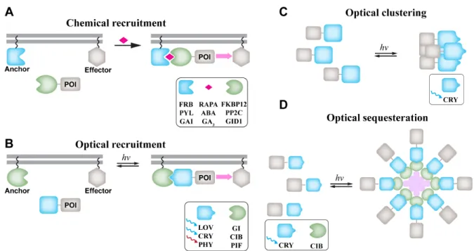
Control of protein localization and interaction
was not widely adopted because of side effects and activity in the absence of hormone. A robust and broadly applied approach harnesses the ability of the small molecule rapamycin to induce tight dimerization of two proteins, FKBP12 and FKBP12-rapamycin binding protein (FRB) (3, 4). The proteins to be controlled are either attached to FKBP12 and FRB so that rapamycin addition drives their dimerization, or one of the rapamycin-binding proteins is anchored at a particular subcellular location (e.g. the plasma membrane), so that small molecule addition drives the other protein to that location (5) (Figure 1.1A).
Protein of interest (POI) is recruited to the region of interest to interact with its effector by dimerization of green and blue proteins in the presence of a small molecule (A) or light (B). Chemical dimerizers include 12 kDa FK506 binding protein (FKBP12):rapamycin (RAPA):FRB, pyrabactin resistance (PYR)/PYR1-like (PYL)/regulatory component:abscisic acid(ABA):protein phosphatase type 2Cs (PP2C), gibberellin insensitive (GAI): gibberellin (GA3): gibberellin insensitive dwarf1 (GID1). Optical dimerizers include light, oxygen, voltage (LOV):GIGANTAE (GI), cryptochrome (CRY)-cryptochrome interacting basic helix-loop-helix (CIB), and phytochrome B (PhyB):phytochrome interacting factor (PIF) pairs. (C) POI-CRY forms clusters upon blue light exposure to amplify the signaling activity. (D) POI is retracted from its localization site into large protein assembly, resulting in inhibition of activity.
Chemical dimerizers have provided a practical means to study a diverse array of cellular phenomena. In Rho-GTPase biology, these include phagocytosis upon recruitment of Rac1 to the plasma membrane (6), Rac1-triggered local positive feedbacks in polarized cells (7, 8), and Cdc42-induced protrusions (9). Other applications include control of clathrin-mediated endocytosis and recycling (10), control of endosome morphology and selective cargo sorting (11), calcium-induced activation of a Ca2+ channel (12, 13), phosphoinositide
regulation of ion conductance (14), actin dynamics (15), mitogen-activated kinase ERK activity at focal adhesions (16), and the collective behaviors of myosinVa and kinesin motors (17). These studies highlight the robustness and broad applicability of chemical dimerizers for rapid and sustained activation of protein activity.
Improvements have included the development of different small molecule/protein systems for orthogonal control, and modified small molecules that avoid the specific side effects of each system (18). Orthogonal control has been achieved through dimerization of the proteins pyrabactin resistance 1 and protein phosphatase type 2C (PP2C) using abscisic acid (19), and with gibberellin-induced dimerization of receptor gibberellin insensitive dwarf1 (GID1) and gibberellin insensitive (GAI) (20). In one example of orthogonal control, histone modification by methylated histone-dependent silencer heterochromatin protein (HP1α) and transcriptional activator VP16 were controlled in the same cells by combining the rapamycin and abscisic acid systems (21). Remarkably, removal of the small molecules did not change the state of histone modification, revealing heritable transmission of histone posttranslational modifications.
diffusion once inside. In animals or complex multicellular systems, the kinetics of delivery to the site of action is also a factor. Spatial resolution is limited by diffusion of the small molecule as well. Therefore, investigators usually target specific cells or tissue regions, rather than subcellular regions. Subcellular localization of activity has been achieved by producing gradients of rapamycin concentration across cells (8). Spatial and temporal resolution has been improved by generating ‘caged’ derivatives of rapamycin, which are inert until irradiated (22, 23). Such molecules can build up at the site of action before they are rapidly triggered with light. Importantly, activation using rapamycin is rapid, but the dimerization they induce is essentially irreversible.
Optogenetics, the use of genetically encoded proteins to control cell behavior with light, offers important advantages over chemical dimerizers, including rapid activation with the spatial resolution of the light microscope. Optogenetics began with the use of light-gated ion channels expressed in specific neuron populations (24). In an outpouring of imaginative work over the last five years, this has been extended to non-channel proteins using a number of different light-responsive proteins (Figure 1.1B). In addition to outstanding temporal and spatial resolution, optogenetics offers reversible activation, and kinetics that can be adjusted using point mutations in the light receptors. An important limitation is the poor penetration and dispersion of light in animals, where chemical triggers can provide access.
light-induced dimerization was sufficient to generate WASP activity. In living cells, the PhyB:PIF pair has been used to control the membrane translocation of guanine nucleotide exchange factors (GEFs), which activated downstream GTPases and produced changes in cell morphology (27, 28). PhyB and PIF dimerize in response to red light (approximately 650 nm) and dissociate in far-red light (approximately 750 nm). Different responses to two wavelengths, and the very rapid kinetics of association and dissociation, enabled unusually precise control of protein activity. Different regions of the cell were bathed in different colors of light; molecules that diffused across the boundary between regions changed state in less than a second (27). Using automated feedback control, precise levels of signaling interactions could be maintained (28). By photo-activating Ras while monitoring nuclear translocation of ERK, Toettcher et al. showed that the kinetics of Ras activation affected differential interaction with downstream pathways. Phy and PIF have been used to drive proteins to different subcellular locations in yeast, including the plasma membrane, nucleus, bud neck, myosin ring, peroxisome, endosome, and spindle pole body (29). As with most photoresponsive proteins, light absorption is due to a small molecule cofactor. PhyB and PIF are plant proteins that do not exist in yeast or mammalian cells. This necessitates either the addition of cell-permeable cofactor phycocyanobilin to the cell media or in situ synthesis of phycocyanobilin (30).
can be turned on in less than a second, but the half-life of CRY:CIB return to the dark state is substantially slower, approximately 5.5 minutes.
Translocation of proteins using CRY:CIB dimerizer has been widely used to control various cellular processes, including nuclear translocation of transcription factors and movement of GTPases to the plasma membrane (31), local production of phosphoinositide (32, 33), transcription and epigenetic regulation (34), and G protein signaling mediating cell migration (35).
The light, oxygen, or voltage (LOV) domain, from Zeitlupe or Phototropin family proteins, responds to light using a different mechanism than the dimerizer proteins described above. This has been exploited both to control dimerization, and for other optogenetic designs to be discussed in later sections. Like Cry:CIB, the LOV domain uses a flavin chromophore that requires no exogenous cofactor addition, and responds to light between 400 and 500 nm (36). The LOV2 domain of the protein flavin-binding/kelch repeat/F-box (FKF1) interacts with the protein gigantea (GI) to control flowering of Arabidopsis thaliana (37, 38). This was exploited to produce optogenetic dimerizers LOV2 and GI, used to control transcription of luciferase (38, 39), and recruitment of Rac1 to the plasma membrane (38). Rac1 remained in the membrane approximately 1.5 hours due to the slow deactivation kinetics of this system.
concentrate β-catenin, Rac1, RhoA, and DNA damage checkpoint protein TopBP1 in living cells (41, 42).
Cry2-mediated protein clustering has also been used to inhibit protein activity, through reversible sequestration of the target protein away from its site of action (Figure 1.1D). Ca2+/calmodulin-dependent protein kinase II
α (CaMKIIα) forms oligomers consisting of twelve subunits. Each subunit of CaMKII was fused to CIB1, and target proteins were fused to CRY2. Upon blue light irradiation, the proteins fused to Cry2 were trapped in CaMKIIα-CIB complexes. To provide broader applicability, CRY2 was fused to an anti-GFP antibody fragment potentially enabling inactivation of any proteins fused to GFP.
Control of active site exposure
(A) Active site accessibility is controlled using PDZ domain (green) and its binding peptide (light purple). An exogenous peptide (dark purple) which has a higher affinity to PDZ is used to diassosiacte PDZ-peptide interaction, resulting an accessible active site (B) A similar strategy was employed using light-sensitive LOV2 (L) and Dronpa (D) proteins.
mDia (55, 56). Natural DNA binding proteins that include LOV2 were engineered to control gene transcription (57, 58).
Depending on the location of amino terminal and the active site, simple fusion of LOV2 to the protein may not be sufficient to block the active site. To overcome this problem, photoactivatable protein Dronpa proteins were fused to each terminal of catalytic domains of GEF intersectin and hepatitis C virus protease (Figure 1.2). Upon exposure of light at 390 nm, Dronpa domains at the terminals associated and blocked the active site. Shifting the light to 490 nm, caused dissociation of the domains and expose the active site (59). Since Dronpa is also a GFP-like fluorescent protein, the approach includes the simultaneous visualization of protein localization.
In addition to proteins, short peptides were also caged using LOV2 domain. Such approach can be useful to control activity of endogenous proteins. Small peptides were embedded into the Jα helix of LOV2. Unfolding of Jα helix render the peptides exposed so they are able to interact with targets. Caged peptides were used to recruit proteins to plasma membrane (60) and nucleus (61). Vinculin binding peptide ipaA, and protease delivery protein SspB binding protein SsrA were caged using molecular modeling (62). Inhibitory peptides of kinases including PKA, MLCK were successfully caged with LOV2 (63).
Allosteric control through domain insertion
offers a robust way to control protein activity. In some exceptional cases in which terminal region functions as an allosteric region, end to end fusion of regulatory domain and the host protein can render the protein allosterically controlled. For example, fusion of LOV2 to N-terminus of truncated transcription factor trp repressor can regulate DNA binding at a remote site in a light dependent manner (64). Similarly, catalytic domain of an unregulated 3-deoxy-D-arabino-heptulosonate 7-phosphate synthase was simply fused to a ligand binding ACT domain to achieve an allosteric chimera (65). However, in the majority of signaling proteins, termini region intrinsically has insignificant effect on the activity that makes the allosteric control through end-to-end fusion infeasible.
(A) Insertable FKBP (iFKBP) domain is inserted into an allosteric site of the kinase domain (gray). This construct is destabilized, as iFKBP is not stable alone. Rapamycin binds to both iFKBP and co-expressed FRB protein, resulting reactivation of the kinase. S denotes the substrate.
have naturally inserted domains (66), and many proteins permit large domain insertions (67, 68), this approach may offer broad applicability. FKBP12 domain was one of the first proteins used as a non-native regulatory domain. FKBP12 was inserted into a flexible loop of DHFR that is inactivated and FKBP12 ligand FK506 rescues the activity of DHFR that leads to growth of the yeast (69). Similarly maltose binding protein was used a regulatory domain inserted into TEM1β-lactamase which response to maltose as it changes lactamase activity 600 fold (68). The insertion site was identified by an iterative in vitro recombination approach.
be challenging to control the stoichiometry of two components, thus a single chain version of RapR switch is required.
Control of active site residues
(A) A key bulky residue important for the activity is substituted with a smaller amino acid, and then rescued with a small molecule. (B) A key residue is modified with a photo-cleavable group.
Modifying a key residue with a covalently bound photocleavable-protecting group is a common way to create a photoactivatable protein. Actin regulatory protein cofilin is caged by converting its serine at position 3 to cysteine and labeling it with the light cleavable group α-bromo-(2-nitrophenyl) acetic. This modified cofilin mimics the inactive phosphorylated state, and upon light activation. It was found that cofilin polymerases actin, leads to protrusions and affects the navigation of cell migration (77). In another application, photolysis of the caging group partially leads to both activation and increase in fluorescence of cAMP-dependent protein kinase (78). Recently photoswitchable cadherin, which is functional when its bound to calcium, was built by conjugating photoisomerizable chromophore BSBCA to a calcium binding loop to control activity of cadherin by controlling its binding to calcium (79). Moreover, photoactivatable key cytoskeleton proteins including paxilin (80), focal adhesion kinase (FAK) (81), myosin light chain kinase (82) were built to investigate their precise roles in cell motility.
challenge, often resulting in cellular stress. On the other hand, genetic modification of key residues with light-cleavable caging groups was enabled through development of engineered orthogonal aminoacyl-tRNA synthetase/tRNA pairs (83). Examples include caged tyrosine of B-galactosidase (84), and DNA polymerase (85) in E.coli, topoisomerase Cre recombinase (86), T7 RNA polymerase (87), zinc finger nuclease (88) in HEK 293T cells, caged serine of transcription factor Pho4 (89) in Saccharomyces cerevisiae, and caged lysine in the active site of MEK1 (90). Further applications of these tools remain as an opportunity to interrogate the signaling pathways and associated cellular behaviors.
Control of protein stability
A destabilized mutant protein A is fused to protein of interest (orange), resulting in degradation of the protein. Binding of a small molecule (pink) to the restabilized protein rescues the protein from the degradation.
CHAPTER 2: RATIONAL DESIGN OF A LIGAND-CONTROLLED PROTEIN
CONFORMATIONAL SWITCH1
2.1 Introduction
Design of a regulatable multi-state protein is a challenge for protein engineering. Here we design a protein with a novel topology, called uniRapR, whose conformation is controlled by the binding of a small molecule. We confirm switching and control ability of uniRapR in silico, in vitro and in vivo. As a proof of concept, uniRapR is used as an artificial regulatory
domain to control activity of kinases. By activating Src kinase using uniRapR in single cells and whole organism, we observe two novel phenotypes consistent with its role in metastasis. Activation of Src kinase leads to rapid induction of protrusion with polarized spreading in HeLa cells, and morphological changes with loss of cell-cell contacts in the epidermal tissue of zebrafish. The rational creation of uniRapR exemplifies the strength of computational protein design, and offers a powerful means for targeted activation of many pathways to study signaling in living organisms. The past two decades have seen a revolution in computational protein design, with remarkable milestones including design of a helical protein from first principles (96), redesign of zinc finger proteins (97), and de novo design of an alpha/beta protein (98).
1 This chapter previously appeared as an article in the Proceeding of the National Academy of Sciences. The
original citation as follows: Dagliyan O. “Rational design of a ligand-controlled protein conformational switch
These studies highlighted, as a proof-of-principle, our ability to rationally control the structure of proteins using basic physical principles and phenomenology. These approaches are based on finding an optimal sequence for a given single structure or ensemble of related states, and do not provide a strategy to construct a protein capable of large on-demand conformational transitions (99, 100). A number of multi-state protein design algorithms (99, 101) have been proposed, however designing an experimentally confirmed, regulatable multistate protein, or a conformational switch (100), still remains as a challenging because of the necessity of engineering and controlling multiple protein states (99, 102, 103).
Such a conformational switch protein has great advantages in cell signaling, because it can be used as a universal regulatory domain (104) for precise, specific and temporal control over rapidly activated signaling proteins (1, 44, 48, 70, 71, 100, 105). Traditional genetically encoded methods for temporal protein control at the protein level have several drawbacks (100, 105). Recently developed protein switches, including derivatives of the LOV domain (36, 106), can provide direct control at the protein level with light, but cannot be readily employed in non-transparent animals. Our previous RapR kinase method (71) can potentially overcome this problem, but it requires expression and control of two proteins (Figure 1.3). The variable stoichiometry of these proteins renders the response more heterogeneous and essentially impractical in animals. Therefore, a single chain, insertable and transferable regulatory domain would be very valuable.
temporally activating Src kinase with uniRapR in living single cells and zebrafish, we reveal two novel phenotypes related to the role of this kinase in metastasis. activation.
2.2 Results and Discussion
Design of uniRapR with desired stability and conformational dynamics
(A) The FKBP12 (blue)/FRB (green) complex was used to build the switch module. While keeping the sequence from ß2 to ß5 of iFKBP, we linked ß5 of subdomain A to the carboxy-terminal α helix (α4) of subdomain-B using an optimized GS linker to permit a hinge-like motion. Because the N-terminus of α1 is relatively close to the C-terminus of α4, we linked these two helices using a PPGPGSG linker. Sequences of helices α2 and α3 were kept as in wild type FRB, and α3 was linked to the C-terminal ß-strand (ß6) of subdomain A, since the N-terminus of ß6 of FKBP is in the vicinity of the ternary complex interface. (B) A model of the holo-uniRapR (blue: subdomain-A, green: subdomain (B) protein was built based on the crystal structure of the FKBP12/FRB complex (pdb: 1fap) using DMD. (C) Heat capacities of apo (red) and holo (black) forms of uniRapR were calculated using WHAM11. (D) RMSDs of apo (red) and holo (black) subdomain A were calculated for different temperatures using WHAM. (E) Distance between Cα atoms of amino and carboxyl termini as a function of temperature for apo (red) and holo (black) forms of uniRapR. (F) Relative positions of uniRapR subdomains compared to the FKBP12/FRB proteins in complex. Distance between centers of masses (CM) of uniRapR subdomains A and B was calculated using multiple molecular dynamics trajectories. In the presence of rapamycin (black), distance of CM of uniRapR subdomains is approximately 24 Å, close to that of FKBP12 and FRB complex (green). In the absence of rapamycin (red), uniRapR subdomains move randomly and they are not in contact.
(Figure 2.1C and D). We observed the stabilization of uniRapR upon rapamycin binding as a shift in Tm to a higher temperature. UniRapR can achieve regulatory function when inserted into a host kinase because its thermal stability and a change in its equilibrium amino (N-) and carboxy (C-) termini distance are dependent on rapamycin binding. Indeed, we observed reduced distance between Cα atoms of N- and C-termini upon binding of rapamycin (Figure 2.1E). Equilibrium simulations confirmed that subdomain-A only interacts with subdomain-B in the presence of rapamycin (Figure 2.1F). These observations suggest that the conformation of uniRapR depends on the presence of rapamycin, where binding of rapamycin to the pocket formed by the two subdomains stabilizes uniRapR.
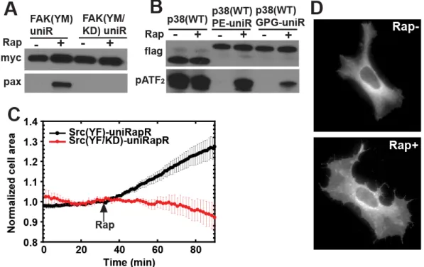
(A) Illustration Schematic representation of activity control with the uniRapR domain. (B) Root mean square fluctuations of the ATP binding site (gray structure) based on multiple equilibrium DMD simulations for wild type Src (black), apo (red), and holo (green) uniRapR inserted Src (p-value < 0.01). (C) HEK293T cells expressing the Src-uniRapR-cerulean-myc construct were treated with different concentrations of rapamycin (0 to 2µM) and lysates were assayed for expression of the construct with Western blotting using anti-GFP. The construct was pulled down with anti-myc and mixed with the paxillin substrate in the presence of ATP for 10 minutes. Reaction suspensions were blotted and probed with anti-myc and anti-pY31-paxillin to confirm binding and phosphorylation of the substrate, respectively. (D) As controls, constitutively active Src (YF) without the uniRapR domain, kinase dead (YF/KD), Y271A and L1polyP Src mutants with the uniRapR domain, and our previous dimerization-based switch were tested. (E) Y271A and L1polyP substitutions shown on the Src-uniRapR modeltime.
UniRapR enables specific and robust control over various kinases in vitro
(A) Testing different lengths of double linkers between uniRapR subdomains. A double linker connecting the subdomains A and B were shown in red and cyan. The linker with asteriks is the one used in the current version of uniRapR. (B) Src-uniRapR-cerulean-myc constructs were expressed in HEK293T cells. Cell lysates were blotted with anti-GFP to confirm the expression of the construct. In dimerization-based switch (RapR), co-expressed FRB was also tested with anti-GFP. Cell lysates were pulled down with anti-myc and mixed with substrate paxillin in the presence ATP for 10 minutes. Reaction suspension was blotted with anti-myc to confirm the binding, with anti pY31-paxilin to confirm the phosphorylation of the substrate. The linker2 is the one required for functional uniRapR. (C) The design with truncated linker 2 (linker 1) does not provide control over Src.
Specific activation of Src kinase leads to polarized spreading in single cells
Signaling cascades containing Src kinase play important roles in cell growth, proliferation, migration, and tumor invasiveness (115). However, the specific roles of Src catalytic activity, especially in cell migration, are unclear due to the limitations of existing chemical and genetic methods, including limited temporal control of activation or inactivation. For example, overexpression of constitutively active Src prevents the observation of events immediately following Src activation, as the cell compensates for Src expression, probably with other Src kinase family members, during gradual increase in expression level. Likewise, blocking Src expression with RNA interference is also a slow process.
(A) Immunoprecipitation, in vitro FAK (A) and p38 (B) assays were performed similarly as for Src kinase. (C) Change in cell area of HeLa cells expressing either Src (YF)-uniRapR (8/8 cells) or Src (YF/KD)-uniRapR (8/8 cells). (D) HeLa cells expressing Src-uniRapR (YF)-cerulean demonstrate spreading after the addition of rapamycin.
We overcome these limitations by using uniRapR Src (YF), which can reach maximal stimulation in less than 3 minutes. To observe the effect of Src activation on cell motility, we expressed uniRapR Src (YF) in HeLa cells. In the absence of rapamycin, we observed only peripheral ruffles near the cell edge, a phenotype also seen in untransfected cells. After rapamycin addition, we observed a statistically significant increase in cell area for all cells examined, relative to those expressing catalytically dead Src (YF/KD)-uniRapR (area increase = 30% ± 5 %, n = 8 cells; Figure 2.4C and D). Control cells showed no statistically significant change (area change = 8% ± 6 %, n = 8 cells). In a control study, rapamycin alone did not have any effect on the phenotype of untransfected cells. Polarized spreading of HeLa cells following Src-activation supports a role for Src in cell invasiveness.
Specific activation of Src kinase causes loss of cell-cell contacts in zebrafish epidermis
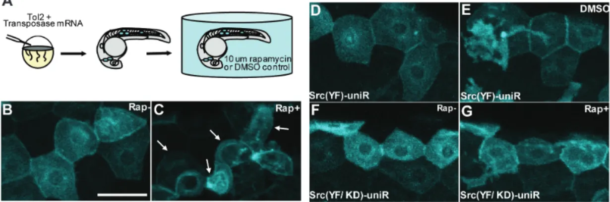
While we observe a profound impact of Src activation in cultured cells, it is crucial to determine whether uniRapR Src (YF) enables Src activation to be studied in the context of a multi-cellular organism. To study the role of Src activation during development, we expressed uniRapR Src (YF) in zebrafish embryos (Figure 2.6A). Epidermal cells expressing uniRapR Src (YF) demonstrated wild-type polygonal shape and formed tight connections with no gapping in the absence of rapamycin (Figure 2.6B). When we activated uniRapR Src (YF) by adding rapamycin, the epidermal cells had extended protrusions and underwent significant morphological changes in 12-16 hours. These morphological changes caused the loss of cell-cell contacts as cell-cells became more rounded (Figure 2.6C).
(A) Synthetic transposase mRNA was co-injected with the Tol2 Krt4 Src (YF)-uniRapR-cerulean plasmid into one-cell zebrafish embryos, resulting in mosaic expression of Src-uniRapR in the epidermis. Epidermal cells with characteristic flat honeycomb morphology were selected and imaged before (B, F) and after (C, G) 16 hours of rapamycin treatment. (C) Epidermal cells expressing Src (YF)-uniRapR-cerulean in zebrafish embryos were exposed to 10 µM rapamycin become rounded and undergo dynamic cell shape changes. White arrows indicate an epidermal cell before and after each treatment. Control epidermal cells expressing Src (YF)-uniRapR-cerulean in vehicle (DMSO) (D) or expressing the kinase-dead construct Src (YF/KD)-uniRapR-cerulean in rapamycin (G) have a static morphology and do not undergo dynamic cell shape changes. Scale bar 30 µm.
In control experiments, we did not observe morphological changes when cells expressing uniRapR Src were treated only with vehicle (Figure 2.6D and E), or when we expressed the catalytically-dead uniRapR Src (YF/KD) construct in zebrafish embryos in the presence of rapamycin (Figure 2.6F and G), demonstrating that the observed effects are due specifically to uniRapR Src activation by rapamycin. This dramatic phenotype of altered cell morphology upon Src activation demonstrates the applicability of uniRapR in studying signaling pathways in whole organisms.
2.3 Conclusion
presence of iRap (Figure 2.7). UniRapR can potentially be applied to a wide range of rapidly activating, allosteric signaling proteins. The dynamic behavior of a particular protein in a signaling pathway can be investigated by using a uniRapR protein analog to activate the proteins with minute resolution. Moreover, by inserting uniRapR into putative allosteric loops in a protein of interest, allosteric sites that are coupled can be experimentally identified.
The velocity of Src (CA)-uniRapR, Src(KD)-uniRapR in the presence of rapamycin, and non-immunosuppressive rapamycin analog iRap.
members including Yes, Fyn, Lyn, Lck, Hck, Blk, Fgr, and Yrk kinases, differential roles of these proteins in cell motility can ultimately be elucidated.
An important advantage of uniRapR is its practical use in animals. Light-dependent control can be useful at the single cell level, but uniRapR can provide activity control with the small molecule rapamycin even within deep tissues, where light cannot penetrate. To investigate the effect of Src kinase activation on a higher order process such as intercellular communication in animals, a significant process involved in tissue development, repair, immune response and homeostasis (115, 120), we tested uniRapR Src in living zebrafish epidermal tissue. We demonstrated that activation of Src kinase leads to a decrease in communication between cells, consistent with its oncogenic role in metastasis. The dynamic behavior of proteins downstream of Src, for example Connexin 43 or Cas that are involved in inter-cellular communication (121), will be the subject of future study.
UniRapR is a unique example of an insertable, transferable, and ligand-controllable protein switch. This switch has widespread potential applications for understanding signaling pathways involving kinases, a glimpse of which is offered by the novel phenotypes we demonstrate in mammalian cells and zebrafish tissue upon Src kinase activation. The potentially wide applicability of uniRapR is underscored by the structural conservation of kinases, and the allosteric properties of many signaling proteins.
2.4 Materials and Methods DNA construction
mutagenesis kit (Stratagene). PCR products were used as mega-primers for QuikChange mutagenesis reactions (Supplementary Table 1). The uniRapR domain was created using PCR such that the 5′- and 3′-end sequences anneal at the desired insertion site within the Src, FAK and p38 genes.
Modeling of uniRapR and Src-uniRapR structure
To model uniRapR, we removed first twenty residues from FKBP12 (PDB code: 1fap) using PyMol (http://www.pymol.org/), so that the two termini are close in space for insertion. We built a circularly permuted FRB, by excising at Asn2093 and joining the N- and C-termini together. We removed first four unstructured residues of FRB and connected the two termini with a peptide linker (PPGPGSG). In order to build a single chain protein, we excised FKBP12 in Pro88 and connected FRB-G2092 to FKBP12-Gly89 and FKBP12-His87 to FRB-Val2094 with corresponding linkers. We optimized the complex structure by minimizing the energy using all-atom DMD simulations. We kept the unmodified regions of FKBP12 and FRB molecules static, whereas linkers were allowed to move to sample conformation and form peptide bonds under the peptide bond constraints1 between FKBP12/FRB and linkers.
Computational analysis of uniRapR
We perform all-atom DMD simulations to study the conformational dynamics of uniRapR. A complete description of the discrete molecular dynamics (DMD) algorithm can be found elsewhere (122, 123). We performed replica exchange DMD simulations to estimate the folding thermodynamics. We used eighteen replicas with temperatures ranging from 0.52 kcal/mol·kB (~260K) to 0.75 kcal/mol·kB (~390K), with an increment of 0.02 kcal/mol·kB
(0.01 kcal/mol·kB between 0.64 kcal/mol·kB and 0.72 kcal/mol·kB). The length of each
simulation is 1 × 10⁶ time units (~50ns). We applied the weighted histogram analysis method (124) (WHAM) to estimate thermodynamic properties of the uniRapR domain. Next, we computed the partition function, Z=∫ρ(E)exp(−E/kBT)dE, which allowed us to derive other thermodynamic parameters, including the specific heat (Cv) at different temperatures. We also calculated RMSD and tail distance of subdomain A as function of temperature using WHAM analysis.
We performed a constant temperature simulation of Src-uniRapR at 0.5 kcal/mol·kB.
Immunoprecipitation and in vitro kinase assay
Anti-myc and anti-FLAG antibodies were purchased from Millipore and Sigma, respectively. Anti-phospho-paxillin-pTyr31, anti-GFP (JL8) antibodies were purchased from Biosource and Clontech, respectively. Anti phospho-ATF2 was purchased from Santa Cruz. Rapamycin was purchased from Sigma. HEK293T cells were transfected with 2 µg of DNA constructs using Fugene6 reagent (Roche). Cells were treated with either rapamycin or an equivalent volume of ethanol (solvent of rapamycin solution). Cells expressing Src or FAK constructs were then washed with cold PBS and lysed with 20 mM HEPES-KOH, pH 7.8, 50 mM KCl, 100 mM NaCl, 1 mM EGTA, 1% NP40, 1mM NaF, 0.1 mM Na3VO4, and Roche
protease inhibitor cocktail. Cells expressing p38 were lysed with 20 mM Tris, pH 7.5, 150 mM NaCl, 1 mM EGTA, 1mM EDTA, 1% Triton X-100, 0.5% NP40, 20mM NaF, 0.2 mM Na3VO4, and Roche protease inhibitor cocktail. Cells treated with rapamycin were lysed with
the appropriate lysis buffer containing the corresponding rapamycin concentration. Cleared lysates were incubated with protein-G beads attached to myc (for Src and FAK) or anti-FLAG (for p38) antibody for 2 hours. The beads were washed three times with wash buffer (20 mM HEPES-KOH, pH 7.8, 50 mM KCl, 100 mM NaCl, 1 mM EGTA, 1% NP40), and then with kinase buffer (25 mM HEPES-KOH, pH 7.5, 5 mM MgCl2, 0.5 mM EGTA, 0.005%
BRIJ-35) for Src and FAK assays. For p38 assays, beads were washed with p38 lysis buffer and then with p38 kinase buffer (25 mM HEPES-KOH, pH 7.4, 10 mM MgCl2, 25 mM
beta-glycerophosphate, 4 mM dithiothreitol (DTT), 0.1 mM Na3VO4). For Src and FAK assays, 20
Live cell imaging
HeLa cells were plated on fibronectin coated coverslips (10 mg/ml fibronectin for 1 hour at 37 ºC, Sigma) and then incubated for 3 hours. DMEM (with 10%FBS) medium was then replaced with L-15 imaging medium (Invitrogen) supplemented with 5% FBS. An open heated chamber (Warner Instruments) was used to maintain the cells. Live cell imaging was performed with an Olympus IX-81 microscope equipped with ZDC focus drift compensator. Images were collected using a PhotometrixCoolSnap ES2 CCD camera (Roper Photometrics). Metamorph software (Molecular Devices) was used to control the microscope, and to acquire and process images at each time point. To calculate cell area, each cell was manually traced around its edge, and a binary mask was created using Metamorph. Values were normalized to be 1 at the time point before addition of rapamycin.
Imaging of live zebrafish
CHAPTER 3: CHEMOGENETIC PAK1 SWITCHES ENABLE SPECIFIC ACTIVATION OF A PAK ISOFORM IN CELL MOTILITY
3.1. Introduction
p21-activated kinases (PAKs), consisting of six members (PAK1-6), are serine/threonine kinases involved in many cellular processes including cell growth, death, survival, proliferation, differentiation, motility and polarity (125). PAK is a key regulator of cytoskeleton networks, as its activity has been associated with protrusions (126-128), dissolution of stress fibers and loss of focal adhesions (129), control of adhesion turnover and strength (10, 129, 130). Such major cytoskeletal roles render PAK particularly relevant in various motility-driven biological processes including cancer metastasis (131), angiogenesis (132), synaptic transmission (24, 133, 134), and neurogenesis (135). However, these studies remain to be correlative; it is unclear how PAK dynamically contributes to cell motility, and the requirement of its kinase activity in motility remains controversial (125, 126, 136).
3.2 Results and Discussion
Engineering and optimization of a dual chain PAK1 switch
(A) Dual chain (rapamycin regulatable or RapR) PAK1 switch was built by inserting insertable FKBP (iFKBP) domain into the catalytic domain. The system requires co-expression of FRB. (B) Single chain (unimolecular rapamycin regulatable or uniRapR) PAK1 switch was built by inserting insertable uniRapR domain into the catalytic domain. This system does not require co-expression of FRB. (C) Crystal structure of PAK1 in inactive state. Auto-inhibitory domain (AI) and p21 binding domain (PBD) are shown with salmon and green, respectively. Insertion sites are red residues shown with black arrows. (D) Rapamycin dependent activity of constitutively active (CA) PAK1, kinase-dead (KD) PAK1, RapR-PAK1, and uniRapR-PAK1. (E) Area and (F) perimeter of MDA-MB-231 cells that stably express uniRapR-PAK1 before and after addition of rapamycin.
(A) Residues used for insertion are not conserved. (B) Conservation of the kinase domain, represented by sequence entropy, mapped on the structure.
insertion of iFKBP domain into the loop between residues 288 and 293 generates a robust PAK1 switch.
(A) Structure of PAK1 dimer and residues in the insertion loop. (B) Biochemical in vitro test of various RapR-PAK constructs.
Single chain PAK1 switch in triple negative breast cancer cells
dynamic range of uniRapR-PAK1 by introducing another mutation L107F (141) that blocks dimerization and enhance activity of PAK1. These results showed that both dual and single chain PAK1 switches could be rapidly activated in living cells by rapamycin.
PAK1 was identified as a breast cancer oncogene that transforms breast epithelial cells (142). Moreover, overexpression of mutant PAK1 was shown to increase migration rates of breast cancer cells (143). In order to demonstrate direct effects of PAK1 kinase activity in breast cancer cells, we expressed uniRapR-PAK1 in highly metastatic triple-negative breast cancer cells MDA-MB-231. We measured the area and perimeter of cells after 5, 20, and 60 minutes of rapamycin addition. Remarkably, cell area and perimeter had a transient increase upon addition of rapamycin (Figure 3.1E and F). After such a rapid spreading, cells went back to their original morphology at the 20-minute time point. Control cells exhibited no response to rapamycin. These results indicated that catalytic activation of PAK1 resulted in transient spreading; suggesting metastasis signaling circuits have additional requirements for persistent spreading in addition to PAK1 activity.
PAK1 activation results in persistent dendritic spine enlargement
(A) Image of RapR-PAK1-Venus in a dendrite. (B) A series of RapR-PAK1-Venus images in a dendrite before and after stimulation with rapamycin (2.5 µM), which was applied in the period between 0 and 15 min. (C) Volume changes of two spines (#1 and #2) indicated in (B). The changes were estimated from the total fluorescence of RapR-PAK1-Venus in the spines. (D) Mean time courses of spine volume obtained from 33 or 13 spines in either RapR-PAK1-Venus or EGFP along with FRB-mCherry transfected neurons. Error bars indicate SEM. (E) Statistical comparisons of the effects of rapamycin (2.5 µM) shown in (D). *p < 0.05, **p < 0.01.
3.4C), there was an overall persistent increase in the spine volume (area of change 60% ± 15%, n = 35 spines), whereas neuron expressing only EGFP did not respond to rapamycin (n = 13 spines) (Figure 3.4D and E). Based on these results, we concluded that activation of PAK1 leads to persistent dendritic spine enlargement.
Allosteric propagation in PAK1 can be maintained through alternative pathways
What are the rules to make a kinase switch? We hypothesize that loops that move in concert with the active site can be an insertion site for the allosteric domain, as perturbation of the insertion site would be propagated to the active site. Additionally, we select loops that are surface exposed or not in contact with the rest of the protein. We analyzed the molecular dynamics of PAK1 kinase using discreet molecular dynamics (111, 123). Indeed, motions of the insertion loop (L1) correlate with the catalytic site, particularly the ATP binding site (Figure 3.5A). Using the same analysis, we also identified another loop (L2) whose motions correlate with the motion of ATP binding site. Similar to L1, this loop is also solvent exposed and is not evolutionarily conserved (Figure 3.5B).
Overall, a PAK1 switch can be generated by inserting uniRapR into the loops whose motions correlate with the motions of catalytic site.
(A) Correlative motions of residues in PAK1 kinase domain. Correlation map of the kinase domain. Red denotes correlation, while blue denotes anti-correlation between two residues. b. Network representation of kinase domain based on contacts (<5.5Å). Gray nodes represent residues; edges represent the contacts. L1 is the canonical insertion site and L2 is newly identified insertion site. Red arrows represent the propogation pathways to ATP binding site (yellow node). (C) Insertion of uniRapR to both insertions sites confer rapamycin-mediated control. In the second version of L2 based switch (uniRapR-PAK1-L2-v2), loop residues from PAK1 removed, resulting in a tighter control. (D) Suggested mechanism of L1 based switch. (E) Suggested mechanism for L2 based switch. (F) Summary of the mechanisms of PAK1 switches.
allosteric path, which can be responsible for the propagation. In order to track potential propagation pathways, we performed a network analysis by converting motion dynamics of residues into an undirected network, in which nodes represent residues, edges represent contacts, and edge weights are assigned based on correlation coefficients calculated by molecular dynamics simulations (Figure 3.5B).
Co-evolved residues of PAK1 kinase domain were identified using MISTIC (148).
D41, and E26-D41 (Figure 3.5D and E). As an alternative approach, we analyzed potential allosteric pathways using evolution of residues (148). Identification of residues that co-evolve with the catalytic site indicated a similar inter-strand pathway (Figure 3.6). All these results showed that perturbation is propagated from our canonical (L1 insertion), which was used in our previous work, and non-canonical (L2 insertion) insertion loop via intra-strand and inter-strand manner, respectively.
3.3 Conclusion
We focus on PAK1 as a target protein, because its role in cytoskeleton remains controversial, and to our knowledge, there is currently no PAK1 switch in the literature. Many of the previous studies included overexpressed constitutively active or dominant negative PAKs. These studies suggest that overexpression of constitutively active PAK leads to large and polarized lamellipodia, whereas expression of dominant negative PAK leads to multiple random lamellipodia in fibroblasts (127). Nevertheless, overexpression of constitutively active kinases can increase intracellular signaling above the physiological basal state. On the other hand, studies that used acute activations through injection of constitutively active PAK1 into the cells report rather different phenotypes (126, 136). Because PAK1 is upregulated in metastatic breast cancer, we first tested phenotypic effects of PAK1 in highly metastatic MDA-MB-231 cells. PAK1 activation caused transient spreading, suggesting that other pathways are required for a persistent metastatic behavior of these cells.
by PAK1 independent mechanisms. These results may give insight into the roles of PAK1 in long-term-potentiation, learning and memory.
Our analysis on propagation mechanisms revealed that the perturbation is allosterically propagated through the beta strands located between G293-Q300 and G282-D289 in our canonical switch designs. This observation is expected as destabilization of a loop in a structural unit such as beta-strand perturbs other residues in the same strand. However, we observed an inter-strand allosteric propagation in our second type of design. Such an allosteric propagation is meaningful, because correlated motions in B-sheets have been suggested to be fundamental for allosteric regulation of protein function (152). Understanding propagation mechanisms in our allosteric switches will be helpful to generate novel switches not only for different kinases but also for other protein families.
3.4 Materials and Methods
DNA constructs
All restriction enzymes were purchased from New England Biolabs. Point mutations and insertions of iFKBP and uniRapR sequences into PAK1 cDNA were done with Quikchange mutagenesis kit (Stratagene). Retroviral plasmids were generated by cloning uniRapR-PAK1-mCherry into pBABE-Tet-Off-puro plasmid (Clontech).
Biochemical characterizations
reagent (Roche). After 24 hours of transfection, cells were treated with either rapamycin or an equivalent volume of ethanol for 30 minutes. Cells were lysed with lysis buffer (20 mM HEPES-KOH, pH 7.8, 50 mM KCl, 100 mM NaCl, 1 mM EGTA, 1% NP40, 1mM NaF, 0.1 mM Na3VO4, and Roche protease inhibitor cocktail). Endogenous phosphorylation of MEK
was detected by western blotting. For immuniprecipitation experiments, cleared lysates were incubated with protein-G beads attached to anti-GFP antibody for 2 hours. The beads were washed three times with wash buffer (20 mM HEPES-KOH, pH 7.8, 50 mM KCl, 100 mM NaCl, 1 mM EGTA, 1% NP40), and then with kinase buffer (25 mM HEPES-KOH, pH 7.5, 5 mM MgCl2, 0.5 mM EGTA, 0.005% BRIJ-35) for phosphorylation assays. Purified ATF2 and
ATP (0.5 mM) in 40 µL of kinase buffer were mixed with bead suspension for the reaction for 10 minutes. Phosphorylated MEK was detected in pull-down samples by western blotting.
Imaging of MDA-MB-231 cells
Imaging of mouse hippocampal cells
Hippocampal slices with a thickness of 350 µm were prepared from 7-day-old Sprague-Dawley rats. Slices were mounted on 0.4-µm culture inserts (Millipore, Billerica, MA) and incubated at 35°C under 5% CO2 in a medium consisting of 50% MEM (Invitrogen, Carlsbad,
CA), 25% Hanks’ balanced salt solution (Invitrogen), 25% horse serum (Nichirei, Tokyo, Japan), and glucose (6.5 g/l) (Nacalai Tesque, Kyoto, Japan). After 4 days in culture, the slices were transfected with a Gene Gun system (PDS-1000; Bio-Rad, Hercules, CA) with RapR-PAK-Venus and FRB-mCherry. Imaging experiments were performed 3 days after the transfection. Each culture insert was transferred to a recording chamber and superfused with a solution (ACSF) that contained 125 mM NaCl, 2.5 mM KCl, 2 mM CaCl2, 1 mM MgCl2,1.25
mM NaH2PO4, 26 mM NaHCO3, 20 mM glucose, 1uM TTX, and 200uM Trolox, and had been
equilibrated with 95% O2 and 5% CO2. Rapamycin (Wako, Tokyo) was applied through bath
Modeling, discrete molecular dynamics, and correlative motion analysis
Crystal structure of the kinase domain was obtained from Protein data bank (PDB id: 1f3m). Medusa Toolkit was used to model the protein (153). All simulations were performed using all-atom discrete molecular dynamics (154). Atomic clashes were corrected and the protein was minimized at heat exchange coefficient 10 at 0.7 kcal/mol·kB with harmonic
potential constant of 1 kcal/mol•A2. The system was then packed without any constraint at 0.3
kcal/mol·kB with heat exchange coefficient 1. Upon packing, the system was simulated at 0.4
kcal/mol·kB for 1 million DMD steps.
Monitoring propagation and dynamic networks
The network was built using the Cα atoms which are represented as nodes of the graph, and edges represent the contacts between residues. Edge weights were assigned based on the correlation coefficient of motions calculated using discreet molecular dynamics. The shortest distance between two selected residues were calculated by Dijkstra’s algorithm (149).
Sequence conservation and co-evolution analysis
CHAPTER 4: COMPUTATIONAL DESIGN OF SPLIT PROTEINS TO CONTROL PROTEIN ACTIVITY
4.1 Introduction
Split protein reassembly methods have been valuable for visualization of protein activity in living cells. However, the design of split proteins for protein control has been challenging due to spontaneous assembly, and inefficient induction of effective reassembly. Here, we introduce a generalized approach, termed split protein reassembly enabled by ligands or light (SPELL), to predict split sites using a novel scoring function (split energy), prevent spontaneous assembly, and facilitate efficientreassembly of the split parts using an engineered domain. The split proteins can be controlled by light or a small molecule. Our analysis of 16 known split proteins shows that functional split sites can be predicted using the split energy. Moreover, our engineered domain enables destabilization of split proteins, thus preventing spontaneous assembly of the split protein before activation. We apply SPELL to a new target, the catalytic domain of the guanine exchange factor Vav2, and demonstrate that its activation modulates the protrusion dynamics of living cells. The SPELL methodology offers new opportunities to interrogate dynamics of previously untargeted proteins.
have been used to control protein localization and interactions (3, 4, 27, 38). However, such approaches have limited effectiveness in that they typically generate activity only at specific subcellular locations. Controlling protein conformation has been accomplished by inactivating the target protein using inducible domains, and then restoring its activity rapidly with light or ligands (36, 46, 48, 59, 139, 156). However, these proteins and those under the control of dimerizers often show some level of activation before they are switched on (43).
Another strategy to achieve protein control has been to inactivate proteins by splitting them into two parts, rendering them inactive, and then inducing reassembly for activation (157). Despite the extensive use of such split-protein approaches to make fluorescent biosensors (158), controlling protein activity through splitting has been limited by several critical challenges. First, it is difficult to predict an appropriate split site. Second, there can be high basal activity of split proteins in the absence of an inducer due to spontaneous protein reassembly, and third, activity can be diminished with respect to wild type protein upon induction, due to misfolding of reassembled protein or low efficiency of reassembly. Most successful split protein designs have been generated by splitting at linkers that connect two separate domains (both necessary for activity) which we will term “easy targets”. Proteins that have single catalytic domains may be considered “hard targets”. Few split protein analogues of such targets have been generated, and these have required extensive experimental screening approaches. For both hard and easy targets, in some cases the interface of the split parts have been further engineered to reduce spontaneous assembly or to increase the efficiency of reassembly (159, 160).
an engineered domain, and efficient ligand- or light-mediated reassembly of the split parts. We first investigated existing split proteins to assess the accuracy of our method, and then applied SPELL to a new “hard” target that has a single catalytic domain.
4.2 Results and Discussion
Split protein reassembly enabled by ligands or light (SPELL)
within cores may unfold the split parts irreversibly. In the multi-well minima case, the protein can be split either at interdomain and subdomain loops or at sites with minimum effect on the core of the major split parts (Figure 4.1A).
(A) Identification of the split site by computing solvent accessible area (surface exposure), sequence conservation, and energy change upon splitting from residue i, where 1≤ i≤ N. The selected loop was marked with a green box. (B) Prevention of spontaneous assembly with insertable FKBP (iFKBP), which destabilizes the core lobe. (C) Structural model of iFKBP-FRB-rapamycin system. The N terminus of iFKBP is fused to the C terminus of target protein’s N-lobe, whereas the C terminus of the FRB is fused to the N terminus of C-lobe of the target protein.
Computational prediction of split sites on GFP
it surprisingly suggested the presence of two hidden subdomains (Figure 4.2A and B), not detectable from visual inspection of the structure.
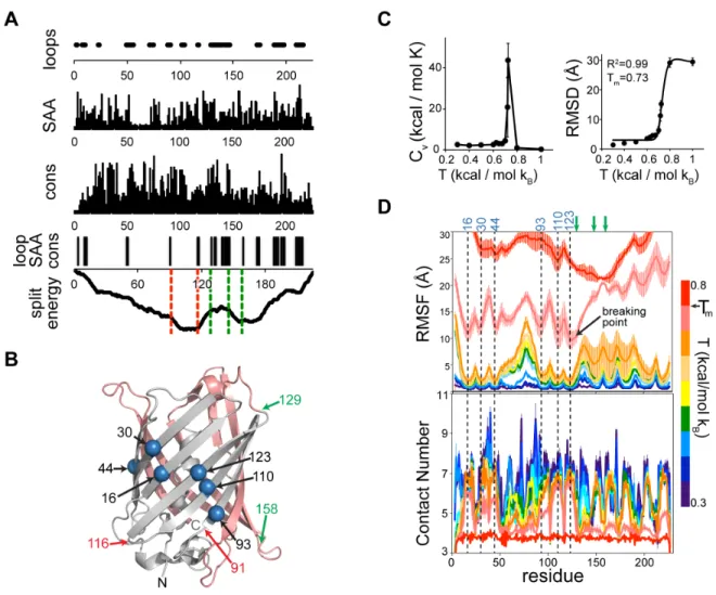
(A) Loops, solvent accessible area (SAA), sequence conservation (cons), consensus of these three parameters (loop, SAA cons), and split energy. X axis = amino acid number. Dashed lines in the split energy profile show the experimentally tested successful (green) and unsuccessful (red) split sites. (B) Prediction of transition temperatures based on heat capacity and average root mean square difference (RMSD) with respect to initial structure. (C) Root means square fluctuations (RMSF) and contact numbers of GFP at different temperatures. Green arrows indicate experimentally tested successful sites. bp: break point. (D) Structure of GFP (pdb id: 20yg) with computed core residues (blue spheres), and the experimentally tested successful (green) and unsuccessful (red) split sites. N-lobe = gray and C-lobe = salmon.
Experimentally successful split sites (at residues 129, 145, 158) (162) are located between these subdomains, supporting our argument about the conditions necessary for a split site (Figure 4.2B). Specifically, residue 158 is in proximity to the second energy well, keeping the first core intact (Figure 4.2A). These results show that the split energy predicts functional split sites in GFP.
The split energy detected split sites on a static structure. We next tested whether the same split sites could be identified by investigating protein dynamics. We performed molecular dynamics simulations (111, 154) of GFP at different temperatures (Figure 4.2C) and estimated protein dynamics using root mean square fluctuations (RMSF) and contact numbers. Local minima in such unfolding energy diagrams suggested that the possible folding core residues are mostly located in the region between the N-terminus and the loop in residues 128-133 (Figure 4.2D). The unfolding curve showed a remarkably sharp transition starting after this loop and reaching to the C terminus, suggesting a less stable region compared to the N-lobe, and a split site after such a breaking point. Indeed, the experimentally validated sites were after this predicted breaking point (Figure 4.2D). These results show that both the split energy profile and unfolding curves obtained by molecular dynamics simulations predict the split sites in GFP.
Split energy predicts critical sites to split proteins
(Figure 4.3). Split sites close to the core of TEV protease and phosphatase result in only 40% and 65% of wild-type activity, respectively (163, 164). The split energy profile of Cre recombinase features two minima. The experimental split was performed within a loop in the first small domain to prevent spontaneous assembly. This site produces only moderate activity upon assembly (165), indicating a potential distortion of the core of the first domain.
Split energy profile for each known split protein. Red lines represent unsuccessful split sites in the original work. Dark green lines represent the most successful split site, whereas light green lines represent less successful (low activity upon reassembly) split sites.
termini. These observations all suggest that the dominant core in the protein should be preserved for an efficient split design.
SPELL Vav2 and control of cellular protrusion signalling
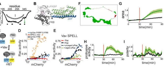
To validate our approach, we targeted the Rho family guanine exchange factor Vav2, a protein that has not been previously controlled by splitting. As a crystal structure of Vav2 is not available, we generated a homology model of Vav2 based on the crystal structure of Vav1 (167). Using the split energy profile, we predicted a single subdomain architecture, making the catalytic Dbl homology (DH) domain of Vav2 a hard target to split (Figure 4.4A). Unchanged split energy on the loop around residue 345 (L4) pointed to a potential split site there. We fused the N-lobe of split Vav2 with iFKBP, and the C lobe with FRB (Figure 4.4B). To test the activity of this split protein in living cells, we used Rac1 FLARE.dc (168), a Rac1 FRET biosensor that should be activated by Vav2 (Figure 4.4C). The Rac1 biosensor switched to its active state upon increasing co-expression of constitutively active Vav2 (Figure 4.4D).
using light, we employed our photo-activatable rapamycin, pRap (23), and showed that Vav2 SPELL was active upon exposure of cells with a brief period of light (1 minute). Activation of Vav2 SPELL in HeLa cells induced protrusions within minutes as visualized by both increased area and non-uniform spreading (Figure 4.4F-I).
(A) Energy profile of DH domain of Vav2 upon splitting from residue i, where 195≤ i≤ 390. Green box and arrow show the least destabilized region and chosen loop for splitting, respectively. (B) Model of Vav SPELL. iFKBP (light gray) was fused to C terminus of N lobe of DH (green), and FRB (dark gray) was fused to N terminus of C lobe of DH (blue) in the presence of rapamycin (purple). (C) Dual chain Rac1 FRET sensor (Rac1 FLARE.dc1g) is in high FRET state when Rac1 is active, in low state when Rac1 is inactive. (D) FRET state (activity) of Rac1 with respect to amount of spVav-FKBP12-FRB (mCherry-DHN-FKBP12 and FRB-DHC-PH-ZnF), Vav2 SPELL (mCherry-DHN-iFKBP and FRB- DHC-PH-ZnF), spVav-FRB (mCherry-DHN and FRB- DHC-PH-ZnF), and Vav (mCherry-DH-PH-ZnF). (E)
Testing Vav2 SPELL with rapamycin and caged rapamycin. (F) A representative HeLa cell expressing Vav2 SPELL after 19 minutes of rapamycin addition. Green and red areas indicate the protrusions and retractions, respectively. Morphological changes of cells expressing Vav2 SPELL (green, mean ± s.e.m., n=19 cells), and cells expressing only membrane marker (black, mean ± s.e.m., n= 36 cells) were quantified for (G) normalized area, (H) protrusive activity, and (I) change in the polarity.
These live cell studies showed activation through no detectable background activity, and robustly activated protrusions either through addition of rapamycin or light-activation of a ligand.
4.3 Conclusion
We developed a novel methodology providing a rational route to control protein activity through protein splitting and reassembly. This approach is substantially different than previous strategies, which relied on experimental screening of random split libraries (170) or insertion of long flexible linkers into candidate split sites (171). In some published cases, such as split ubiquitin, substitution of key amino acids at the interface of the split parts was used to prevent spontaneous assembly (159); such extensive engineering has hindered broad applicability.
advantage reduces the effort required to optimize the linkers between the ligand/light gated domains and the split protein.
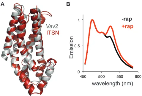
(A) Crystal structure of ITSN (red) and homology model of Vav2 (gray). (B) Normalized emission of Cdc42 sensor co-expressed with SPELL ITSN.