INVESTIGATION OF FLT1 AND VEGF SIGNALING IN CONNECTIONS DURING DEVELOPMENTAL SPROUTING ANGIOGENESIS
Jessica E. Nesmith
A dissertation submitted to the faculty at the University of North Carolina at Chapel Hill in a partial fulfillment of the requirements for the degree of Doctorate of Philosophy in the
Curriculum in Genetics and Molecular Biology in the School of Medicine.
Chapel Hill 2016
ABSTRACT
Jessica Nesmith: Investigation of Flt1 and VEGF signaling in connections during sprouting angiogenesis
(Under the Direction of Victoria L. Bautch)
To my family, those I was lucky enough to be born with and the ones I have chosen. You have provided support and love during the highs and lows of this work, willingly listened to my scientific excitement and helped me figure out my career.
ACKNOWLEDGEMENTS
This work was accomplished with the help of a great many people over the years, without whom this work and my success would not be possible.
First I want to thank my advisor Vicki Bautch for the guidance and critical analysis of this project since its generation. Your suggestions and help have shaped this story and my scientific goals. The laboratory environment you fostered has helped me find the courage and strength to continue questioning and investigating scientific research. With your mentorship I feel prepared to enter the scientific community as a contributing, well-informed, and active member.
To the members of the Bautch lab, past and present, your input has been invaluable. Specifically, I want to thank John Chappell who was available as a conceptual sound board, for countless moments of troubleshooting, and provided invaluable input in designing realistic experiments and shaping the body of this work. Many discussions with the lab members in our regular meetings and the sounding board you offered for conclusions on data, presentation techniques, and career goals were instrumental in my time here. You all have seen me through tough personal and academic challenges that I faced these past years. Each of you contributed to the rigorous scientific environment which challenged and kept me on my toes and kept a wonderful balance with levity and humor that truly made this a great group to be a part of.
PREFACE
TABLE OF CONTENTS
ABSTRACT... iii
ACKNOWLEDGEMENTS ...v
PREFACE ... vii
LIST OF FIGURES ... xi
LIST OF TABLES ... xii
LIST OF ABBREVIATIONS ... xiii
CHAPTER 1: Introduction ...1
I. Angiogenic blood vessel network formation during development ...1
II. VEGF signal propagation and ligand specific effects ...3
a. Flk1 during developmental angiogenesis ...4
b. Flt1 during developmental angiogenesis ...7
c. VEGF ligand effects in animal models ...9
III. Sprouting angiogenesis during development ... 12
a. Sprout initiation from quiescent vessels ... 12
b. Sprout migration and extension ... 13
c. Anastomosis to create connections and new vessel conduits ... 15
d. Formation of a new lumen ... 17
V. REFERENCES ... 20
CHAPTER 2: Materials and Methods ... 28
I. Experimental Materials and Methods ... 28
a. Retinal Angiogenesis Imaging and Analysis... 28
b. Cell Lines and Culture ... 29
c. HUVEC Sprouting Angiogenesis Assay Imaging and Analysis ... 29
d. ES Cell-derived Vessel Imaging and Analysis ... 30
e. Zebrafish Experiments and Husbandry ... 31
f. Protein Quantification ... 31
g. Lentivirus and siRNA knockdown ... 32
h. Fluorescence-activated Cell Sorting (FACS) Analysis... 33
i. mRNA Preparation and Quantification ... 33
j. Statistical Analysis ... 33
II. Tables ... 34
III. REFERENCES ... 37
CHAPTER 3: Blood Vessel Anastomosis is Spatially Regulated by ... Flt1 During Angiogenesis ... 38
I. Introduction ... 38
II. Results ... 40
a. Flt1 Influences Retinal Vessel Interactions ... 40
b. Transient Contacts Precede Stable Blood Vessel Connections ... 40
c. Flt1 Regulates the Number of Transient Contacts ... 41
IV. Figures ... 47
V. REFERENCES ... 61
CHAPTER 4: Flt1 During Sprouting Angiogenesis ... 63
I. Results ... 63
a. Connection stability and success requires Flt1 ... 63
b. Flt1 isoforms differently affect mitosis and protein localization ... 66
c. Flt1 and VEGF signaling influence anastomosis within zebrafish vessels ... 69
II. Figures ... 73
III. REFERENCES ... 79
CHAPTER 5: Conclusions and Future Directions ... 81
I. Summary of Flt1 regulation of blood vessel anastomosis ... 81
II. Future directions for investigating Flt1 regulation of anastomosis ... 83
a. Mechanism of mFlt1 influence on vessel interaction outcome ... 83
b. mFlt1 regulation influence on non-endothelial anastomotic regulation ... 86
c. Applications for anastomotic regulation in disease states... 89
III. Perspective ... 90
IV. Figures ... 92
LIST OF FIGURES
Figure 3.1: Flt1 Influences Retinal Vessel Interactions In Vivo. ... 48
Figure 3.2: Blood Vessel Stable Connections are Preceded by Transient Contacts. ... 50
Figure 3.3: Transient Contact Dynamics during Vessel Anastomosis are Regulated by Flt1... 52
Figure 3.4: Both Flt1 Isoforms Reduce Transient Contacts Prior to Connection. ... 54
Figure 3.5: Target Vessel Connection Site is Biased by Flt1. ... 56
Supplementary Figure 3.1: Flt1 shRNA Validation. ... 57
Supplementary Figure 3.2: Transient Contacts and Stable Connections ... in Mosaic Target Vessels... 60
Figure 4.1: Vessel connection outcomes in ES cell-derived sprouting angiogenesis ... is influenced by Flt1. ... 74
Figure 4.2: Isoform specific effects on sprouting human endothelial venous endothelial cells. .. 76
Figure 4.3: Flt1 and VEGF influence connections in zebrafish intersegmental vessels. ... 78
LIST OF TABLES
Table 1: Major Phosphorylation Sites of Flk1 and Cellular Effects/Interactions. ...4
Table 2.1: Antibodies and Nuclear Stains. ... 34
Table 2.2: siRNA sequences targeting Flt1 isoforms. ... 35
LIST OF ABBREVIATIONS
AJ adherens junction
AKT AK-thymoma/protein kinase B ANOVA analysis of variance
BMP bone morphogenetic protein Cdc42 Cell division control protein 42 cDNA complementary deoxyribonucleic acid CI confidence interval
CVP caudal vein plexus
DAPI 4',6-Diamidino -2-Phenylindo le DIC differential interference contrast DLAV dorsal longitudinal anastomotic vessel Dll4 Delta-like 4
DMEM Dulbecco modified eagle medium dpf days post-fertilization
EC endothelial cells ECM extracellular matrix EPC endothelial precursor cells ES embryonic stem cell
FACS fluorescence-activated cell sorting FAK focal adhesion kinase
Flk1 fetal liver kinase 1 (see VEGFR2/Kdr) Flt1 fms-like kinase 1 (see VEGFR1)
Flt4 fms-related tyrosine kinase 1 (see VEGFR3) GAPDH glyceraldehyde 3-phosphate dehydrogenase GFP green fluorescent protein
GOF gain of function
HB hindbrain
hpf hours post-fertilization
HS heparan sulfate
hsp heatshock protein
HUVEC human umbilical venous endothelial cells
InDels insertion or deletion of deoxyribonucleic acid bases
IP intraperitoneal
ISVs intersegmental vessels JAM junctional adhesion molecule
KD knockdown
Kdr kinase insert domain receptor (see Flk1/VEGFR2) LOF loss of function
MAPK p38 mitogen-activated protein kinase mFlt1 membrane-bound Flt1
MO morpholino
NRP neuropilin
Par3 portioning defective homolog 3 PAK1 p21 activated kinase 1
PBS phosphate buffered saline
PECAM platelet endothelial cell adhesion molecule 1
PFA paraformaldehyde
pFlk1 phosphorylated fetal liver kinase 1 PI3K phosphoinositide 3-kinase
PKC protein kinase C
PLCγ phospholipase C gamma
PlGF plancental growth factor PODXL podocalyxin
PVDF polyvinylidene fluoride
qRT-PCR quantitative real-time polymerase chain reaction Rac1 Ras-related C3 botulinum toxin substrate 1 RFP red fluorescent protein
RIPA Radiommunoprecipitation assay
RNAi ribonucleic acid interference
RT room temperature
sFlt1 soluble/secreted Flt1
Src Proto-oncogene tyrosine-protein kinase Src TBS Tris buffered saline
TJ tight junction
TSAd T-cell specific adapter VE-Cad VE-Cadherin
VEGF vascular endothelial growth factor
VEGFR1 vascular endothelial growth factor receptor 1 (see Flt1) VEGFR2 vascular endothelial growth factor receptor 2 (see Kdr/Flk1) VPF vascular permeability factor
VEGFR3 vascular endothelial growth factor receptor 3 (see Flt4) Wnt Wingless-related integration site
WT wildtype
CHAPTER 1: Introduction
The body of work presented here seeks to understand the impact of regulatory signaling on the formation of a complex, structured blood vessel network. There are a large number of growth factors implicated in the process; however, the vascular endothelial growth factor ligand family is a major main growth factor implicated in directing blood vessel growth and the focus of these studies. Blood vessel expansion into a hierarchical network of arteries and veins connected by capillaries is accomplished through angiogenesis. The varying interactions and interplay of signaling cascades that direct the initiation and coordinated migration of angiogenesis is highly researched but these same cascades are not evaluated during anastomosis, the process of two vessel connecting. Therefore, this work addresses the regulation anastomosis of sprouting angiogenesis through spatio-temporal restriction of growth factor signaling.
I. Angiogenic blood vessel network formation during development
forming early, the vascular system is also necessary for continued development. Zebrafish embryos with mutations which cause defective heart muscle, can survive for 5 days post-fertilization (dpf) without a functioning circulatory system using passive oxygen diffusion from the water, but subsequent development requires cardiovascular function (J. N. Chen et al. 1996). Similarly, mouse embryos cannot survive past E9.5 without a functional circulatory system, as is observed in various genetic mutants which fail to form blood vessels (Carmeliet et al. 1996; Ferrara et al. 1996). Thus, in diverse vertebrate lineages embryonic survival and appropriate development relies upon the formation and expansion of an effective vascular network.
Blood vessel development during embryogenesis is a multistep process that begins with primitive vessel formation from endothelial progenitor cells (EPCs) through a process known as vasculogenesis (Risau & Flamme 1995; Drake & Fleming 2000). Vasculogenesis creates the large, primary arteries and veins within the embryo and generates the initial vessel network. Blood vessels are composed of multiple cell types: 1) the endothelium is composed of endothelial cells (EC) that line the luminal side of the vessel; and 2) support cells, including pericytes and the smooth muscle cells of arteries that constrict to pump blood. The initial EPC undergo directed migration and coalesce into a cord that is then remodeled into a tubular structure which later becomes a lumenized vessel (Xu & Cleaver 2011). The vessels that form the initial arteries and veins carry blood throughout the developing embryo.
pathways. The primary growth factor implicated in both processes is vascular endothelial growth factor (VEGF) ligand family. VEGF was first described as vascular permeability factor (VPF) and is a secreted protein first identified by its ability to increase vessel density and permeability in tumors and upon exogenous expression (Folkman et al. 1971; Senger et al. 1983). Since its initial description and cloning (Senger et al. 1983; Leung et al. 1989; Ferrara & Henzel 1989), an extensive body of research has investigated the VEGF ligand family and signaling.
II. VEGF signal propagation and ligand specific effects
The VEGF ligand family induces responses in the endothelium that are complex and influenced by a variety of factors, including ligand identity and cofactors. There are 4 known VEGF ligands in the endothelial cells, VEGFA-D, that are differentially expressed and bind with varying affinities to three tyrosine kinase receptors (VEGFR1-3) (Lohela et al. 2009).
In regards to EC, VEGF-A ligand binds to two tyrosine kinase receptors, Flk1 (KDR/VEGFR2) and Flt1 (VEGFR1) which initiate and dampen its signaling cascade,
a. Flk1 during developmental angiogenesis
Flk1 contains a conserved selective binding domain for the VEGF family of ligands, and is capable of binding VEGF-A with a Kd of 750 pM (Terman et al. 1992; Quinn et al. 1993; Shinkai et al. 1998). Flk1 bound to VEGF-A triggers mitogenic signaling that increases EC proliferation, chemotaxis, and promotes cell survival; thereby initiating and sustaining sprouting angiogenesis (Koch et al. 2011).
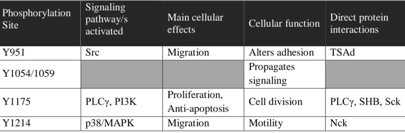
To understand the role Flk1 plays within EC of growing vessels, mechanistic studies investigating the main phosphorylation sites that trigger signaling were mapped. The four main tyrosine/Tyr/Y phosphorylation sites on Flk1 were found to be Y951, Y1054/1059, Y1175, and Y1214 (Olsson et al. 2006). The receptor tyrosine kinase activity is initiated through a
phosphorylation cascade, largely downstream of phospholipase C gamma (PLCγ) and phosphoinositide 3-kinase (PI3K). The sites and their effects are summarized in Table 1 and explained in further detail below.
Table 1: Major Phosphorylation Sites of Flk1 and Cellular Effects/Interactions.
Phosphorylation Site Signaling pathway/s activated Main cellular
effects Cellular function
Direct protein interactions
Y951 Src Migration Alters adhesion TSAd
Y1054/1059 Propagates
signaling Y1175 PLCγ, PI3K Proliferation,
Anti-apoptosis Cell division PLCγ, SHB, Sck
Y1214 p38/MAPK Migration Motility Nck
the activating phosphorylation sites on the Flk1 receptor. Phosphorylation at Y951 alters cell surface localization of VE-Cadherin and cytoskeletal rearrangements that are needed for cellular migration through T-cell-specific adapter molecule (TSAd) (Wu et al. 2000; Matsumoto et al. 2005; Gavard & Gutkind 2006). This phosphorylation event is therefore associated with chemotaxis and an increase in migratory potential. In fact, recent studies demonstrated TSAd is directly linked to junction adhesiveness and migration (Gordon et al. 2016). Phosphorylation at Y1214 also induces migration through activating p38 mitogen-activated protein kinase (MAPK) when phosphorylated (Lamalice et al. 2004; Lamalice et al. 2006). Unlike the other two sites, when Y1175 is phosphorylated it recruits and phosphorylates PLCγ, which then induces activation of protein kinase C (PKC) to increase the proliferation
(thereby also blocking apoptosis) (Takahashi et al. 1999; Meadows et al. 2001). Active PKC downstream of Flk1 signaling initiates a signaling cascade that is directly linked to and increases DNA synthesis (Xia et al. 1996; Takahashi et al. 1999). This signaling serves to increase the number of EC, generating new EC that can be used to expand the vessel network. Phosphorylated Y1175 can also activate migration through FAK/Cdc42 downstream of PI3K signaling and through Src activation (Xia et al. 1996; Wong & Jin 2005; Holmqvist et al. 2004). The combination of increasing both proliferation and migration results in more cells which are motile within the vessel, the two key components to angiogenesis.
receptor deletion phenotype (Sakurai et al. 2005). These two deletion experiments indicate that Flk1 is required for proliferation and survival of the endothelium, and support a critical role for the Y1175 phosphorylation site. Flk1-/- cells within chimeric mouse embryos, generated with Flk1-deficient stem cells, did not contribute to early blood islands which generate the
hematopoietic lineage or nascent vascular beds; indicating VEGF-A signaling through Flk1 is essential for ECs to contribute to the growing vasculature (Shalaby et al. 1997). Similarly, the two zebrafish isoforms of Flk1, kdra and kdrb, are both expressed and essential in vessel growth (Bahary et al. 2007). Depletion of either kdr in zebrafish embryos using MO injection results in angiogenic failure, specifically the intersegmental vessels (ISVs) from the common artery do not sprout (Jin et al. 2005; Wiley et al. 2011). In fact, the EC restricted expression of this receptor is specific, such that the kdr promoter driving GFP is commonly used as a
transgenic fish line for visualization of blood vessels from as early as 20 hpf (Lawson &
Weinstein 2002; Wiley et al. 2011). These in vivo studies demonstrate an intimate link between blood vessel growth and VEGF-A signaling through Flk1.
Binding of VEGF-A to Flk1 causes a series of intracellular signaling events resulting in blood vessel growth through angiogenesis. The combined data from biochemical studies and in vivo analyses indicate how appropriate activation and regulation of this receptor is essential for endothelial growth and quiescence, thus implicating this ligand-receptor cascade in
b. Flt1 during developmental angiogenesis
VEGFR1/Flt1 was identified as a tyrosine kinase receptor for members of the VEGF ligand family. VEGF-A binds to the Flt1 receptor with a Kd of 15 pM, a 10-fold higher affinity than Flk1, but exhibits weak kinase activity(Kendall & Thomas 1993; Ito et al. 1998). Additionally, Flt1 is expressed as two alternatively spliced isoforms, a membrane-bound tyrosine kinase transmembrane receptor (mFlt) and a soluble isoform lacking the transmembrane and tyrosine kinase domains (sFlt) (de Vries et al. 1992; Kendall & K. A. Thomas 1993; C. P. Thomas et al. 2010).
Flt1 is expressed selectively in the endothelial lineage of mice, both developmentally and postnatally (Peters et al. 1993). Complete Flt1 deletion in mice (flt1-/-) is embryonic lethal by E9.5 due to a poorly organized vasculature and over-proliferation of hemangioblasts over ECs (Fong et al. 1995; Fong et al. 1999). Interestingly, inactivation of the tyrosine kinase domain, which presumably blocks signaling activity while preserves ligand binding activity, results in a developmentally normal mouse; although the mice are more susceptible to pathological
that despite lacking a strong signaling role during development, Flt1 is required to generate and pattern EC to expand the vasculature during both vasculogenesis and angiogenesis in mice.
In addition to genetic mouse studies, recent work has explored the role of Flt1 in zebrafish angiogenesis. Flt1 is expressed in the embryonic fish vasculature, in both the arterial lineage of the intersegmental vessels (ISVs) and venous in the caudal vein plexus (CVP) (Krueger et al. 2011). Analysis of loss-of-function (LOF) and gain-of-function (GOF) manipulations in the sub-intestinal vessels (SIVs) demonstrated a regulatory role for Flt1 in angiogenesis.
Specifically, excess and precocious sprouting was in SIVs without Flt1 and dampened after reintroduction of Flt1 by mRNA injection (Avraham-Davidi et al. 2012). LOF MO and GOF mRNA injections of VEGF-A and Flt1 also demonstrated that Flt1 influences branching via tip cell formation in the ISVs; thus mimicking the phenotypes in the postnatal retinal vessels of mice (Chappell et al. 2009; Krueger et al. 2011). Again these data suggest that Flt1 restricts angiogenesis, as its deletion results in excess blood vessel growth.
Finally, previous work from our group examined the role of Flt1 and its isoforms in vitro using mouse embryonic stem (ES) cells that undergo endogenously directed differentiation and form a multi-cell type bed from which an EC network sprouts and expands (Rylova et al. 2008). The normal pattern of complex vessel branching is lost and proliferation is increased in flt1-/- mouse ES cell-derived vessels (Kearney et al. 2002; Kearney et al. 2004). The
proliferative effects in vessels are present when either isoform is absent, although the
branching is largely regulated by sFlt1 within these vessels (Kappas et al. 2008). We recently discovered that flt1-/- mouse ES cell-derived vessels show increased sprout initiations
(Kappas et al. 2008; Chappell et al. 2009). These discrepant effects were resolved by the fact that flt1-/- vessel connections are less stable over time (Chappell et al. 2016). In total, these experiments suggest a reliance on Flt1, acting as a VEGF-A ligand sink in sprouting
angiogenesis, to fine-tune VEGF-A levels during angiogenesis. This role supported by in vivo and in vitro studies in model systems.
Flt1 has been demonstrated to be a ligand-sink for VEGF-A in many contexts, both vasculogenic and angiogenic. As the angiogenic and developmental role of the receptor has been demonstrated to function outside of its signaling capabilities, the phosphorylation sites and signaling cascades, while understood at a basic level (Koch et al. 2011), are not discussed here. Rather this receptor is of greater interest for its potential use by the EC to determine local levels of VEGF-A signaling, previously hypothesized as a method by which anastomosis is directed during angiogenesis.
c. VEGF ligand effects in animal models
Genetic deletion of even a single VEGFA allele, vegfa+/-, causes embryonic lethality by E10.5 in mouse embryos (Carmeliet et al. 1996; Ferrara et al. 1996). VEGFA hemizygous embryos exhibit pronounced endothelial apoptosis and compromised vessel integrity,
characterized by a failure in dorsal aorta closure and decreased EC numbers within the vessels of many organ systems (Carmeliet et al. 1996; Ferrara et al. 1996); thus implicating VEGFA in the embryonic vascular system. Following the initial characterization of VEGFA in animal models, a large body of work has explored the role of VEGF ligands in multiple contexts and animal systems.
three most prevalent isoforms in angiogenic vessels and many of their distinct roles in vessel growth have been elucidated. Mice genetically engineered to express only VEGF-120 survive birth but die by two weeks of age due to cardiorespiratory failure (Carmeliet et al. 1999). Both VEGF-164 and VEGF-188 isoform expressing mice survive to fertility, although expression of only VEGF-188 exhibits reduced survival and non-Mendelian ratios (Stalmans et al. 2002). VEGF-120 and VEGF-188 expressing mice have decreased and disrupted vessel morphology in the retinal of postnatal mice, although the VEGF-120 mice show a more severe phenotype than the VEGF-188 (Stalmans et al. 2002). Thus, regardless of the isoform, VEGF-A ligand interacts with EC to promote vessel growth. In fact, the shared vessel phenotypes in these rescue models suggest an overlapping and conserved ability of VEGF-A to regulate ECs. This conclusion is supported by parallel experiments in zebrafish that express multiple isoforms of VEGF-A, including both VEGF-120 and VEGF-164, within the endothelial lineage of embryos as determined by in situ hybridization (Liang et al. 1998; Liang et al. 2001; Bahary et al. 2007). Over-expression of either of these isoforms, through mRNA injection, results in excessive vessel growth and an increased commitment of cells to the hematopoietic lineage (Liang et al. 2001). Morpholino (MO) injection to block all VEGF-A ligand causes a decrease in vessels throughout the embryo (Nasevicius et al. 2000). Combined, the data from these model systems support a conserved role the VEGF-A ligand in appropriate proliferation and health of the endothelium.
Jakobsson et al. 2006; Kawamura et al. 2008). VEGF-A bound to heparan sulfate in the ECM confers the spatial patterning of the blood vessels via ligand sequestration in ECM that generates a growth factor gradient to direct angiogenesis (Ruhrberg et al. 2002; Jakobsson et al. 2010). Complexing of VEGF-A and Flk1 or Flt1 at the cell surface with NRP-1 promotes phosphorylation that augments downstream signaling and increases migration and sprouting of endothelial cells (Wang et al. 2003; Pan et al. 2007). Further increases in signal propagation are accomplished by αVβ3-integrin, which complex with VEGF-A/Flk1/NRP to amplify the signal cascade (Ruhrberg et al. 2002; Robinson et al. 2009). These data demonstrate that VEGF-A signaling activity within EC is not only a product of ligand binding at receptors but also impacted by the complexing of additional cofactors, an intricate and complicated series of binding.
III. Sprouting angiogenesis during development
Sprouting angiogenesis is initiated by mature ECs within established vessels. After exposure to new growth factors these EC activate proliferative and migratory programs in order to extend processes and initiate a new sprout, followed by migration into the extravascular space along guidance cues, then connection or anastomosis with another vessel, and finally create a continual luminal space to carry blood flow (Chappell & Bautch 2010). Each of these processes is
described in more detail below.
a. Sprout initiation from quiescent vessels
Sprouts form in response to a complex interplay of growth factors within the vascularized tissue. The vessels start as stable and quiescent, with the EC contributing to the vessel wall but not actively migratory or proliferative, and surrounding by the supportive pericytes and
macrophages along with the basement membrane. Only upon a change in growth factors in the local environment do the EC activate and undergo structural changes.
A wide array of growth factors have been implicated in sprout initiations, including VEGF, Notch, and bone morphogenetic protein (BMP) (Kappas et al. 2008; Wiley et al. 2011;
Hellström et al. 2007). Notably, EC with low Notch are primed to respond to growth factor ligands, such as BMP and VEGF, and contribute to a new sprout while their immediate
individual ECs. Sprout initiation is a result of the combined effects of these signaling
pathways, leading to the EC exiting quiescence. Exit from quiescence requires suppression of Notch signaling, demonstrated by multiple LOF Notch manipulations in mouse vessels that show increased sprout formation (del Toro et al. 2010). For the EC in the sprout to migrate away from the parent vessel and into the ECM, the EC loosens established EC-EC junctions, alter cellular structures and extends filopodia (Chen et al. 2010; Cruys et al. 2016; Gordon et al. 2016; Kushner et al. 2014; Wright et al. 2015), both effects known to occur downstream of VEGF-A signaling (Senger et al. 1983; Keck et al. 1989; Dvorak et al. 1995; Taylor et al. 2010; Zeng & Bautch 2009).
From these data it becomes apparent that the cumulative growth factor effects within each cell contribute to sprout initiation within a vessel. Thus understanding growth factor
interactions and EC within the context of whole vessels is essential for a complete understanding of how these signaling pathways initiate sprouting angiogenesis. Once an individual cell has initiated a migratory program and exits the axis of the parent vessel it is a part of the new, active sprout.
b. Sprout migration and extension
To expand the vessel network, the sprout must migrate away from the parent vessel and towards another existing vessel. Cell migration is characterized by the extension of filopodia and selectively EC activation, which are both observed when Notch or VEGF-A signaling are altered (Gerhardt et al. 2003; Hellström et al. 2007; Bentley et al. 2014). This sprouting process requires two components, guidance cues to direct sprout growth and coordinated cell
The signaling and growth cues which direct migration function both locally and over long distances. The local growth factor environment surrounding the cell is influenced by secreted proteins by the EC; for example, Flt1 modulation of the VEGF-A gradient in the ECM directs sprouts to migrate away from the parent vessel (Chappell et al. 2009). Similar to neuronal migration, EC are predicted to use filopodia to sense directional migration along local and long distance cues within the targeted tissue (Kater & Rehder 1995). As they encounter these
guidance factors, the sprouts either alter their directional migration to either persist or redirect the trajectory of the vessel. The EC within the sprout undergo collective cell migration and individual cells within the vessel change positions frequently as the sprout extends into the ECM along guidance cues (Arima et al. 2011; Jakobsson et al. 2010). Migration and EC
movement within the sprout is facilitated by extended junctions at the sprout front (Pelton et al. 2014), resulting in cells which move in parallel and providing a border that permits cell-cell communication and presumably coordinates EC activity. The ability of EC to communicate and migrate is dependent on multiple signaling pathways, including Notch and VEGF-A. Increased Notch signaling, through genetic deletion of Dll4, or loss of VEGF-A signaling, due to heterozygously deleted Flk1, reduces migratory potential; conversely, increasing VEGF-A signaling, through loss of Flt1, increases migratory potential (Jakobsson et al. 2010).
Through a combination of these external cues and cell communications, the sprout migrates in a directional manner within the extracellular space. From these data a picture of sprout migration emerges. The ECM and existing vessels contain factors to direct the nascent sprout, which is able to follow these sets of cues by continually extending sensory processes,
c. Anastomosis to create connections and new vessel conduits
At the most basic level, anastomosis requires that two cells generate a new, stable, cell junction; appropriately, junction components have been the main consideration in anastomotic studies to date. Within the endothelial cells the creation of new junctions occurs in a
stereotyped order. The order is as follows: first is homophilic binding of platelet endothelial cell adhesion molecule 1 (PECAM) on the surface of two EC; second is establishment of the adherens junctions (AJs), composed of multiple proteins including vascular endothelial cadherin (VE-Cad), β-catenin, p120, and plakoglobin; and finally the tight junctions (TJs), comprising proteins like zona occludins 1 (ZO1), claudins, nectins, and junctional adhesion molecules (JAMS) (Dejana 2004). The formation of each of these junctions is briefly discussed below.
protein at the cell surface and set up the basis for AJ formation. Normal and full expression of VE-Cadherin is essential for new connections (Lenard et al. 2013; Montero-Balaguer et al. 2009). The AJ components, including β-catenin and p120, are then actively recruited and maintained at EC junctions thus stabilizing the new EC-EC interaction through a positive feedback loop of AJ recruitment (Williams et al. 2000; Gaengel et al. 2012). Finally, stable scaffolding junctional proteins, namely ZO1, claudins and occludins, create TJs at the basal side of the junction. These junctions provide structural integrity to the cell in a tissue specific manner, for example the lung vessels have loosely organized TJs to allow exchange but the neurovascular interface has rigid and complex TJ (Gallicano et al. 2001; Saitou et al. 2000). All three types of junctions are detected between EC in established vessels, indicating their essential nature to blood vessels and linking their deposition at sites of connection to anastomosis.
but are not an obligate component of anastomosis. Another non-obligate contributor was found in the F4/80+ macrophages in angiogenic retinal vessels of postnatal mice. F4/80+
macrophages were coincident with many sprout fronts and branch points, suggesting these cells support the vessel network, and potentially connections, during angiogenesis (Fantin et al. 2010; Outtz et al. 2011).
Combined, these studies suggest that the ability of vessels to form new, stable connections depends on creating new junctions and is assisted by non-EC support cells. However, a large number of questions remain from these data. For instance, whether new connections between EC are directed or stochastic, the signaling changes within sprouts and existing vessels as the two discrete EC form a new junction and which signaling components are most critical for permitting or blocking connections.
As the physical aspects involved in the anastomosis have been described, at least in regards to the generation of new junctions, studying the regulatory and signaling events that direct these physical changes is a high priority. The signaling that directs endothelial cells during terminal guidance and anastomosis is a pressing question which is currently unaddressed by existing studies. The fluctuations between high Notch and high VEGF-A are conducive for coordinated migration of EC as a sprout but no information on how these signaling cascades influence and direct connections is available.
d. Formation of a new lumen
main processes thought to be critical to lumen formation are apical-basal polarity and blood flow.
Apical-basal polarity is established within a cell to define a ‘top’ and ‘bottom’,
respectively, in tissues that require this additional structural information for each cell, most notably in epithelium (Datta et al. 2011). This polarity is firmly established within vessels, such as the arterioles, and observed by unequal distribution of proteins like portioning
defective homolog 3 (Par3), podocalyxin, and β1 integrin (Horvat et al. 1986; Iruela-Arispe &
Davis 2009; Zovein et al. 2010). However, the distinct sidedness must be created or transferred when a new vessel is formed. These components are actively shuttled to and maintained at the appropriate domains of the cell surface by assistive proteins. Cell division control protein 42 (Cdc42) and Ras-related C3 botulinum toxin substrate 1 (Rac1) are known to establish apical-basal polarity in epithelial cells and are also required for EC to create lumens (Koh et al. 2008). Expression of podocalyxin (PODXL) and β1 integrin, the established markers in lumens for
apical and basal polarity, respectively, can be found between the leading cells in a sprout (Horvat et al. 1986; Pelton et al. 2014), suggesting EC are primed for lumen formation early on during sprouting. The final stage of lumenization requires joining two formed lumens in
Once the new, continual lumen is formed, exchange of blood flow between the pre-existing vessels is found. At this point the vessels return to a quiescent state, and sprouting angiogenesis considered completed.
IV. Perspective
V. REFERENCES
Aase, K. et al., 1999. Localization of VEGF‐ B in the mouse embryo suggests a paracrine role of the growth factor in the developing vasculature. Developmental Dynamics, 215(1), pp.12– 25.
Arima, S. et al., 2011. Angiogenic morphogenesis driven by dynamic and heterogeneous collective endothelial cell movement. Development, 138(21), pp.4763–4776.
Avraham-Davidi, I. et al., 2012. ApoB-containing lipoproteins regulate angiogenesis by modulating expression of VEGF receptor 1. Nature Medicine, 18(6), pp.967–973.
Bahary, N. et al., 2007. Duplicate VegfA genes and orthologues of the KDR receptor tyrosine kinase family mediate vascular development in the zebrafish. Blood, 110(10), pp.3627–3636. Bellomo, D. et al., 2000. Mice lacking the vascular endothelial growth factor-B gene (Vegfb)
have smaller hearts, dysfunctional coronary vasculature, and impaired recovery from cardiac ischemia. Circulation Research, 86(2), pp.E29–35.
Benedito, R. et al., 2009. The Notch Ligands Dll4 and Jagged1 Have Opposing Effects on Angiogenesis. Cell, 137(6), pp.1124–1135.
Bentley, K. et al., 2014. The role of differential VE-cadherin dynamics in cell rearrangement during angiogenesis. Nature Cell Biology, 16(4), pp.309–321.
Carmeliet, P. et al., 1996. Abnormal blood vessel development and lethality in embryos lacking a single VEGF allele. Nature, 380(6573), pp.435–439.
Carmeliet, P. et al., 1999. Impaired myocardial angiogenesis and ischemic cardiomyopathy in mice lacking the vascular endothelial growth factor isoforms VEGF164 and VEGF188. Nature Medicine, 5(5), pp.495–502.
Chappell, J.C. & Bautch, V.L., 2010. Vascular development: genetic mechanisms and links to vascular disease. Current Topics in Developmental Biology, 90, pp.43–72.
Chappell, J.C. et al., 2016. Flt-1 (VEGFR-1) coordinates discrete stages of blood vessel formation. Cardiovascular research, 111(1), pp.84–93.
Chappell, J.C. et al., 2009. Local guidance of emerging vessel sprouts requires soluble Flt-1. Developmental Cell, 17(3), pp.377–386.
Chen, J.N. et al., 1996. Mutations affecting the cardiovascular system and other internal organs in zebrafish. Development, 123, pp.293–302.
Communications, 7, p.12240.
Datta, A., Bryant, D.M. & Mostov, K.E., 2011. Molecular regulation of lumen morphogenesis. Current biology: CB, 21(3), pp.R126–36.
de Vries, C. et al., 1992. The fms-like tyrosine kinase, a receptor for vascular endothelial growth factor. Science, 255(5047), pp.989–991.
Dejana, E., 2004. Endothelial cell–cell junctions: happy together. Nature Reviews Molecular Cell Biology, 5(4), pp.261–270.
del Toro, R. et al., 2010. Identification and functional analysis of endothelial tip cell-enriched genes. Blood, 116(19), pp.4025–4033.
Drake, C.J. & Fleming, P.A., 2000. Vasculogenesis in the day 6.5 to 9.5 mouse embryo. Blood, 95(5), pp.1671–1679.
Dvorak, H.F. et al., 1995. Vascular permeability factor/vascular endothelial growth factor, microvascular hyperpermeability, and angiogenesis. American Journal of Pathology, 146(5), pp.1029–1039.
Esser, S. et al., 1998. Vascular endothelial growth factor induces VE-cadherin tyrosine phosphorylation in endothelial cells. Journal of Cell Science, 111(13), pp.1853–1865. Fantin, A. et al., 2010. Tissue macrophages act as cellular chaperones for vascular anastomosis
downstream of VEGF-mediated endothelial tip cell induction. Blood, 116(5), pp.829–840. Feng, D. et al., 2004. Ultrastructural localization of platelet endothelial cell adhesion molecule
(PECAM-1, CD31) in vascular endothelium. The Journal of Histochemistry and Cytochemistry, 52(1), pp.87–101.
Ferrara, N. & Henzel, W.J., 1989. Pituitary follicular cells secrete a novel heparin-binding growth factor specific for vascular endothelial cells. Biochemical and Biophysical Research Communications, 161(2), pp.851–858.
Ferrara, N. et al., 1996. Heterozygous embryonic lethality induced by targeted inactivation of the VEGF gene. Nature, 380(6573), pp.439–442.
Folkman, J. et al., 1971. Isolation of a tumor factor responsible for angiogenesis. The Journal of Experimental Medicine, 133(2), pp.275–288.
Fong, G.-H. et al., 1999. Increased hemangioblast commitment, not vascular disorganization, is the primary defect in flt-1 knock-out mice. Development, 126(13), pp.3015–3025.
Fong, G.H. et al., 1995. Role of the Flt-1 receptor tyrosine kinase in regulating the assembly of vascular endothelium. Nature, 376(6535), pp.66–70.
Angiogenesis by Regulating the Interplay between VE-Cadherin and VEGFR2. Developmental Cell, 23(3), pp.587–599.
Gallicano, G.I., Bauer, C. & Fuchs, E., 2001. Rescuing desmoplakin function in extra-embryonic ectoderm reveals the importance of this protein in embryonic heart, neuroepithelium, skin and vasculature. Development, 128(6), pp.929–941.
Gavard, J. & Gutkind, J.S., 2006. VEGF controls endothelial-cell permeability by promoting the β-arrestin-dependent endocytosis of VE-cadherin. Nature Cell Biology, 8(11), pp.1223– 1234.
Gebala, V. et al., 2016. Blood flow drives lumen formation by inverse membrane blebbing during angiogenesis in vivo. Nature Cell Biology, 18, pp.443-450.
Gerhardt, H. et al., 2003. VEGF guides angiogenic sprouting utilizing endothelial tip cell filopodia. The Journal of Cell Biology, 161(6), pp.1163–1177.
Gordon, E.J. et al., 2016. The endothelial adaptor molecule TSAd is required for VEGF-induced angiogenic sprouting through junctional c-Src activation. Science Signaling, 9(437),
pp.ra72–ra72.
Hellström, M. et al., 2007. Dll4 signalling through Notch1 regulates formation of tip cells during angiogenesis. Nature, 445(7129), pp.776–780.
Herwig, L. et al., 2011. Distinct cellular mechanisms of blood vessel fusion in the zebrafish embryo. Current Biology, 21(22), pp.1942–1948.
Hiratsuka, S. et al., 1998. Flt-1 lacking the tyrosine kinase domain is sufficient for normal development and angiogenesis in mice. Proceedings of the National Academy of Sciences, 95(16), pp.9349–9354.
Hiratsuka, S. et al., 2001. Involvement of Flt-1 tyrosine kinase (vascular endothelial growth factor receptor-1) in pathological angiogenesis. Cancer Research, 61(3), pp.1207–1213. Hogan, B.M. et al., 2009. Vegfc/Flt4 signalling is suppressed by Dll4 in developing zebrafish
intersegmental arteries. Development, 136(23), pp.4001–4009.
Holmqvist, K. et al., 2004. The Adaptor Protein Shb Binds to Tyrosine 1175 in Vascular Endothelial Growth Factor (VEGF) Receptor-2 and Regulates VEGF-dependent Cellular Migration. Journal of Biological Chemistry, 279(21), pp.22267–22275.
Horvat, R. et al., 1986. Endothelial cell membranes contain podocalyxin--the major sialoprotein of visceral glomerular epithelial cells. The Journal of Cell Biology, 102(2), pp.484–491. Houck, K.A. et al., 1992. Dual regulation of vascular endothelial growth factor bioavailability by
Iruela-Arispe, M.L. & Davis, G.E., 2009. Cellular and Molecular Mechanisms of Vascular Lumen Formation. Developmental Cell, 16(2), pp.222–231.
Isogai, S., Horiguchi, M. & Weinstein, B.M., 2001. The Vascular Anatomy of the Developing Zebrafish: An Atlas of Embryonic and Early Larval Development. Developmental Biology, 230(2), pp.278–301.
Ito, N. et al., 1998. Identification of Vascular Endothelial Growth Factor Receptor-1 Tyrosine Phosphorylation Sites and Binding of SH2 Domain-containing Molecules. Journal of Biological Chemistry, 273(36), pp.23410–23418.
Jakobsson, L. et al., 2010. Endothelial cells dynamically compete for the tip cell position during angiogenic sprouting. Nature Cell Biology, 12(10), pp.943–953.
Jakobsson, L. et al., 2006. Heparan Sulfate in trans Potentiates VEGFR-Mediated Angiogenesis. Developmental Cell, 10(5), pp.625–634.
Jeltsch, M. et al., 1997. Hyperplasia of lymphatic vessels in VEGF-C transgenic mice. Science, 276(5317), pp.1423–1425.
Jin, S.-W. et al., 2005. Cellular and molecular analyses of vascular tube and lumen formation in zebrafish. Development, 132(23), pp.5199–5209.
Kappas, N.C. et al., 2008. The VEGF receptor Flt-1 spatially modulates Flk-1 signaling and blood vessel branching. The Journal of Cell Biology, 181(5), pp.847–858.
Kater, S.B. & Rehder, V., 1995. The sensory-motor role of growth cone filopodia. Current Opinion in Neurobiology, 5(1), pp.68–74.
Kawamura, H. et al., 2008. Neuropilin-1 in regulation of VEGF-induced activation of p38MAPK and endothelial cell organization. Blood, 112(9), pp.3638–3649.
Kearney, J.B. et al., 2004. The VEGF receptor flt-1 (VEGFR-1) is a positive modulator of vascular sprout formation and branching morphogenesis. Blood, 103(12), pp.4527–4535. Kearney, J.B. et al., 2002. Vascular endothelial growth factor receptor Flt-1 negatively regulates
developmental blood vessel formation by modulating endothelial cell division. Blood, 99(7), pp.2397–2407.
Keck, P.J. et al., 1989. Vascular permeability factor, an endothelial cell mitogen related to PDGF. Science, 246(4935), pp.1309–1312.
Kendall, R.L. & Thomas, K.A., 1993. Inhibition of vascular endothelial cell growth factor activity by an endogenously encoded soluble receptor. Proceedings of the National Academy of Sciences, 90(22), pp.10705–10709.
Journal of Biological Chemistry, 274(10), pp.6453–6460.
Koch, S. et al., 2011. Signal transduction by vascular endothelial growth factor receptors. Biochemical Journal, 437(2), pp.169–183.
Koh, W., Mahan, R.D. & Davis, G.E., 2008. Cdc42- and Rac1-mediated endothelial lumen formation requires Pak2, Pak4 and Par3, and PKC-dependent signaling. Journal of Cell Science, 121(Pt 7), pp.989–1001.
Krueger, J. et al., 2011. Flt1 acts as a negative regulator of tip cell formation and branching morphogenesis in the zebrafish embryo. Development, 138(10), pp.2111–2120.
Kushner, E.J. et al., 2014. Excess centrosomes disrupt endothelial cell migration via centrosome scattering. The Journal of Cell Biology, 206(2), pp.257–272.
Lamalice, L. et al., 2004. Phosphorylation of tyrosine 1214 on VEGFR2 is required for VEGF-induced activation of Cdc42 upstream of SAPK2/p38. Oncogene, 23(2), pp.434–445.
Lamalice, L., Houle, F. & Huot, J., 2006. Phosphorylation of Tyr 1214within VEGFR-2 Triggers the Recruitment of Nck and Activation of Fyn Leading to SAPK2/p38 Activation and
Endothelial Cell Migration in Response to VEGF. Journal of Biological Chemistry, 281(45), pp.34009–34020.
Lawson, N.D. & Weinstein, B.M., 2002. Arteries and veins: making a difference with zebrafish. Nature Reviews Genetics, 3(9), pp.674–682.
Lenard, A. et al., 2013. In vivo analysis reveals a highly stereotypic morphogenetic pathway of vascular anastomosis. Developmental Cell, 25(5), pp.492–506.
Leung, D.W. et al., 1989. Vascular endothelial growth factor is a secreted angiogenic mitogen. Science, 246(4935), pp.1306–1309.
Liang, D. et al., 1998. Cloning and characterization of vascular endothelial growth factor (VEGF) from zebrafish, Danio rerio. Biochimica et Biophysica Acta, 1397(1), pp.14–20. Liang, D. et al., 2001. The role of vascular endothelial growth factor (VEGF) in vasculogenesis,
angiogenesis, and hematopoiesis in zebrafish development. Mechanisms of Development, 108(1-2), pp.29–43.
Lobov, I.B. et al., 2007. Delta-like ligand 4 (Dll4) is induced by VEGF as a negative regulator of angiogenic sprouting. Proceedings of the National Academy of Sciences, 104(9), pp.3219– 3224.
Lohela, M. et al., 2009. VEGFs and receptors involved in angiogenesis versus lymphangiogenesis. Current Opinion in Cell Biology, 21(2), pp.154–165.
Meadows, K.N., Bryant, P. & Pumiglia, K., 2001. Vascular endothelial growth factor induction of the angiogenic phenotype requires Ras activation. The Journal of Biological Chemistry, 276(52), pp.49289–49298.
Montero-Balaguer, M. et al., 2009. Stable Vascular Connections and Remodeling Require Full Expression of VE-Cadherin in Zebrafish Embryos. PLoS ONE, 4(6), p.e5772.
Nasevicius, A., Larson, J. & Ekker, S.C., 2000. Distinct requirements for zebrafish angiogenesis revealed by a VEGF-A morphant. Yeast, 17(4), pp.294–301.
Olsson, A.-K. et al., 2006. VEGF receptor signalling — in control of vascular function. Nature Reviews Molecular Cell Biology, 7(5), pp.359–371.
Outtz, H.H. et al., 2011. Notch1 controls macrophage recruitment and Notch signaling is
activated at sites of endothelial cell anastomosis during retinal angiogenesis in mice. Blood, 118(12), pp.3436–3439.
Pan, Q. et al., 2007. Neuropilin-1 Binds to VEGF 121and Regulates Endothelial Cell Migration and Sprouting. Journal of Biological Chemistry, 282(33), pp.24049–24056.
Pelton, J.C. et al., 2014. Multiple endothelial cells constitute the tip of developing blood vessels and polarize to promote lumen formation. Development, 141(21), pp.4121–4126.
Peters, K.G., de Vries, C. & Williams, L.T., 1993. Vascular endothelial growth factor receptor expression during embryogenesis and tissue repair suggests a role in endothelial
differentiation and blood vessel growth. Proceedings of the National Academy of Sciences, 90(19), pp.8915–8919.
Phng, L.-K., Stanchi, F. & Gerhardt, H., 2013. Filopodia are dispensable for endothelial tip cell guidance. Development, 140(19), pp.4031–4040.
Privratsky, J.R. et al., 2011. Relative contribution of PECAM-1 adhesion and signaling to the maintenance of vascular integrity. Journal of Cell Science, 124(9), pp.1477–1485.
Quinn, T.P. et al., 1993. Fetal liver kinase 1 is a receptor for vascular endothelial growth factor and is selectively expressed in vascular endothelium. Proceedings of the National Academy of Sciences, 90(16), pp.7533–7537.
Risau, W. & Flamme, I., 1995. Vasculogenesis. Annual Review of Cell and Developmental Biology, 11, pp.73–91.
Robinson, S.D. et al., 2009. Alphav beta3 integrin limits the contribution of neuropilin-1 to vascular endothelial growth factor-induced angiogenesis. Journal of Biological Chemistry, 284(49), pp.33966–33981.
Rylova, S.N., Randhawa, P.K. & Bautch, V.L., 2008. Chapter 6: In Vitro Differentiation of Mouse Embryonic Stem Cells Into Primitive Blood Vessels. Methods in Enzymology, pp. 103–117.
Saitou, M. et al., 2000. Complex phenotype of mice lacking occludin, a component of tight junction strands. Molecular Biology of the Cell, 11(12), pp.4131–4142.
Sakurai, Y. et al., 2005. Essential role of Flk-1 (VEGF receptor 2) tyrosine residue 1173 in vasculogenesis in mice. Proceedings of the National Academy of Sciences, 102(4), pp.1076– 1081.
Sauteur, L. et al., 2014. Cdh5/VE-cadherin promotes endothelial cell interface elongation via cortical actin polymerization during angiogenic sprouting. Cell Reports, 9(2), pp.504–513. Schoppmann, S.F. et al., 2010. Tumor-Associated Macrophages Express Lymphatic Endothelial
Growth Factors and Are Related to Peritumoral Lymphangiogenesis. American Journal of Pathology, 161(3), pp.947–956.
Senger, D.R. et al., 1983. Tumor cells secrete a vascular permeability factor that promotes accumulation of ascites fluid. Science, 219(4587), pp.983–985.
Shalaby, F. et al., 1997. A requirement for Flk1 in primitive and definitive hematopoiesis and vasculogenesis. Cell, 89(6), pp.981–990.
Shinkai, A. et al., 1998. Mapping of the Sites Involved in Ligand Association and Dissociation at the Extracellular Domain of the Kinase Insert Domain-containing Receptor for Vascular Endothelial Growth Factor. Journal of Biological Chemistry, 273(47), pp.31283–31288. Stalmans, I. et al., 2002. Arteriolar and venular patterning in retinas of mice selectively
expressing VEGF isoforms. Journal of Clinical Investigation, 109(3), pp.327–336.
Suchting, S. et al., 2007. The Notch ligand Delta-like 4 negatively regulates endothelial tip cell formation and vessel branching. Proceedings of the National Academy of Sciences, 104(9), pp.3225–3230.
Sun, J. et al., 2000. Contributions of the extracellular and cytoplasmic domains of platelet-endothelial cell adhesion molecule-1 (PECAM-1/CD31) in regulating cell-cell localization. Journal of Cell Science, 113 ( Pt 8), pp.1459–1469.
Takahashi, T. et al., 2001. A single autophosphorylation site on KDR/Flk-1 is essential for VEGF-A-dependent activation of PLC-gamma and DNA synthesis in vascular endothelial cells. The EMBO Journal, 20(11), pp.2768–2778.
Takahashi, T., Ueno, H. & Shibuya, M., 1999. VEGF activates protein kinase C-dependent, but Ras-independent Raf-MEK-MAP kinase pathway for DNA synthesis in primary endothelial cells. Oncogene, 18(13), pp.2221–2230.
endothelial cells of developing blood vessels. Blood, 116(16), pp.3108–3117.
Terman, B.I. et al., 1992. Identification of the KDR tyrosine kinase as a receptor for vascular endothelial cell growth factor. Biochemical and Biophysical Research Communications, 187(3), pp.1579–1586.
Thomas, C.P. et al., 2010. Alternate processing of Flt1 transcripts is directed by conserved cis-elements within an intronic region of FLT1 that reciprocally regulates splicing and
polyadenylation. Nucleic Acids Research, 38(15), pp.5130–5140.
Tischer, E. et al., 1991. The human gene for vascular endothelial growth factor. Multiple protein forms are encoded through alternative exon splicing. The Journal of Biological Chemistry, 266(18), pp.11947–11954.
Wang, L. et al., 2003. Neuropilin-1-mediated vascular permeability factor/vascular endothelial growth factor-dependent endothelial cell migration. The Journal of Biological Chemistry, 278(49), pp.48848–48860.
Wiley, D.M. et al., 2011. Distinct signalling pathways regulate sprouting angiogenesis from the dorsal aorta and the axial vein. Nature Cell Biology, 13(6), pp.686–692.
Williams, B.O. et al., 2000. A comparative evaluation of beta-catenin and plakoglobin signaling activity. Oncogene, 19, pp.5720–5728.
Wong, C. & Jin, Z.G., 2005. Protein kinase C-dependent protein kinase D activation modulates ERK signal pathway and endothelial cell proliferation by vascular endothelial growth factor. Journal of Biological Chemistry, 280(39), pp.33262–33269.
Wright, C.E. et al., 2015. LGN Directs Interphase Endothelial Cell Behavior via the Microtubule Network. PLoS ONE, 10(9), p.e0138763.
Wu, L.W. et al., 2000. VRAP is an adaptor protein that binds KDR, a receptor for vascular endothelial cell growth factor. The Journal of Biological Chemistry, 275(9), pp.6059–6062. Xia, P. et al., 1996. Characterization of vascular endothelial growth factor's effect on the
activation of protein kinase C, its isoforms, and endothelial cell growth. Journal of Clinical Investigation, 98(9), pp.2018–2026.
Xu, K. & Cleaver, O., 2011. Tubulogenesis during blood vessel formation. Seminars in Cell and Developmental Biology, 22(9), pp.993–1004.
Zeng, G. & Bautch, V.L., 2009. Differentiation and Dynamic Analysis of Primitive Vessels from Embryonic Stem Cells. Methods in Molecular Biology, pp. 333–344.
CHAPTER 2: Materials and Methods1
I. Experimental Materials and Methods
a. Retinal Angiogenesis Imaging and Analysis
Mice (Mus musculus) with flt-1flox/flox alleles (Genentech; San Francisco, CA) were bred with [Tg(UBC-cre/ERT2), JAX #007001] mice that also carried the Cre-mediated
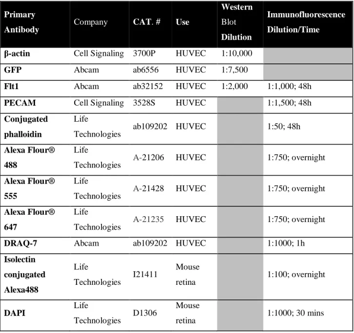
recombination reporter gene R26R TdTomato [Gt(ROSA)26Sortm14(CAG-tdTomato)Hze, JAX #007914]. Mice were maintained in accordance with the University of North Carolina, Chapel Hill Institutional Animal Care and Use Committee. Mosaic Cre excision of vessels was accomplished by IP injection of 0.5 mg/ml tamoxifen (MP Biomedicals; Santa Ana, CA) at P2. P5 retinas were perfusion fixed with 0.5% PFA/PBS, harvested, and fixed for 2h with 2% PFA. Retinas were rinsed, counterstained for isolectinB4 and DAPI, and mounted using established protocols (Chappell et al. 2009). Images were acquired on a Leica DMI 6000B or Zeiss LSM 880 confocal microscope at 40× magnification, with optimal z-stacks compressed post-acquisition using ImageJ software. All antibody manufacturers and concentrations are listed in Table 2.1.
Sprouts were identified in the angiogenic front using previously established methods (Chappell et al. 2009). Cellular genetic identity was classified from reporter expression during analysis and interactions between sprouts and other sprouts/vessels were identified as overlap of cellular extensions with other extensions/cells in the isolectinB4 channel.
1 Chapter 2 is adapted in part from Nesmith, J.E. et al. Blood Vessel Anastomosis is Spatially
b. Cell Lines and Culture
Human umbilical venous endothelial cells (HUVEC; Lonza, Portsmouth, NH) were maintained in EBM-2 media with the EGM Bullet Kit and 1X antibiotic/antimycotic (Sigma; St Louis MO) according to manufacturer’s directions. Normal human lung fibroblasts
(NHLF; Lonza, Portsmouth, NH) were maintained in high-glucose DMEM supplemented with 10% FBS and 1X antibiotic/antimycotic. Mouse embryonic stem cells isolated from WT mice and flt-1-/- mice (gift from G.H. Fong) were maintained and differentiated as previously described (Rylova et al. 2008).
c. HUVEC Sprouting Angiogenesis Assay Imaging and Analysis
The HUVEC sprouting angiogenesis assay was set up as described (Nakatsu & Hughes 2008). Briefly, HUVEC were detached, combined with Cytodex microcarrier beads (Sigma; St Louis, MO), then incubated with periodic agitation for 4h. Following overnight growth, the HUVEC-coated microcarrier beads were embedded in a 1.5% fibrinogen-thrombin gel that was allowed to polymerize at 37°C for 15-45 min, and then a layer of normal lung fibroblasts were seeded on top of the solidified fibrinogen gel.
4°C with either goat anti-mouse conjugated secondary or goat anti-rabbit conjugated
secondary in TBST or PBST (Life Technologies; Grand Island, NY). Confocal images were acquired on an Olympus FV1200 system using a 10× objective (NA 0.40) and optimal Z-stacks, compressed post-acquisition for analysis.
Vessel interactions were scored when two cell bodies overlapped in either the DIC or fluorescent channels, depending upon the experimental set-up. Interactions classified as transient contacts were defined as overlap in a single time frame, therefore lasting <10 minutes. Stable connections were defined as overlap that persisted for >3 time frames, at least 20 min. Target vessel activity was classified as static or protrusive based upon the endothelial cells within 30 µm of the interaction site. Static vessels exhibited no LifeAct protrusions extending away from the vessel axis, while protrusive vessels contained a minimum of 3 extensions, defined as fluorescent protrusions >4 m.
d. ES Cell-derived Vessel Imaging and Analysis
e. Zebrafish Experiments and Husbandry
Zebrafish (Danio rerio) lines were carried on an AB strain background with transgenic insertion of either Tg(kdrl:eGFP) or Tg(kdrl:eGFP); Tg(hsp70l:vegfaa121;cmlc2:GFP) that
were maintained as heterozygotes and genotyped through test crosses (Jin 2005; Wiley et al. 2011). Heat shock inducible expression was activated in embryos 20 hpf by incubation for 30 mins at 30°C. All zebrafish maintained in accordance with the University of North Carolina, Chapel Hill Institutional Animal Care and Use Committee.
The Flt1 morpholino was previously designed, published, and verified for vessel defects and effectiveness (Krueger et al. 2011). Flt1 morpholino (3 ng) and control (PBS of equivalent volume) injections were performed on one-cell fertilized embryos. Genotyping of experimental embryos was identified using presence or absence of GFP expression in the cardiac muscle. Embryos were fixed at 48 hpf in 4% PFA for 15 mins for end point analysis. Imaging was performed on an Olympus FV1200 using 10× magnification and optimal z-stacks, approximately 5 µm in width. Analysis was performed using Image J-Fiji software.
All embryos with gross morphological defects were excluded from quantification. ISVs at the base of the yolk were imaged as described above. The confocal images were binned according to their connections along the DLAV. Those vessels which connected with a vessel not at the DLAV or formed multiple connection at the dorsal aspect were considered
‘inappropriate’ or outside of the typical somite boundaries.
f. Protein Quantification
was performed 48h after lentivirus infection or siRNA transfection. Briefly, protein concentration was quantified by the Bradford reaction (BioRad; Berkeley, CA) and
equivalent protein amounts were loaded onto a 7.5% sodium dodecyl sulfate polyacrylamide gel for electrophoresis. Protein was transferred to a polyvinylidene fluoride (PVDF)
membrane, stained with Ponceau S solution to visualize transferred protein and then incubated with the appropriate primary antibody in TBSor PBS with 0.5% Triton-100 (Sigma; St Louis, MO). Horseradish peroxidase-conjugated secondary antibody was
incubated with membranes for 1h at RT and chemiluminescent detection was performed. All antibody manufacturers, and concentrations are listed in Table 2.1.
g. Lentivirus and siRNA knockdown
The Flt1 shRNA construct (RHS3979-201732907; Dharmacon, Lafayette, CO) was modified to include GFP in addition to shFlt1. Lentivirus was produced by the UNC
LentiCore Facility. Lentivirus was incubated with endothelial cells for 6-8h with addition of 0.25 µg/mL of polybrene (EMD Millipore; Billerica, MA). Lentiviral infection with LifeAct-GFP or LifeAct-RFP (gifts from Rusty Lansford [Addgene plasmid #51010] and Weiping Han [Addgene plasmid #64048], respectively) allowed visualization of F-actin.
h. Fluorescence-activated Cell Sorting (FACS) Analysis
HUVEC were washed with PBS, detached using 1X accutase (Sigma; St. Louis, MO) and
then fixed in FACS buffer containing 1% PFA. Cells were analyzed by flow cytometry using
a BD AccuriTM C6 flow cytometer and CFlow Plus Analysis software (BD Biosciences; San
Jose, CA). Samples were manually gated and analyzed using the FloJo v10 software package.
i. mRNA Preparation and Quantification
RNA was collected 48h post-treatment using TRIzol® (Life Technologies; Grand Island, NY), according to the manufacturer’s protocol, and converted to cDNA using iScript
(BioRad; Berkeley, CA), according to the manufacturer’s protocol. Quantitative real-time polymerase chain reaction (qRT-PCR) ΔΔCT analysis was performed on a 7900HT Fast Real-Time PCR system (Applied Biosystems; Grand Island, NY). Primers for Flt1 and glyceraldehyde 3-phosphate dehydrogenase (GAPDH) were used to quantify mRNA. Primer sequences are listed in Table 2.3. Data are reported normalized to GAPDH.
j. Statistical Analysis
II. Tables
Table 2.1: Antibodies and Nuclear Stains.
Primary
Antibody Company CAT. # Use
Western
Blot
Dilution
Immunofluorescence
Dilution/Time
β-actin Cell Signaling 3700P HUVEC 1:10,000
GFP Abcam ab6556 HUVEC 1:7,500
Flt1 Abcam ab32152 HUVEC 1:2,000 1:1,000; 48h
PECAM Cell Signaling 3528S HUVEC 1:1,500; 48h
Conjugated phalloidin
Life
Technologies ab109202 HUVEC 1:50; 48h Alexa Flour®
488
Life
Technologies A-21206 HUVEC 1:750; overnight Alexa Flour®
555
Life
Technologies A-21428 HUVEC 1:750; overnight Alexa Flour®
647
Life
Technologies A-21235 HUVEC 1:750; overnight
DRAQ-7 Abcam ab109202 HUVEC 1:1000; 1h
Isolectin conjugated Alexa488
Life
Technologies I21411
Mouse
retina 1:100; overnight
DAPI Life
Technologies D1306
Mouse
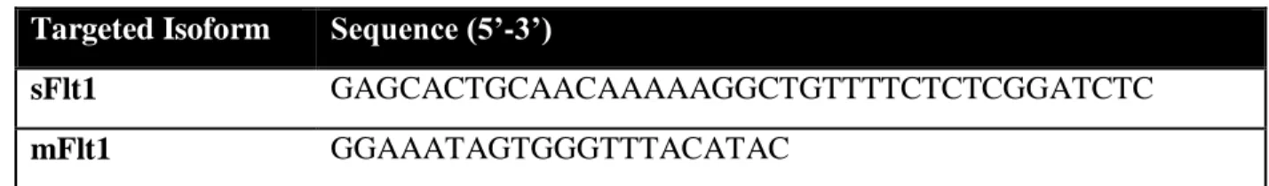
Table 2.2: siRNA sequences targeting Flt1 isoforms. Targeted Isoform Sequence (5’-3’)
sFlt1 GAGCACTGCAACAAAAAGGCTGTTTTCTCTCGGATCTC
Table 2.3: qRT-PCR primers.
Gene Forward Primer (5’-3’) Reverse Primer (5’-3’)
GAPDH CCTCAAGATCATCAGCAATGCCTCCT TTGGTATCGTGGAAGGACTCATGACC
Flt1 AGGGCCTCTGATGGTGATTGTTGA ATGCAGCACTACACATGGAGCCTA
III. REFERENCES
Chappell, J.C. et al., 2009. Local guidance of emerging vessel sprouts requires soluble Flt-1. Developmental Cell, 17(3), pp.377–386.
Jin, S.W., 2005. Cellular and molecular analyses of vascular tube and lumen formation in zebrafish. Development, 132(23), pp.5199–5209.
Kearney, J.B., 2004. The VEGF receptor flt-1 (VEGFR-1) is a positive modulator of vascular sprout formation and branching morphogenesis. Blood, 103(12), pp.4527–4535.
Krueger, J. et al., 2011. Flt1 acts as a negative regulator of tip cell formation and branching morphogenesis in the zebrafish embryo. Development, 138(10), pp.2111–2120.
Nakatsu, M.N. & Hughes, C.C.W., 2008. Chapter 4 An Optimized Three‐Dimensional In Vitro Model for the Analysis of Angiogenesis. Methods in Enzymology, pp.65–82.
Rylova, S.N., Randhawa, P.K. & Bautch, V.L., 2008. Chapter 6 In Vitro Differentiation of Mouse Embryonic Stem Cells Into Primitive Blood Vessels. Methods in Enzymology, pp.103–117.
CHAPTER 3: Blood Vessel Anastomosis is Spatially Regulated by Flt1 During Angiogenesis2
I. Introduction
As described in CHAPTER 1, blood vessel formation is an essential, conserved process in vertebrates that provides oxygen and nutrients to tissues and organs (Carmeliet 2005; Adams & Alitalo 2007; Chappell & Bautch 2010). Aberrant angiogenesis is associated with disease; for example, tumor angiogenesis is one of the hallmarks of cancer (Khurana 2005; Hanahan & Weinberg 2011). Blood vessel development during embryogenesis is a multistep process that begins with primitive vessel formation from endothelial progenitor cells through vasculogenesis (Risau & Flamme 1995; Drake & Fleming 2000; Xu & Cleaver 2011), and the subsequent formation of branched vessel networks is called sprouting angiogenesis. Sprouting angiogenesis is initiated by endothelial cells that proliferate, extend processes, migrate into extravascular space, and finally connect, or anastomose, with another vessel (Betz et al. 2016; Blanco & Gerhardt 2013; Larrivee et al. 2009).
Among numerous regulatory signaling pathways, Vascular Endothelial Growth Factor-A (VEGF) signaling is required for sprouting angiogenesis (Shibuya 2013; Simons et al. 2016). VEGF binds to the endothelial cell-expressed receptor tyrosine kinases Flk1 (VEGFR2) and Flt1 (VEGFR1). VEGF-bound Flk1 triggers a signaling cascade that promotes endothelial cell proliferation, chemotaxis, and cell survival, thereby initiating and sustaining sprouting
2 Chapter 2 is adapted in part from Nesmith, J.E. et al. Blood Vessel Anastomosis is Spatially
angiogenesis (Khurana 2005; Koch et al. 2011; Hanahan & Weinberg 2011). Flt1 is alternatively spliced to form two isoforms, a membrane-localized tyrosine kinase receptor (mFlt1) and a soluble isoform lacking the transmembrane and tyrosine kinase domains (sFlt1) (Kendall & Thomas 1993). Both isoforms of Flt1 have a 10-fold higher binding affinity for VEGF-A ligand than Flk1 and complete genetic deletion is embryonic lethal in mice (Kendall & Thomas 1993; Fong et al. 1995). Nonetheless, sFlt1 cannot independently signal, and mFlt1 has weak kinase activity that is not required for developmental angiogenesis (Ito et al. 1998; Hiratsuka et al. 1998). Thus Flt1 functions as an endothelial cell-intrinsic decoy receptor or ligand sink to negatively modulate VEGF signaling amplitude during angiogenesis.
Stages of sprouting angiogenesis include sprout initiation, extension, anastomosis, and lumenization (Chappell et al. 2011; Geudens & Gerhardt 2011). Sprout initiation, extension and lumen formation are relatively well understood processes. Recent zebrafish studies revealed a role for endothelial cell filopodia in vessel anastomosis, and found that adherens junction-mediated cell rearrangements subsequent to connection promote lumen formation (Lenard et al. 2013; Phng et al. 2013). However, it is unknown whether the site or timing of sprout anastomosis is regulated.
II. Results
a. Flt1 Influences Retinal Vessel Interactions
Global or vascular-selective deletion of flt1 in mouse post-natal retinal vessels increased overall sprouting and filopodia (Chappell et al. 2009; Ho et al. 2012; Chappell et al, in prep). To better understand the role of negative modulation of VEGF-A signaling in sprout anastomosis, we used low-dose tamoxifen to induce mosaic excision of flt1 in retinal vessels, with an excision reporter (Fig 3.3.1).
Sprouts were defined as previously described (Chappell et al. 2009) and interactions between vessels at the angiogenic front were identified (Fig 3.3.1A). The interacting endothelial cells were classified based on the cytoplasmic reporter expression in the endothelial cell from which filopodia extended (Fig 3.3.1B-C). Interestingly, wildtype (WT) sprouts were linked to flt1-/- endothelial cells significantly more often than to WT cells (Fig 3.3.1D), suggesting that Flt1 influences sprout connection parameters. However, further analysis of sprout anastomosis in mouse retinas was hampered by our inability to follow this dynamic process over extended time periods to determine what precedes and follows the static interactions. Moreover, most retinal interactions are at the front and consist of two sprouts intersecting, which does not allow for analysis of target site selectivity.
b. Transient Contacts Precede Stable Blood Vessel Connections