EVALUATION OF MARGINAL INTEGRITY AS A RESULT OF DIFFERENT FINISHING INSTRUMENTATION BASED ON RESTORATIVE MATERIAL AND
MARGIN LOCATION
Alejandro J. Delgado
A thesis submitted to the Faculty of The University of North Carolina at Chapel Hill in partial fulfillment of the requirements for the degree of Master on Science in the Department of
Operative Dentistry of the School of Dentistry
Chapel Hill 2014
ii © 2014
ABSTRACT
ALEJANDRO J. DELGADO: Evaluation of the Marginal Integrity as a Result of Different Finishing Instruments Based on Restorative Material and Margin Location
(Under the direction of Harald O. Heymann)
Purpose: The purpose of this study was to assess the marginal integrity of composite and glass-ionomer
restorations as a function of finishing technique, restorative material and margin location. Materials and
Methods: Forty extracted third molars free of defects were assigned to four groups (N=10) according to
finishing instruments (aluminum oxide discs, fluted carbides, fine diamonds, and coarse diamond). Each
specimen received standardized Class V preparations on the facial and lingual surfaces with occlusal
margins on enamel and gingival margins on dentin. Each preparation was randomly assigned to be
restored with either resin-based composite (RBC) or resin-modified glass ionomer cement (RMGIC).
Specimens were finished with standardized pressure at approximately 0.15 N and evaluated at a
magnification of 600X using an environmental scanning electron microscope (ESEM). Occlusal and
gingival margins were analyzed using an imaging software and means for all measured gaps were
calculated. Data were analyzed using a linear regression using generalized estimating model.
Result: There were no statistically significant differences among the four types of finishing instruments
used in the study. . RBC-restored specimens exhibited significantly smaller mean marginal gaps (1.70
µm, 7.56 µm) than RMGI-restored specimens (5.24 µm, 14.24 µm) in enamel and dentin margins,
respectively. There was a statistically significant difference between enamel and dentin with regards to
marginal gap formation. Conclusion: Under the conditions of this study, marginal gap formation was not
affected by finishing technique. Resin-based composite margins exhibited significantly less marginal gap
than did resin-modified glass ionomer margins, while enamel margins resulted in significantly less
iv
To my wife Maria Gabriela “Peca” Delgado and my children Cammie and Lucas, is your unconditional support and love that inspired me.
v
ACKNOWLEDGEMENTS
I would like to express my sincere appreciation to my mentor Dr. Harald O. Heymann and the committee members, Dr. Terence E. Donovan, Dr. André V. Ritter and Dr. Thomas Ziemiecki, for their invaluable guidance, patience, time and effort.
I would also like to thank Mr. Wallace Ambrose and Chapel Hill Analytical and Nanofabrication Laboratory Institute for Advanced Materials for having taught me and allowing me to use the environmental scanning electron microscope for this project. Also I would like to extend my gratitude to Dr. Ceib Phillips and Dr. Chaitanya Puranik for their extensive help in the data analysis and Mrs. Kate McGraw for her advice with the manuscript.
Special thanks to Dr. Ed Swift, Dr. Lee Boushell, Dr. John Sturdevant, Dr. Andrea Zandona, Dr. Ken May, Dr. Rick Walter, Dr. James Parker and Dr. Scott Eidson, it has been an honor to be part of the Operative Dentistry Family.
I thank the staff of the Department of Operative Dentistry, Mrs. Shannon Tate, Mrs. Dayna McNaught, Mrs. Jamie Desoto, Mrs. Barbara Walton, Mrs. Cynthia Lambert and Mrs. Rosanna Arrington, for all your devotion and commitment.
I would like to thank 3M ESPE and Brasseler USA for the donation of the materials and their assistance.
vi
vii
TABLE OF CONTENTS
LIST OF TABLES………... ... x
LIST OF FIGURES………. ... xi
LIST OF ABREVIATIONS……… ... xii
1. CHAPTER 1: INTRODUCTION ...1
LITERATURE REVIEW……….………….…. ...2
1.1 Enamel………..……….……….…… ...2
1.2 Dentin………. ...2
1.3 Adhesion to Enamel and Dentin………..….. ...3
1.3.1 Adhesion to Enamel……….…….. ...4
1.3.2 Adhesion to Dentin……… ...5
1.4 Resin Composite………..………….…….. ...7
1.4.1 Resin Matrix………...7
1.4.2 Filler Particles……… ...8
1.4.3 Coupling Agent……….….……… ...8
1.4.4 Classification of Resin Composites……….…..…… ...8
1.4.5 Polymerization Shrinkage………. ...9
1.5 Glass Ionomers……….…. ...10
1.5.1 Resin Modified Glass Ionomer Cements….……….…… ...11
viii
1.6 Finishing and Polishing Adhesive Restorations……….. ...12
1.7 Marginal Integrity………..…… ...13
1.8 Finishing and Polishing Instrumentation………...… ...14
1.8.1 Impregnated Aluminum Oxide Discs………...… ...14
1.8.2 Fluted Carbide Finishing Burs………..… ...14
1.8.3 Diamond Finishing Burs………..… ...15
1.9 Scanning Electron Microscope……….… ...16
1.9.1 Environmental Scanning Electron Microscope………..…..… ...16
REFERENCES……… ... 17
2. CHAPTER 2: MANUSCRIPT ...20
2.1 Introduction………...…...20
2.2 Material and Methods……….……… ...22
2.2.1 Specimens Preparation……….….… ...22
2.2.2 Specimens Finishing and Polishing………..………… ...24
2.2.3 Scanning Electron Microscope Evaluation……….….. ...25
2.3 Statistical Analysis……….….…...26
2.4 Results……… ...26
2.4.1 Inter-examiner Reliability………..…..… ...26
2.4.2 Descriptive Statistic for Marginal Integrity………. ...27
2.4.3 Average of Finishing and Polishing Pressure………... ...27
2.5 Discussion……….. ...27
2.6 Limitations………. ...33
ix
3. TABLES ...34
x
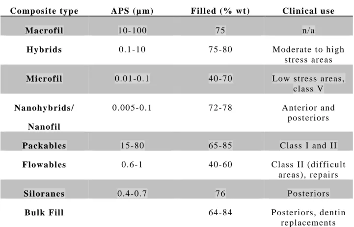
LIST OF TABLES
Table 1. Classification of resin-based composites according
to the average particle size (APS) ...35 Table 2. Finishing and polishing instruments by group ...36 Table 3. Intraclass correlation coefficient (reliability) showing
concordance of 0.9 ...37 Table 4. Descriptive statistical analysis of the mean value for
each marginal gap calculated individually ...38 Table 5 - Analysis of the Generalized Estimation Equation parameter
and final model for the enamel ...39 Table 6. Analysis of the Generalized Estimation Equation parameter
and final model for the dentin ...40 Table 7. Mean pressure (in N) applied for individual instruments, series and complete study
xi
LIST OF FIGURES
Figure 1. Standarized class V preparation with occlusal
margin on enamel and gingival margin on dentin ...42 Figure 2. Restorative materials resin-based composite (RBC)
and resin-modified glass ionomer (RMGIC) ...43 Figure 3. Finishing and polishing instrument by groups. Group 1: Sof-Lex discs,
Group 2: Fluted carbide finishing burs, Group 3: Fine diamond finishing burs
and Group 4: Regular coarse diamond...44 Figure 4. Finishing procedure using pressure/abrassion
deviced at approximadetely 0.15 N. ...45 Figure 5.1. Environmental Scanning Electron Microscopy
at the Chapel Hill Analytical and Nanofabrication Laboratory ...46 Figure 5.2. Environmental Scanning Electron Microscopy
at the Chapel Hill Analytical and Nanofabrication Laboratory ...47 Figure 6. The sample was affixed to an aluminum planchet
and held it with a copper tape for evaluation on environmental
scanning electron microscopy. ...48 Figure 7. Image at 25X magnification to localize restoration and
subdivisions of occlusal and gingival margin ...49 Figure 8.Enamel margin at 600 X magnification showing
no gap formation between restoration and substrate ...50 Figure 9. Dentin margin showing marginal gap formation...51 Figure 10. Flow chart of the images taken per specimen ...52 Figure 11. Marginal integrity at the enamel showed statistically significant difference
between restorative materials, but no significant differences among instruments ...53 Figure 12. Marginal integrity at the dentin resulted in statistically
significant difference between RMGI and RC. No statistically significant
xii
LIST OF ABREVIATION
4-META: 4-methacryloxyethyl trimellitate anhydrate APS: Average particle size
BIS-GMA: Bisphenol A glycidyl methacrylate BPDM: biphenyl dimethacrylate
DEJ: Dentin-enamel junction
ESEM: Environmental scanning electron microscope Gpa: Giga pascal
GIC: Glass ionomer cement HEMA: hydroxyethil methacrylate kV: Kilovolts
mm: millimeters Mpa: Mega pascal N: Newton
PENTA: penta acrylate monophosphate RBC: Resin-based composite
RMGIC: Resin-modified glass ionomer cement SEM: Scanning electron microscope
CHAPTER 1: INTRODUCTION
Over the past decades the importance of esthetic dentistry has become more evident. In current clinical treatment many types of restorations are available for the replacement of tooth structure. The introduction of adhesive restorations introduced a new concept in operative dentistry. These tooth-colored materials are not only esthetic, but also, more importantly, are very conservative of tooth structure. Today, adhesive restorations provide numerous potential treatment options to the patients. Moreover, adhesive restorations have become one of the most popular materials for the restoration of both anterior and posterior teeth.
2
LITERATURE REVIEW
1.1. Enamel
Enamel is a highly mineralized crystalline structure containing 95% to 98% inorganic matter by weight in which hydroxyapatite is the main constituent in the form of crystals (90%-92%). Structurally, enamel is composed of enamel rods and prisms, which vary in number from five million to twelve million depending on the location.1,2 In general, they are aligned perpendicular to the dentin-enamel junction (DEJ) and they are separated by an interrod substance.3
Enamel is the hardest substance in the human body. It is a brittle structure with a high elastic modulus of 40-80 Gpa and low tensile strength. Enamel is relatively translucent; its translucency is related to the degree of mineralization. The color of enamel is primarily a function of its thickness and that of the underlying dentin.4
For maximal strength in tooth preparation, all enamel rods should be supported by dentin. Enamel rods not supported by a dentin base are subject to fracture. Due to enamel’s inherent
brittleness, it relies on a dentin substrate to supply it with toughness.1,4 Enamel is a non-vital and non-sensitive tissue that cannot repair itself.1,3,4
1.2. Dentin
3
DEJ to the pulp. The dentinal tubules contain cytoplasmic cell processes from pulpal odontoblasts, known as Tomes fibers. Each dentinal tubule is surrounded by two main types of dentin that are present: (1) Peritubular dentin, which is the mineralized wall of the tubules and (2): Intertubular dentin, is the dentin around and between dentinal tubules and exhibits the greatest surface area at which primary resin/dentin bonding occurs. Because the odontoblasts form dentin while progressing inward towards the pulp, the tubules are forced closer together.1 The number of tubules at the pulp varies from 45,000/mm2 to 65,000/mm2 and decrease to from 15,000/mm2 to 20,000/mm2 when approaching to the DEJ.5
After the removal of caries by an operative procedure, the majority of odontoblasts die. The remaining odontoblasts can, however, repair the remaining dentin and form reparative dentin. When 1 mm2 of dentin is exposed, about 30,000 living cells are damaged. It is recommended to subsequently seal the exposed dentin with a non-irritating material. The sensitivity of teeth is widely accepted to be related to the “hydrodynamic theory” of dental
pain developed in the 1960’s.6 Brannstrom also stated that bacteria can leak into the dentinal tubules if a gap exists between the tooth and the restoration, causing an insult to the pulpal tissues.
1.3 Adhesion to Enamel and Dentin
The remarkable introduction of the enamel acid-etching technique by Buonocore in 1955 made adhesive dentistry truly possible, and later revolutionized esthetic dentistry. Buonocore’s discovery, coupled with the introduction of fluoride-containing restorative
4
challenge.7,8 The reason why dentin bonding is less predictable is mainly because of its organic content and permeability.9
1.3.1 Adhesion to Enamel
The success of the acid-etched enamel bond is well established. This technique creates a micro-mechanical bond between the restorative material and the enamel. In his study, Buonocore10 found that the used of 85% phosphoric acid for 30 seconds on the enamel enhanced the bond strength of acrylic resin to the tooth. It was reported that reducing the concentration and the time did not affect the shear bond strength, and the etched enamel displayed a similar microporosity pattern.11 An optimal concentration of acid-etchant should produce a minimal loss of enamel surface while creating a strong bond. Acid etching techniques removes about 10 µm of the enamel surface and creates a porous layer ranging from 5-50 µm.8 There are three patterns in enamel-etching that have been described. Type I,
is the dissolution of the prism cores without affecting the prism peripheries; Type II, is characterized by a predominance of dissolution of prism peripheries while leaving the cores intact; and Type III, in which no prism structures are evident, and in some cases the above patterns are resembled.1,4 Enamel dissolution results in the formation of resin tags; in which the monomer polymerizes into the demineralization pattern of the crystals. Two types have been described, macrotags that are formed circularly between the enamel prism peripheries; and microtags, that are formed at the core of the prisms. Microtags probably contribute most to the bond strength because of a greater quantity and larger surface area.4
5
stress, to prevent marginal openings and to overcome shear stress. Thorough etching and bonding techniques are critical to enhance the bond strength. Alternative acids for etching enamel have been studied, reporting a significant decrease in bond strength when weaker etchants are used.12
1.3.2 Adhesion to Dentin
Successful bonding to enamel is a predictable procedure achieved with a simple technique, but bonding to dentin has been a more challenging procedure. Dentin is an intrinsically moist organic substrate with a dense network of tubules containing the odontoblastic processes, which communicate with the pulp. Moreover, these tubules become wider and denser close to the pulp. It has been reported that tubules occupy about 22% of the surface closer to the pulp and only about 1% when approaching the DEJ.13
The fundamental principle of adhesion to a tooth is mostly based upon an exchange of inorganic substrate for resin. This process involves two phases. The first one consists of removing the organic tissue and exposing microporosities. The second phase is called the hybridization phase that involves the infiltration of a monomer resin within the microporosities. This results in micro-mechanical interlocking. This is believed to be the first step in reliable bonding, with potential benefits from additional chemical interaction.14
6
Conditioning the dentin can be defined as any chemical alteration of the dentinal surface by acids with the objective of removing the smear layer and simultaneously demineralizing the surface.4 The smear layer constitutes a barrier; and it must be removed, or made permeable to let the resin monomers penetrate and contact the dentin surface directly. In addition to removing the smear layer and the majority of hydroxyapatite crystals, the substrate exposed by dentin conditioning is a mesh of collagen fibrils that when dried can collapse and shrink because of the loss of inorganic support.8
In 1982, Nakabayashi published a classic paper on how resin infiltrates into acid-etched dentin, transforming the surface from being crystalline, acid-sensitive, and hydrophilic to an organic, acid-resistant, and relative hydrophobic surface. This new surface was coined the “hybrid layer”. These resins or primers contain hydrophilic monomers dissolved in organic
solvents, such as acetone or ethanol. The primer molecules such as hydroxyethil methacrylate (HEMA), biphenyl dimethacrylate (BPDM), penta acrylate monophosphate (PENTA) and 4-methacryloxyethyl trimellitate anhydrate (4-META) contain two functional groups- a hydrophilic group that has affinity for the exposed collagen fibril and the hydrophobic group for copolymerization with the adhesive resin. The primers wet the collagen, increase the surface energy and hence the wettability of the dentinal surface.8
7 1.4 Resin Composites
The first resin composite was introduced a year after the findings of Buonocore. In 1956, Dr. Rafael Bowen published an article describing the development of a new epoxy resin.16 This material was developed after the earlier failures of silicates and acrylic resin. Silicates had solubility problems and eroded within few years, while acrylic resins had poor color stability due to water sorption, poor wear resistance, and polymerization shrinkage causing leakage around the margins and compromising the adhesion. Newer materials with improved properties have overcome most of those problems.17
The chemical formula for resin composites consist of three main structural components: 1) The resin matrix which is an organic polymer material that forms a continuous phase and binds to the fillers, 2) The inorganic filler particles that reinforce the fibers that are dispersed in the matrix and 3) A coupling agent, that promotes adhesion between the filler particles and the matrix, after being activated with an initiator accelerator.17 Each of these components is necessary for the mechanical and physical properties of the material.
1.4.1 Resin Matrix
8 1.4.2 Filler Particles
The inorganic filler particles are most commonly produced by finer particles of quartz, glass, silica and more. These particles range in sizes from 0.04 to 100 µm. Depending of the type or particle and how they are processed; their shapes can be spherical or irregular. The above mentioned inorganic particles are added into the matrix to greatly improve the material properties. According to Ralph Phillips, the primary purposes of the fillers are to strengthen a composite and to reduce the amount of matrix. Several other properties are improved, like reinforcement of the matrix, decreased wear, reduction of polymerization shrinkage, and the associated shrinkage stress, reduction of thermal expansion, improved workability, reduction of water sorption and increased radiopacity. The filler particles can only provide reinforcement if they are well bonded to the matrix. Because of the importance of well-bonded filler particles, the use of an effective coupling agent is extremely important to the success of a composite material.
1.4.3 Coupling Agent
The coupling agent is a silane whose role, as previously explained, is to maintain the filler particles and the resin matrix together. The purpose of the well applied coupling agent is to transfer stresses to the higher modulus filler particles to improve physical and mechanical properties and inhibit leaching by preventing water from penetrating the interface.17
1.4.4 Classification of Resin Composites
9
distribution. Classes of contemporary composites are outlined in Table 1. Composites with a large APS are called macrofills, and composites with small APS are called microfills. Many composites used mixtures of different APSs, and are collectively called “hybrids.” Any resin
with fillers from two or more size ranges can, in principle, be considered a hybrid. Nanofilled and nanohybrid resin composites were recently introduced. These composites are highly polishable, provide esthetics, have excellent mechanical properties and present good handling. 18,19 Silorane, a novel composite was developed to reduce polymerization shrinkage and the associated stress. Siloranes use a monomeric system based in openings of cationic rings on radical oxiranes. A 36 month clinical trial reported no statistically significant difference between this new composite compared with a nanohybrid, resulting in a similar performance in the clinical setting.20 Another study reported similar results when compared with another nanohybrid over a period of 2 years.21 The advantage of low-shrinkage stress materials, such as this one needs to be further studied and, at this moment, a conclusive statement cannot be drawn from the available data.
1.4.5 Polymerization Shrinkage
Resin-based composites have gone through several changes since their original formulation. Current changes are focused principally on reducing polymerization shrinkage, and perhaps more importantly, reducing and/or counteracting the polymerization shrinkage stress. Recent research has addressed this issue of polymerization shrinkage, which may have a deleterious effect on the interface between tooth and resin composite.22
10
filler particles in the matrix. When more fillers are present, less shrinkage occurs due to a reduced matrix volume. Although shrinkage varies from one composite to another, it ranges from 0.7-5.5 vol% within 24 hours after curing.17,18
Stress resultant from polymerization shrinkage can be affected by the cavity preparation size and configuration. In 1987, Albert Feilzer described the Configuration Factor, also known as the C-factor. The C-factor is the ratio of bonded to unbonded surfaces.4,18 The higher the C-factor, the higher the contraction risk and the potential for bond disruption from polymerization shrinkage stress.
To overcome the problem associated with a material pulling away from the tooth, clinicians must carefully control the insertion technique of the resin composite, appropriate use of the dental bonding agents, control of curing light irradiance and proper isolation. The reality is that the polymerization shrinkage phenomenon cannot be avoided.
Polymerization shrinkage usually results in gap formation at the interface of the restoration and the tooth. The clinical significance of the margin gap is not fully known.
1.5 Glass Ionomer
11
GICs were developed by Wilson and Kent in 1972. Many liquid and powder modifications have been incorporated since the first commercial product emerged, to improve the physical, chemical and mechanical properties. GICs are hydrophilic, and dental composites are hydrophobic, therefore the presence of water makes it difficult to obtain esthetic results as well as mechanical strength with GICs.
1.5.1 Resin-modified glass ionomer cements
Resin-modified glass ionomer cement (RMGIC) was an evolution in glass ionomer technology introduced in the 1980’s by Sumita Mitra. It was produced by adding
methacrylate resin to polyacrilate acid. RMGICs have the same ion-releasing glass and filler particles used in conventional glass ionomers, but their sizes are smaller. They are light-cured, which is supplementary to the acid-base reaction. The initial setting is triggered by the light, which is followed by the chemical reaction.23
Fluoride release from the RMGI is the highest during the first 24 hours. The amount of fluoride released decreases tremendously after 24 hours. The mean concentration for the first 6 hours ranges from 22-65 ppm, which drops to 3-20 ppm after 18-24 hours. Daily release drops from 8-15 ppm on the 1st day to 1-2 ppm on the 7th day.23
1.5.2 Volumetric shrinkage in RMGIC
Volumetric changes due to curing shrinkage can form marginal gaps that may affect the longevity of the restoration. This shrinkage is however counteracted by hygroscopic expansion. A study reported that after 24 hours RMGICs exhibit volumetric shrinkage of 3.2 – 4.5%, and after 28 days of water storage the hygroscopic expansion ranged from 0.3 –
12
It is important to understand the extent of shrinkage, since it can create marginal gaps if the filling material does not have a sufficiently strong bond to the tooth structure.24,25
1.6 Finishing and Polishing Adhesives Restorations
Finishing is an extremely important procedure for the longevity of the restoration as well as the tooth.26-29 A well contoured, finished and polished restoration will promote oral health. Finishing is the gross reduction of the material to obtain the anatomical contour of the restoration, while polishing is making the surface smooth and lustrous.17 The goal of finishing and polishing are to obtain the desired anatomy, proper occlusion, and reduced roughness.
Appropriate rotatory instruments must be selected according to the specific surface being contoured.1 Since lack of proper finishing and polishing procedures can compromise the marginal integrity, therefore leading to staining, discoloration of the restoration, gingival irritation and recurrent caries due to plaque accumulation.26 Proper pressure should be applied during finishing avoiding introduction of stress. This stress can affect the interface creating an opening of the restoration margin that can result in a marginal gap formation that will compromise the restoration.
13
different degrees of abrasiveness, come in sets and should be used in the proper sequence, working gradually toward the finest grits.26,27
It is important to know the effect of polishing direction on the marginal adaptation of the restoration. A study demonstrated that there is a significant difference in the marginal adaptation when polishing is accomplished from resin-composite to tooth structure as opposed to from tooth to resin composite.30 They used flattened enamel and restored it with a nanofilled composite and a microhybrid composite and used polishing discs and rubber points. The margins were polished from resin- composite to tooth and from tooth to resin composite.
1.7 Margin Integrity
Finishing and polishing procedures can be detrimental to the marginal integrity of the restoration and may lead to microcracks in the enamel.31 Polymerization shrinkage is also a factor that can initiate a micro gap at the tooth-restoration interface, when the stress of polymerization shrinkage exceeds the cohesive strength of the tooth structure. Marginal gap can result in secondary caries and pulpal irritation.32 Therefore, it is important for the longevity of the restoration and the vitality of the tooth that the formation of the marginal gaps be prevented or, at the very least, controlled. Finishing and polishing techniques are under the clinician’s control, and are essential to achieve good marginal integrity.
14 1.8 Finishing and Polishing Instrumentation
1.8.1 Impregnated Aluminum Oxide Discs
Impregnated aluminum oxide discs are fabricated by securing abrasive particles of a chemical compound of aluminum and oxygen to a flexible backing material (Mylar or paper). These particles are retained on the disc by a polymeric adhesive coating layer. Aluminum oxide has sufficient hardness (9 on Mohs’ hardness scale) for polishing
composites and ceramics.27 The most common examples of impregnated aluminum oxide discs include Sof-Lex discs (3M ESPE, St Paul, MN) and Super Snap discs (Shofu Dental Corp, Menlo Park, CA). Sof-Lex discs, which are coated with aluminum oxide particles (grit 150, 360, 600, 1200) have a four discs sequence that include coarse (100 µm), medium (40 µm), fine (24 µm) and superfine (8 µm) discs.27 Sof-Lex discs have a great ability to reduce the gross contour with the coarse disc, whereas the medium does it at a much slower rate. These first two discs are used for finishing. Once the ideal anatomy and contour are attained the fine and superfine discs can be used to polish. One of the advantages of Sof-Lex discs is easy access to incisal edges and embrasures due to how thin and flexible the discs re. A major drawback is that they tend to flatten surfaces and cannot be used in concave areas.26
1.8.2 Fluted Carbide Finishing Burs
15
series (Brasseler USA, Savannah, GA) and the H48L Spiral-fluted (Brasseler USA), SE trimming and finishing (SS White, Lakewood, NJ) and Trimming and Finishing Taper T-Series (Midwest-Dentsply, York, PA). These burs kit include two or three specific burs and are available in different subcategories as fine, extra-fine and ultra-fine.
1.8.3 Diamond Finishing Burs
Diamond burs consist of a blank surface that is coated with powdered diamond abrasives bonded by a metallic adhesive. Unlike carbides, diamonds rely on the grinding by abrasive particles rather than the cutting action of blades. Diamond particles vary in shapes and sizes. They range from coarse (50-150 µm), medium (40 µm), fine (25-30 µm), extra-fine (15 µm) and super-fine (7-8 µm).27 The primary intent for diamonds is to contour, adjust, and smooth restorative materials. The clinical performance depends of the size and shape distribution of the particles. The hardness of the diamond particles measures about 10 on Mohs’ scale,
sufficient for polishing resin composites and ceramics.17 It is recommended by manufactures to use these instruments with gentle wiping strokes and under water to avoid heat.
16 1.9 Scanning Electron Microscope
The use of organic dyes has been the oldest method to assess the sealing effectiveness of the margin. The main problem with this methodology is the different molecules size of the dies being too small, and is a qualitative evaluation method.14
The measurement of sealing effectiveness should be semi-quantitative by using scanning electron microscope (SEM). This publication describes a method to quantify the quality of dental restorations.33 The restoration margins are traced on the SEM screen with a digitizer and an interface to measure the margin's length. Simultaneously the margin quality is assessed and assigned to the corresponding lengths.36 There have been many studies evaluating marginal gaps with SEM in vitro. 2,36-38 This method assumes that the stress induced by the polymerization shrinkage and the thermal-mechanical strain exceeds the bond strength and observable gaps will form.14
1.9.1 Environmental Scanning Electron Microscope
17
REFERENCES
1. Roberson TMH, H.O. Swift, E.J. Sturdevants's Art & Science of Operative Dentistry. 5 ed. St. Louis, Missouri: Mosby Elsevier; 2006.
2. Maresca C, Pimenta LAF, Heymann HO, Ziemiecki TL, Ritter AV. Effect of Finishing Instrumentation on the Marginal Integrity of Resin-based Composite Restorations.
Journal of Esthetic and Restorative Dentistry. 2010;22(2):104-112.
3. Nanci A. Ten Cate's Oral Histology: Development, Structure and Function. 7th ed. St. Loius: Elsevier; 2008.
4. Summitt JB. Fundamentals of Operative Dentistry: A Comtemporary Approach. 3 ed. Chicago: Quintessence Pub; 2006.
5. Garberoglio R, Brannstrom M. Scanning electron microscopic investigation of human dentinal tubules. Archives of oral biology. 1976;21(6):355-362.
6. Brannstrom M, Astrom A. The hydrodynamics of the dentine; its possible relationship to dentinal pain. International dental journal. Jun 1972;22(2):219-227.
7. Pashley DH. The effects of acid etching on the pulpodentin complex. Operative dentistry.
Nov-Dec 1992;17(6):229-242.
8. Swift EJ, Jr., Perdigao J, Heymann HO. Bonding to enamel and dentin: a brief history and state of the art, 1995. Quintessence Int. Feb 1995;26(2):95-110.
9. Perdigao J. New developments in dental adhesion. Dental clinics of North America. Apr 2007;51(2):333-357, viii.
10. Buonocore MG. A simple method of increasing the adhesion of acrylic filling materials to enamel surfaces. Journal of dental research. Dec 1955;34(6):849-853.
11. Barkmeier WW, Shaffer SE, Gwinnett AJ. Effects of 15 vs 60 second enamel acid conditioning on adhesion and morphology. Operative dentistry. Summer 1986;11(3):111-116.
12. Swift EJ, Jr., Cloe BC. Shear bond strengths of new enamel etchants. American journal of dentistry. Jun 1993;6(3):162-164.
13. Pashley DH. Clinical correlations of dentin structure and function. The Journal of prosthetic dentistry. Dec 1991;66(6):777-781.
18
15. Pashley DH. Smear layer: physiological considerations. Operative dentistry. Supplement.
1984;3:13-29.
16. Bowen RL. Use of epoxy resins in restorative materials. Journal of dental research. Jun 1956;35(3):360-369.
17. Anusavice K. Phillips' Science of Dental Materials 12 ed: Elsevier; 2013.
18. Heymann HO, Swift E.J, Ritter, A.V. Sturdevant's Art and Science of Operative Dentistry. 6th ed: Elsevier; 2013.
19. Ritter AV. Direct resin-based composites: current recommendations for optimal clinical results. Compend Contin Educ Dent. Jul 2005;26(7):481-482, 484-490; quiz 492, 527. 20. Walter R, Boushell LW, Heymann HO, et al. Three-Year Clinical Evaluation of a
Silorane Composite Resin. Journal of esthetic and restorative dentistry : official publication of the American Academy of Esthetic Dentistry ... [et al.]. Dec 18 2013. 21. Baracco B, Perdigao J, Cabrera E, Ceballos L. Two-year clinical performance of a
low-shrinkage composite in posterior restorations. Operative dentistry. Nov-Dec 2013;38(6):591-600.
22. Ferracane JL. Resin composite--state of the art. Dental materials : official publication of the Academy of Dental Materials. Jan 2011;27(1):29-38.
23. Wiegand A, Buchalla W, Attin T. Review on fluoride-releasing restorative materials--fluoride release and uptake characteristics, antibacterial activity and influence on caries formation. Dental materials : official publication of the Academy of Dental Materials.
Mar 2007;23(3):343-362.
24. Attin T, Buchalla W, Kielbassa AM, Helwig E. Curing shrinkage and volumetric changes of resin-modified glass ionomer restorative materials. Dental materials : official publication of the Academy of Dental Materials. Nov 1995;11(6):359-362.
25. Bausch JR, de Lange K, Davidson CL, Peters A, de Gee AJ. Clinical significance of polymerization shrinkage of composite resins. The Journal of prosthetic dentistry. Jul 1982;48(1):59-67.
26. Jefferies SR. The art and science of abrasive finishing and polishing in restorative dentistry. Dental clinics of North America. Oct 1998;42(4):613-627.
27. Jefferies SR. Abrasive finishing and polishing in restorative dentistry: a state-of-the-art review. Dental clinics of North America. Apr 2007;51(2):379-397, ix.
19
29. Yap AU, Ang HQ, Chong KC. Influence of finishing time on marginal sealing ability of new generation composite bonding systems. Journal of oral rehabilitation. Nov 1998;25(11):871-876.
30. St-Pierre L, Bergeron C, Qian F, et al. Effect of polishing direction on the marginal adaptation of composite resin restorations. Journal of esthetic and restorative dentistry : official publication of the American Academy of Esthetic Dentistry ... [et al.].
2013;25(2):125-138.
31. Statement on posterior resin-based composites. ADA Council on Scientific Affairs; ADA Council on Dental Benefit Programs. J Am Dent Assoc. Nov 1998;129(11):1627-1628. 32. Brannstrom M. Communication between the oral cavity and the dental pulp associated
with restorative treatment. Operative dentistry. 1984;9(2):57-68.
33. Taylor MJ, Lynch E. Marginal adaptation. J Dent. Oct 1993;21(5):265-273.
34. Bernardo M, Luis H, Martin MD, et al. Survival and reasons for failure of amalgam versus composite posterior restorations placed in a randomized clinical trial. J Am Dent Assoc. Jun 2007;138(6):775-783.
35. Soncini JA, Maserejian NN, Trachtenberg F, Tavares M, Hayes C. The longevity of amalgam versus compomer/composite restorations in posterior primary and permanent teeth: findings From the New England Children's Amalgam Trial. J Am Dent Assoc. Jun 2007;138(6):763-772.
36. Roulet JF, Reich T, Blunck U, Noack M. Quantitative margin analysis in the scanning electron microscope. Scanning microscopy. 1989;3(1):147-158.
37. Asmussen E, Jorgensen KD. A microscopic investigation of the adaptation of some plastic filling materials to dental cavity walls. Acta odontologica Scandinavica. Mar 1972;30(1):3-21.
20
CHAPTER 2: MANUSCRIPT
Evaluation of the Margin Integrity as a Result of Different Finishing Instrumentation Based on Restorative Material and Margin Location
2.1 Introduction
Over the past several decades an impressive improvement in adhesive technology has occurred. Adhesive restorations have gained considerable importance due to increasing demand for conservative and esthetic dentistry. However, adhesive restorations are technique sensitive, and achieving an ideal restoration can be challenging. Multiple factors can affect the marginal integrity and the longevity of direct restorations, including the margin location and geometry, restorative material, quality of isolation, polymerization variables, C-factor, insertion technique, finishing technique, and polishing technique. From these, the finishing and polishing techniques are critical steps that are under the clinician’s control, and are essential to achieve marginal
21
Selecting the appropriate finishing instrumentation can be confusing because of the wide range of commercially available products. Other factors that affect the finishing efficiency include the hardness of the abrasive and the composition of the substrate, the size and shape of the abrasive instrument, the pressure applied, the speed, the polishing condition, and direction.7 Proper finishing and polishing techniques should be applied for avoiding introduction of stress. This stress can adversely affect the interface creating disruption at the interface between the restoration and the tooth that can result in a marginal gap formation that will compromise the restoration and the tooth. Proper finishing and polishing of adhesive restorations enhance the esthetic outcome and longevity of the tooth.5,8 Clinically, the main cause of failure of adhesive restorations is related to marginal leakage, which eventually leads to secondary caries, and/or subsequent loss of retention. 9-15
There are many different types of finishing techniques and instruments, and if they are not carefully used they may lead to gap or crevice formation and poor marginal adaptation.15 As noted in Chapter 1, instruments for finishing and polishing include carbide burs, diamonds, rubber cups, points, abrasive discs, stones, strips and pastes.4,10 Fluted carbides burs, fine diamonds and aluminum oxide discs are the most popular instrumentation for finishing and contouring.
Although finishing and polishing procedures for adhesive materials are well documented, there are not many studies reporting the effectiveness of the various finishing instruments regarding the quantitative evaluation of the marginal integrity.
22
many studies reporting quantitative information on marginal gaps. One such study reported that the best results were achieved with the 30 fluted carbide finishing bur.1 Another study presented that carbide finishing burs exhibit the greatest incidence in marginal gap formation.3 A more recent study reported the largest gaps were obtained with regular coarse diamonds, and the smallest gaps with fine diamonds.12 To date, there is not a predictable approach for finishing adhesive restorations that maintains marginal integrity and reduces gap formation. Therefore,
the purpose of this study was to compare the effect of different finishing techniques on the
marginal integrity of resin-based composite (RBC) and resin-modified glass ionomer cement
(RMGIC) restorations in vitro.
2.2 Materials and Methods
2.2.1 Sp eci mens p reparation
Fort y extract ed fres h human third mol ars free of defect s were coll ect ed, and stored i n 0.5% Th ym ol for disi nfection. Each specim en received t wo st andardiz ed Class V preparations (approximat el y 3 mm x 2 mm x 2 mm; m esiodist all y, inci sogi ngivall y, and depth wis e respecti vel y), one on the faci al s urface and one on t he lingual s urface. Each preparati on had an occl usal m argi n i n enam el and a gi ngi val m argin i n denti n (Fi gure 1). Each preparatio n was done wit h a new No. 271 carbide bur (H26M Bras sel er USA) using a wat er -cool ed hi gh -s peed handpi ece. The dim ensions of all preparations were veri fi ed with a di git al caliper.
23
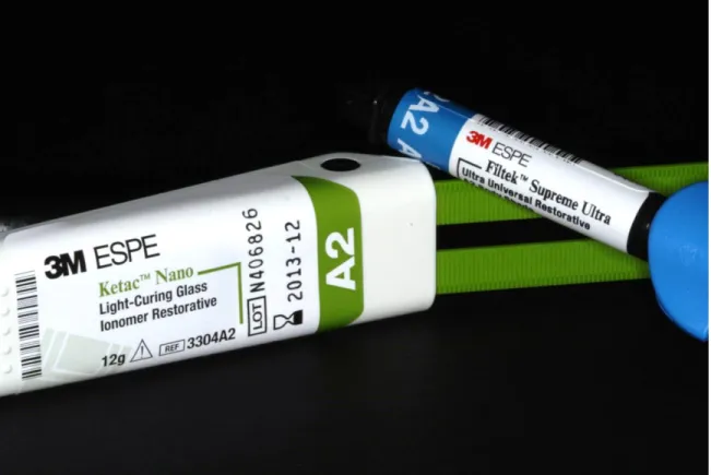
2). Both materials were used following manufacturer’s recommendations. For
the R BC , the preparat ion was etched with 35% phosphori c aci d (S cot chbond Etchand Pl us, 3M ES PE) for 15 s econds and then thoroughl y rinsed with water for 15 s econds. An ethanol and wat er bas ed dent al adhesive agent was us ed (Adper Singl e Bond, 3M ES PE) and appli ed with a microbrush and rubbed for 15 s econds, sli ghtl y dri ed t o evaporat e the s olvent and then coat ed again and repeat ed the sam e protocol as before and then was li ght cured for 20 s econds.
The R BC was i ns ert ed b y t wo increments; the firs t increm ent was placed from t he axial wall t o the gingi val m argi n and the s econd increm ent from t he fi rst increment t o the occl usal m argin. Each i ncrem ent was li ght -pol ym eriz ed with a li ght -curi ng unit ( LED Demetron A.2, Kerr Corporation, Orange, C A) for 20 seconds. The average of the curing int ensi t y of the li ght curing unit was 1150 mW/cm2.
24
Aft er pol ym erizati on, the s pecim ens (n=10) were assi gned randoml y to one of the four experim ental groups (Fi gure 3) accordi ng to t he finis h ing technique.
Group 1, was finis hed with impregnat ed al uminum oxide dis cs (Sof -Lex , 3M ESPE), Group 2 was finis hed with l ong flame spiral fl ut ed carbi de bur s eri es (H48L Brass el er US A,), Group 3 was fi nished with fine di amonds seri es (DET 9 Brass eler USA ), and Group 4 served as a negative cont rol , whi ch was fini shed with regular coars e diamond (888 Brass el er US A).
2.2.2 Specimens Finishing and Polishing
The specim ens assi gned to one of four finishi ng t echni ques were fi nished foll owi ng the respective m anu fact urer’s recomm ended sequence. The fi nis hing sequence and t he particl e s izes (grit s) are expres sed in Tabl e 2 for each group.
25
cycle. The specimens were fixed to the device by using polyvinyl siloxane bite registration material (Regisil PB Bite Registration, Dentsply Caulk, Milford, DE).
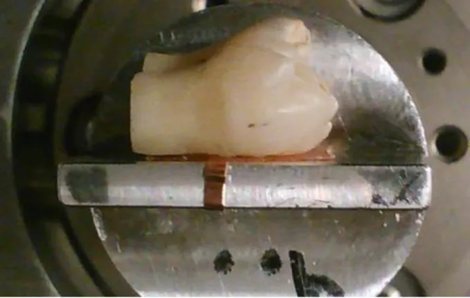
2.2.3 Scanning Electron Microscope Evaluation
After the finishing procedure, all specimens were stored in a moist environment for 24 hours to avoid dehydration before the evaluation of the marginal integrity. An environmental scanning electron microscope (ESEM) (Fei Quanta 200 ESEM Hillsboro, OR) was used to observe the specimens (Figures 5.1 and 5.2). This microscope has the capability of imaging uncoated (Low vacuum mode) and nearly wet (Environmental mode) specimens, along with the conventional high vacuum Scanning Electron Microscope (SEM) imaging. The microscope has secondary and back scattered electron detectors, and a cooling stage. The instrument can be operated at variable pressure modes and hydrated specimens can be viewed without much preparation. It was used at the low vacuum mode with a 10.00 kV accelerating voltage, a working distance between 10-12 mm. and a chamber pressure of 0.4 Torr.
The specimen was affixed to an aluminum planchet with a copper tape and viewed without conductive coating (Figure 6). In a low vacuum mode a small amount of humidity is introduced into the chamber. As the electron beam passes through the chamber, the water vapor ionizes providing a source of ions to passivate the sample surfaces, thereby reducing beam induced charging effects.
26
both margins was acquired at a magnification of 600X (Figures 8 and 9). The margin gap obtained in the photograph was measured by using an imaging software (ImageJ 1.34 software, NIH, Bethesda, MD) to measure from the substrate to the restorative material from each subdivision.
Three measurements obtained from every margin were recorded, totaling six measurements per restoration, twelve measurements per specimen. A total of 480 marginal sites were evaluated and the gap measurements were averaged for each specimen. The mean was obtained by averaging the three greatest observed values for each margin (Figure 10).
2.3 Statistical Analysis
The main purpose of the statistical analysis was to assess whether the outcomes were significantly affected by the instrument, material used or margin location (enamel vs. dentin). Material and surface variables were assessed within subject tooth (facial and lingual, n=40). Finishing instruments were assessed independently between subject variable. Since each tooth has multiple observations there is an expectation of correlation among these observations. For this reason, a linear regression using Generalized Estimating Equations with an unstructured working correlation was used separately for each outcome to specify both the within and between subject variation. All the possible two way interactions were included in the initial model and removed from the final model if not statistically significant present.
2.4 Results
27
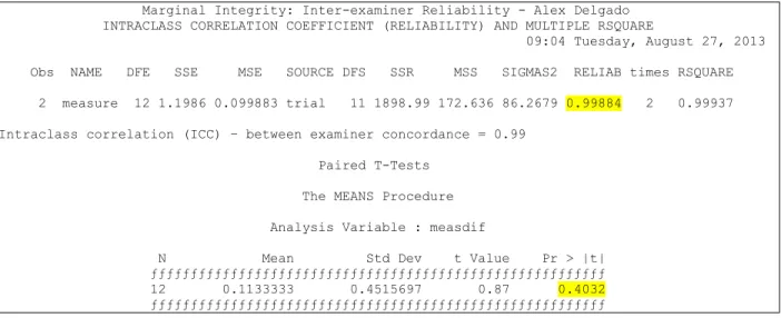
One hundred and sixty margins, representing 480 measurements were included in the study. An inter-examiner reliability test was analyzed to determine the concordance among examiners. The inter-examiner reliability was computed with correlation coefficients of 0.99, indicating a strong agreement between the two examiners (Table 3).
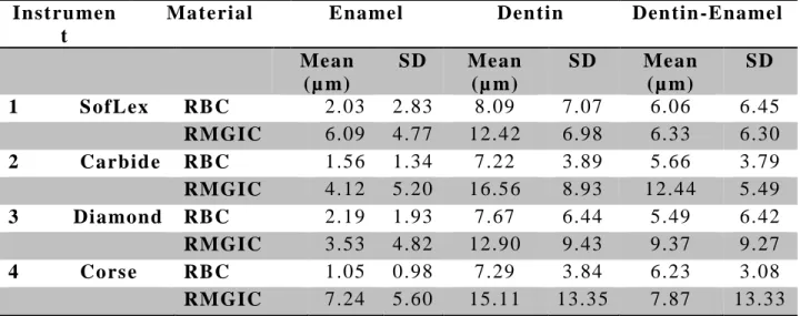
2.4.2 Descriptive Statistics for Margin Integrity
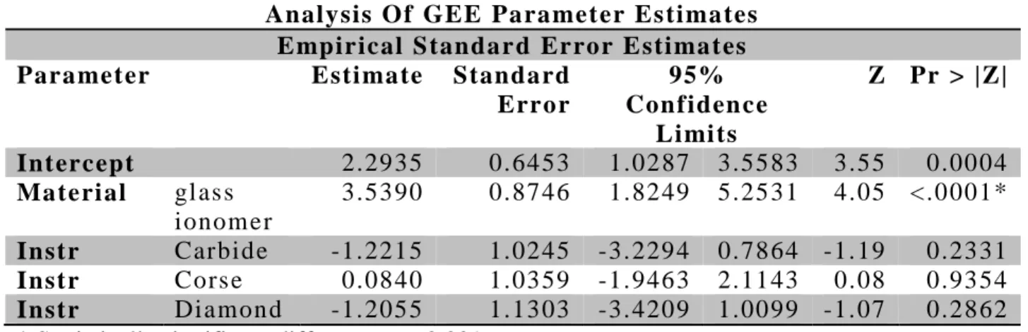
The mean value for each marginal gap was calculated individually and presented in Table 4. When observing the enamel margin there was statistically significant difference between the means of the restorative materials (Figure 11). There is no statistically significance difference among the mean values of the four instruments controlling for material or surface. Regarding the dentin marginal gaps, there were no statistically significant differences among the mean values for the 4 instruments evaluated (Figure 12). There was a statistically significant difference in the mean values of the two restorative materials adjusted for instrument. The final model and the analysis of estimation for both enamel and dentin are expressed in Table 5 and 6.
2.4.3 Average of Finishing Pressure
The average of pressure used throughout the study is showed in Table 7. All the restorations were finished and polished using the pressure/abrasion device and values recorded by the computer software. The mean average of the pressure applied was 0.16N.
2.5 Discussion
28
to the occurrence of marginal leakage, which eventually lead to marginal staining, secondary caries, and subsequent loss of retention.
Three variables related to the marginal integrity were investigated in this study: the finishing instrumentation technique used, the restorative material placed and the margin location on the tooth.
Regarding the first variable, selecting the appropriate finishing instrumentation can be challenging because of the wide range of commercial products. Also, there is not a well-documented, predictable approach for finishing adhesive restorations that may maintain marginal integrity and reduce gap formation. One similar in-vitro study recommended superfine diamond (8 µm) and 40 fluted carbide as finishing instruments, because they resulted in less finishing-line destruction than with other instruments.15 Another study suggested that finishing diamonds were best suited for gross removal and contouring due to their high cutting efficiency, while carbide finishing burs were best suited for smoothing and finishing as a result of their low cutting efficiency.17 A microleakage study comparing fluted carbides with diamonds and finishing discs used in different substrates (enamel and dentin) demonstrated no microleakage occurred in the enamel. Therefore, there was a significant difference when the margin was placed in dentin/cementum. A 30-fluted reported no microleakage involving dentin margins.1
29
stones, rubber points and abrasive pastes reported that a sequence of discs produced the smoothest surface than individual instruments.18 The problem with this article and most of the finishing and polishing studies is the dissimilar approach regarding finishing instruments. It is not appropriate to compare instruments with different abrasive particle sizes, grits or blade resulting in bias to the reader.
Another study stated that finishing with burs alone produced a rougher surface and therefore recommended using subsequent finishing instruments to improve the surface quality.19 They concluded that degree of the generation of enamel damage induced during instrumentation can be influenced by the type of bur. Finishing with fine diamond burs was effective in crack removals. This conclusion supports the fact that clinicians should always finish with a fine instrument.20
The findings of the present study demonstrated that after using a sequence of finishing instruments as a system in a defined series, there is no statistical significant difference on the marginal integrity. In other words, most studies evaluate individual instruments and often inappropriately compare results to other instruments used in series. That is akin to comparing “apples to oranges.” In this thesis study, instruments were evaluated as they were recommended:
as a system or series of finishing instruments. Under these conditions, as noted above, no statistically significant differences were noted with regards to their potential to generate marginal gaps.
30
pressure applied 0.16 N by using a pressure/abrasion device that monitors every second and recorded the pressure in Newton on a computer software. A previous study also used the same device with 0.5 N when polishing composite.12 Additionally, yet another study used a device that held the handpiece and regulated the speed while polishing, and maintained a pressure of 0.2- 0.3 N.21
One manufacturer recommended 0.3-0.6 N for proper use of their discs sequence. A reason for the lack of recording or standardizing finishing procedures is because manufacturers’
recommendations are not clear to the clinician. Light strokes, light touch, or light pressure are some of the recommendations that you find in the instructions or technical guides of the manufactures. The question is how light is light? Such ambiguous recommendations can result in widely different clinical applications and results.
31
Similar to resin composites, RMGICs go through curing shrinkage and volumetric changes. These volumetric changes can create marginal gaps that may contribute to the failure of the restoration.4,7 An in-vitro study that measured the volumetric changes on RMGIC blocks demonstrated that curing and water storage of RMGIC resulted in marked volumetric changes. Moreover, they also expressed that these materials might behave differently if they are bonded to cavity walls, but further studies are needed.22
Resin composites also retain water and that can influence the physical and mechanical properties of the material. A study showed that different types of resin composites can react particularly to the filler size and matrix. The authors compared the water sorption and solubility of 10 discs of a nanofill, microhybrid and microfill, and concluded that there was a significant difference in low solubility of nanofill than microhybrid and microfill23. Hygroscopic expansion causes swelling of the resin composite and may improve the marginal seal.24,25
It is not completely true that the margin gap is formed as a consequence only from the trauma induced by various finishing and polishing instrumentation. Some materials have demonstrated a preference for certain polishing methods.7,9 A study reported no significant difference in microleakage of enamel margins with various types of materials (nanofill, nanohybrid and microhybrid) and polishing systems (Super-Snap disks, Astropol/Astrobrush polishing system).26 Dentin margins, however, showed significant differences with more leakage occurring in the microhybrids followed by the nanohybrids and then the nanofills.26 An in-vitro
32
for finishing composite resin must be selected in accordance with the type of composite resin used.27
Another study using the same methodology of the present study in regards to the tooth preparation compared the marginal sealing ability of two types of composites a microfilled and microhybrid with two finishing protocols (immediate or delay) and two different finishing and polishing systems (aluminum oxide discs and diamonds finishing burs). The results revealed that significantly lower leakage scores were recorded for teeth restored with microfilled resins in delay mode.28
Also, there is a study that suggested placing a thin layer of low viscosity, low elastic modulus flowable resin composite between the adhesive layer and the composite to diminish the negative effects of the polymerization shrinkage. They evaluated 4 different groups: 1) enamel/RBC, 2) enamel/flowable /RBC, 3) dentin/RBC and 4) dentin/flowable/RBC and measured the margin gap with SEM. Their results showed that in the enamel groups it is not necessary to use this layer, but in the radicular dentin the use of a flowable layer reduced the marginal gap in 77%.29 It seems that resin composite placed in root dentin, cannot guarantee an ideal marginal seal in the cementum. A study that evaluated the microleakage on RMGIC with two different materials, with margin in enamel and dentin and polishing with Sof-Lex disc (wet/dry) reported that was a significant less leakage in enamel than dentin with RMGIC.30 Polymerization shrinkage usually does not significantly affect the margin when preparations are in enamel.
33
present study is non-destructive, and microleakage tests could be biased because the sizes of the molecules dye are small and it might over-leak.31,32 The restoration margins are measured on the SEM screen with a digitizer and an interface to measure the margin's length.32,33 Also this method has been proved to be reliable in intra-examiner reliability as was proved in the present study.
Regarding the third variable, it is scientifically proved in the literature that bonding to enamel is stronger and more reliable that bonding to dentin. 9,10,13,14,34-41 It has been showed that deep dentin and radicular dentin have more dentinal tubules and the sizes of those tubules are bigger in diameter. This increase in size and number of tubules leaves minimum surface area for intertubular dentin bonding to occur. This study reported a significant difference between the substrates. Enamel resulted in less marginal gaps than dentin/cementum.
2.6 Limitations
This in vitro study included some manipulations that are not normally performed in a clinical situation. For this study, only Filtek Supreme Ultra and Ketac Nano were evaluated. Results should be interpreted with caution and may not apply to other materials. Individual instrumentation for each technique was not assessed independently. Finally, only margin integrity was assessed; no attempts were made do evaluate surface roughness of the materials which can also be affected by finishing techniques.
2.7 Conclusions
Under the conditions of this study:
• Finishing instruments generated comparable results with regards to marginal gap
34
• Resin-based composite margins exhibited significantly less marginal gap than
resin-modified glass ionomer margins.
• Enamel margins resulted in significantly less marginal gap than dentin/cementum
35
Table 1. Classification of resin-based composites according to the average particle size (APS).
Comp osi te typ e APS (µ m) Fill ed (% wt) Clini cal u se
Macrofil 10-100 75 n/a
Hybrids 0.1 -10 75-80 Moderat e t o hi gh
stress areas
Mi crofil 0.01-0.1 40-70 Low st ress areas ,
cl ass V Nanohyb rids /
Nanofil
0.005-0.1 72-78 Ant eri or and
post eriors
Pack abl es 15-80 65-85 Class I and II
Flowabl es 0.6 -1 40-60 Class II (di ffi cult
areas ), repai rs
Siloranes 0.4 -0.7 76 Posteriors
Bulk Fill 64-84 Posteriors , dentin
36 Table 2. Finishing and polishing instruments by group.
Group Instru men ts Specifi cation s
(µ m) parti cl e si ze
1 Sof -L ex Di scs Coars e
Medi um Fine
Extra Fine
100 µm / 150 grit 40 µm / 360 grit 24 µm / 600 grit 8 µm / 1200 grit 2 H48 Flu ted Carbi de Finishing Burs Fine
Extra Fine Ultra Fine
12 flut es bl ade 20 flut es bl ade 30 flut es bl ade 3 ET Fine Di amond Finishing Burs Fine
Extra Fine Super Fi ne
30 µm 15 µm 8 µm
37
Table 3. Intraclass correlation coefficient (reliability) showing a concordance of 0.99.
Marginal Integrity: Inter-examiner Reliability - Alex Delgado INTRACLASS CORRELATION COEFFICIENT (RELIABILITY) AND MULTIPLE RSQUARE
09:04 Tuesday, August 27, 2013
Obs NAME DFE SSE MSE SOURCE DFS SSR MSS SIGMAS2 RELIAB times RSQUARE
2 measure 12 1.1986 0.099883 trial 11 1898.99 172.636 86.2679 0.99884 2 0.99937
Intraclass correlation (ICC) – between examiner concordance = 0.99
Paired T-Tests
The MEANS Procedure
Analysis Variable : measdif
38
Table 4. Mean gaps values for each finishing technique per type of material and margin location.
Instru men t
Materi al Enamel Den tin Den tin -Enamel
Mean
(µ m)
SD Mean
(µ m)
SD Mean
(µ m)
SD 1 S ofLex RBC 2.03 2.83 8.09 7.07 6.06 6.45
RMGIC 6.09 4.77 12.42 6.98 6.33 6.30
2 Carbid e RBC 1.56 1.34 7.22 3.89 5.66 3.79
RMGIC 4.12 5.20 16.56 8.93 12.44 5.49
3 Diamond RBC 2.19 1.93 7.67 6.44 5.49 6.42
RMGIC 3.53 4.82 12.90 9.43 9.37 9.27
4 Cors e RBC 1.05 0.98 7.29 3.84 6.23 3.08
RMGIC 7.24 5.60 15.11 13.35 7.87 13.33
39
Table 5. Analysis of the Generalized Estimation Equation parameter and final model for the enamel.
Analysis Of GEE Parameter Es ti mates E mpirical S tandar d Error E sti mates Parameter Esti mate Standard
Error
95% Confiden ce
Li mi ts
Z Pr > |Z|
Intercept 2.2935 0.6453 1.0287 3.5583 3.55 0.0004 Materi al gl as s
ionom er
3.5390 0.8746 1.8249 5.2531 4.05 <.0001* Instr Carbide -1.2215 1.0245 -3.2294 0.7864 -1.19 0.2331 Instr Cors e 0.0840 1.0359 -1.9463 2.1143 0.08 0.9354 Instr Diamond -1.2055 1.1303 -3.4209 1.0099 -1.07 0.2862 * Statistically significant difference p < 0.001
( ) ( )
40
Table 6. Analysis of the Generalized Estimation Equation parameter and final model for the dentin.
Analysis Of GEE Parameter Es ti mates E mpirical S tandard Error E sti mates Parameter Esti mate Standard
Error
95% Confid ence Li mi ts
Z Pr > |Z| Intercept 6.9176 1.8410 3.3093 10.5259 3.76 0.0002 Materi al gl as s
ionom er
6.6788 1.7966 3.1575 10.2000 3.72 0.0002* Instr Carbide 1.6315 2.0751 -2.4356 5.6986 0.79 0.4317 Instr Cors e 0.9400 2.6524 -4.2585 6.1385 0.35 0.7230 Instr Diamond 0.0270 2.1325 -4.1527 4.2067 0.01 0.9899 * Statistically significant difference p < 0.001
( ) ( )
41
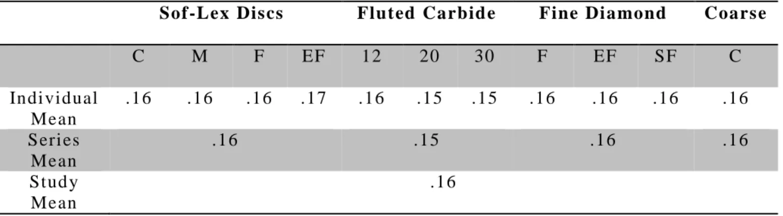
Table 7. Mean pressure (in N) applied for individual instruments, series and complete study reporting 0.16 N.
Sof-Lex Dis cs Fluted Carbid e Fine Di amond Coars e
C M F EF 12 20 30 F EF SF C
Indi vidual Mean
.16 .16 .16 .17 .16 .15 .15 .16 .16 .16 .16 Seri es
Mean
.16 .15 .16 .16
Stud y Mean
.16
42
43
44
45
46
47
48
49
50
51
52
53
54
REFERENCES
1. Yu XY, Wieczkowski G, Davis EL, Joynt RB. Influence of finishing technique on microleakage. Journal of esthetic dentistry. Sep-Oct 1990;2(5):142-144.
2. Yap AU, Ang HQ, Chong KC. Influence of finishing time on marginal sealing ability of new generation composite bonding systems. Journal of oral rehabilitation. Nov 1998;25(11):871-876.
3. Brackett WW, Gilpatrick RO, Gunnin TD. Effect of finishing method on the microleakage of Class V resin composite restorations. American journal of dentistry. Aug 1997;10(4):189-191.
4. Anusavice K. Phillips' Science of Dental Materials 12 ed: Elsevier; 2013.
5. Jefferies SR. The art and science of abrasive finishing and polishing in restorative dentistry. Dental clinics of North America. Oct 1998;42(4):613-627.
6. Powers JM CR, Sakaguchi RL. Craig's Restorative Dental Materials. . 2nd ed2006. 7. Ferracane JL. Materials in Dentistry: Principles and Applications. 2 ed2001.
8. Hoelscher DC, Neme AM, Pink FE, Hughes PJ. The effect of three finishing systems on four esthetic restorative materials. Operative dentistry. Jan-Feb 1998;23(1):36-42.
9. Ferracane JL. Resin composite--state of the art. Dental materials : official publication of the Academy of Dental Materials. Jan 2011;27(1):29-38.
10. Heymann HOS, E.J. Ritter, A.V. Sturdevant's Art and Science of Operative Dentistry. 6th ed: Elsevier; 2013.
11. Lutz F, Setcos JC, Phillips RW. New finishing instruments for composite resins. J Am Dent Assoc. Oct 1983;107(4):575-580.
12. Maresca C, Pimenta LAF, Heymann HO, Ziemiecki TL, Ritter AV. Effect of Finishing Instrumentation on the Marginal Integrity of Resin-based Composite Restorations.
Journal of Esthetic and Restorative Dentistry. 2010;22(2):104-112.
13. Summitt JB. Fundamentals of Operative Dentistry: A Comtemporary Approach. 3 ed. Chicago: Quintessence Pub; 2006.
14. Swift EJ, Jr., Perdigao J, Heymann HO. Bonding to enamel and dentin: a brief history and state of the art, 1995. Quintessence Int. Feb 1995;26(2):95-110.